A method for the ultrastructural preservation of tiny percutaneous needle biopsy material from skeletal muscle
- Authors:
- Tiziana Pietrangelo
- Stefano Perni
- Guglielmo Di Tano
- Giorgio Fanò-Illic
- Clara Franzini-Armstrong
View Affiliations
Affiliations: Department of Neuroscience and Imaging, Section of Physiology and Physiopathology, ‘G. d’Annunzio’ University of Chieti-Pescara and Clinical Research Center (CRC) of G. d'Annunzio Foundation, Chieti I-66100, Italy, Department of Cell and Developmental Biology, University of Pennsylvania School of Medicine, Philadelphia, PA 19104, USA
- Published online on: July 23, 2013 https://doi.org/10.3892/ijmm.2013.1454
-
Pages:
965-970
Metrics: Total
Views: 0 (Spandidos Publications: | PMC Statistics: )
Metrics: Total PDF Downloads: 0 (Spandidos Publications: | PMC Statistics: )
This article is mentioned in:
Abstract
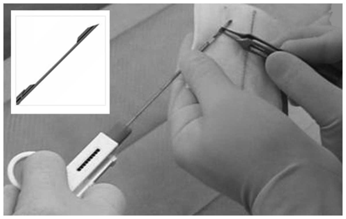
Skeletal muscle biopsies require transecting the muscle fibers resulting, in structural damage near the cut ends. Classically, the optimal ultrastructural preservation has been obtained by the use of relatively large biopsies in which the tissue fibers are restrained by ligating to a suitable retaining support prior to excision, and by examining regions at some distance from the cut ends. However, these methods require invasive surgical procedures. In the present study, we present and substantiate an alternative approach that allows for the excellent ultrastructural preservation of needle biopsy samples, even the very small samples obtained through tiny percutaneous needle biopsy (TPNB). TPNB represents an advantage, relative to standard muscle biopsy techniques and to other needle biopsies currently in use, as in addition to not requiring a skin incision, it leaves no scars in the muscle and requires an extremely brief recovery period. It is most appropriate for obtaining repeated samples in horizontal studies, e.g., in order to follow changes with athletic training and/or aging in a single individual and for studies of sarcopenic muscles in elderly patients. Due to the small size of the sample, TPNB may present limited usefulness for classical pathology diagnostics. However, it offers the major advantage of allowing multiple samples within a single session and this may be useful under specific circumstances.
View References
1
|
Pietrangelo T, D’Amelio L, Doria C,
Mancinelli R, Fulle S and Fano G: Tiny percutaneous needle biopsy:
an efficient method for studying cellular and molecular aspects of
skeletal muscle in humans. Int J Mol Med. 27:361–367. 2011.
View Article : Google Scholar : PubMed/NCBI
|
2
|
Bergstrom J: Muscle electrolytes in man.
Scand J Clin Lab Invest. 14:5111962.
|
3
|
Bergstrom J: Percutaneous needle biopsy of
skeletal muscle in physiological and clinical research. Scand J
Clin Lab Invest. 35:609–616. 1975. View Article : Google Scholar : PubMed/NCBI
|
4
|
Mancinelli R, Pietrangelo T, La Rovere R,
et al: Cellular and molecular responses of human skeletal muscle
exposed to hypoxic environment. J Biol Regul Homeost Agents.
25:635–645. 2011.PubMed/NCBI
|
5
|
Pietrangelo T, Mancinelli R, Toniolo L, et
al: Effects of local vibrations on skeletal muscle trophism in
elderly people: mechanical, cellular, and molecular events. Int J
Mol Med. 24:503–512. 2009. View Article : Google Scholar : PubMed/NCBI
|
6
|
Pietrangelo T, Mancinelli R, Toniolo L, et
al: Transcription profile analysis of vastus lateralis muscle from
patients with chronic fatigue syndrome. Int J Immunopathol
Pharmacol. 22:795–807. 2009.PubMed/NCBI
|
7
|
Engel AG: The muscle biopsy. Myology.
Engel AG and Franzini-Armstrong C: 1. 2nd edition. McGraw-Hill; New
York, NY: pp. 822–831. 1994
|
8
|
Hodgkin AL and Horowicz P: The influence
of potassium and chloride ions on the membrane potential of single
muscle fibres. J Physiol. 148:127–160. 1959. View Article : Google Scholar : PubMed/NCBI
|
9
|
Boncompagni S, Rossi AE, Micaroni M, et
al: Mitochondria are linked to calcium stores in striated muscle by
developmentally regulated tethering structures. Mol Biol Cell.
20:1058–1067. 2009. View Article : Google Scholar : PubMed/NCBI
|
10
|
Boncompagni S, Loy RE, Dirksen RT and
Franzini-Armstrong C: The I4895T mutation in the type 1 ryanodine
receptor induces fiber-type specific alterations in skeletal muscle
that mimic premature aging. Aging Cell. 9:958–970. 2010. View Article : Google Scholar : PubMed/NCBI
|
11
|
Kushmerick MJ and Podolsky RJ: Ionic
mobility in muscle cells. Science. 166:1297–1298. 1969. View Article : Google Scholar : PubMed/NCBI
|
12
|
Fulle S, Protasi F, Di Tano G, et al: The
contribution of reactive oxygen species to sarcopenia and muscle
ageing. Exp Gerontol. 39:17–24. 2004. View Article : Google Scholar : PubMed/NCBI
|