Isolation, culture and phenotypic characterization of human sweat gland epithelial cells
- Authors:
- Yunhe Gao
- Meiying Li
- Xueyan Zhang
- Tingting Bai
- Guanfan Chi
- Jin Yu Liu
- Yulin Li
View Affiliations
Affiliations: The Key Laboratory of Pathobiology, Ministry of Education, College of Basic Medical Sciences, Jilin University, Changchun, Jilin 130021, P.R. China
- Published online on: July 14, 2014 https://doi.org/10.3892/ijmm.2014.1851
-
Pages:
997-1003
-
Copyright: © Gao
et al. This is an open access article distributed under the
terms of Creative
Commons Attribution License [CC BY_NC 3.0].
Metrics: Total
Views: 0 (Spandidos Publications: | PMC Statistics: )
Metrics: Total PDF Downloads: 0 (Spandidos Publications: | PMC Statistics: )
This article is mentioned in:
Abstract
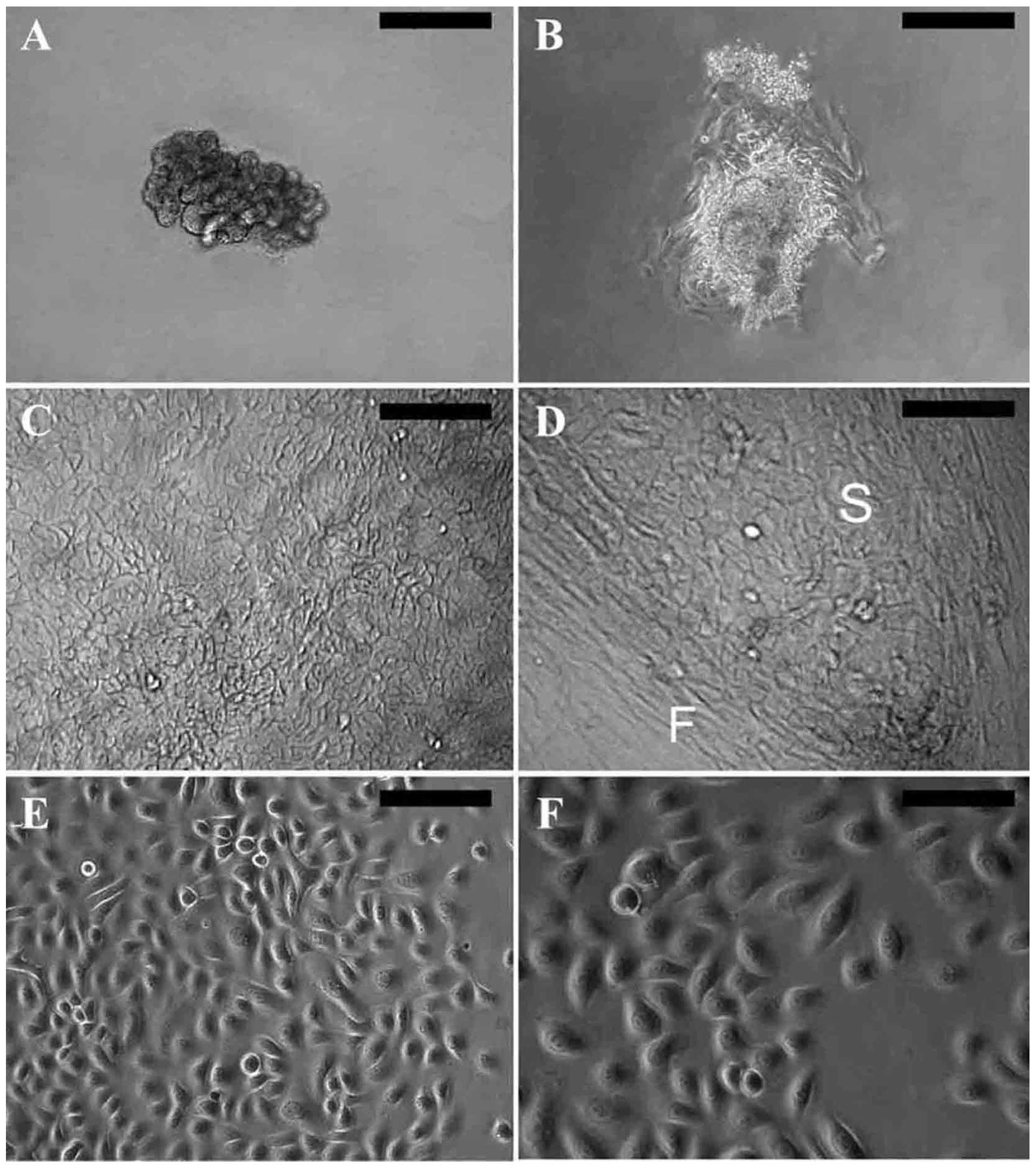
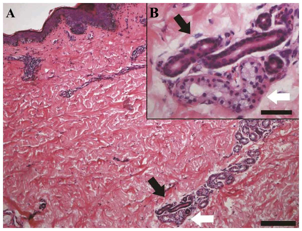
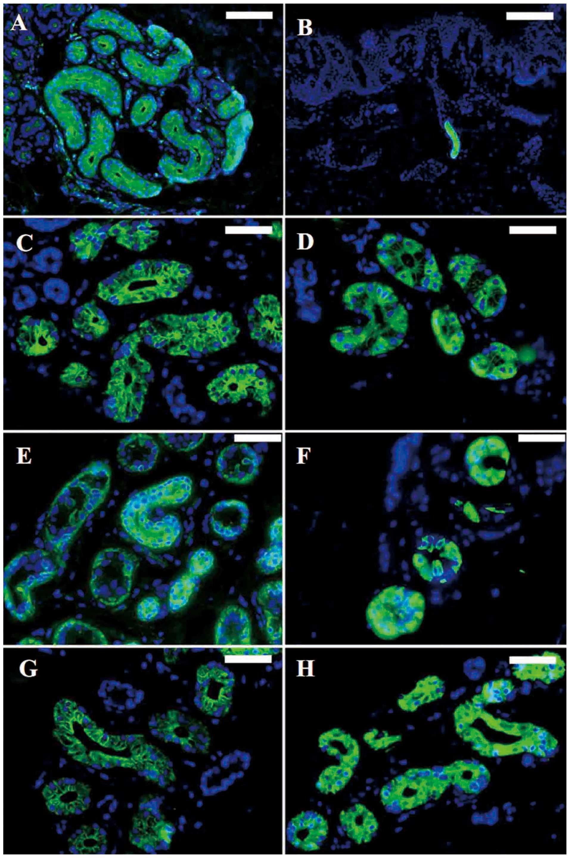
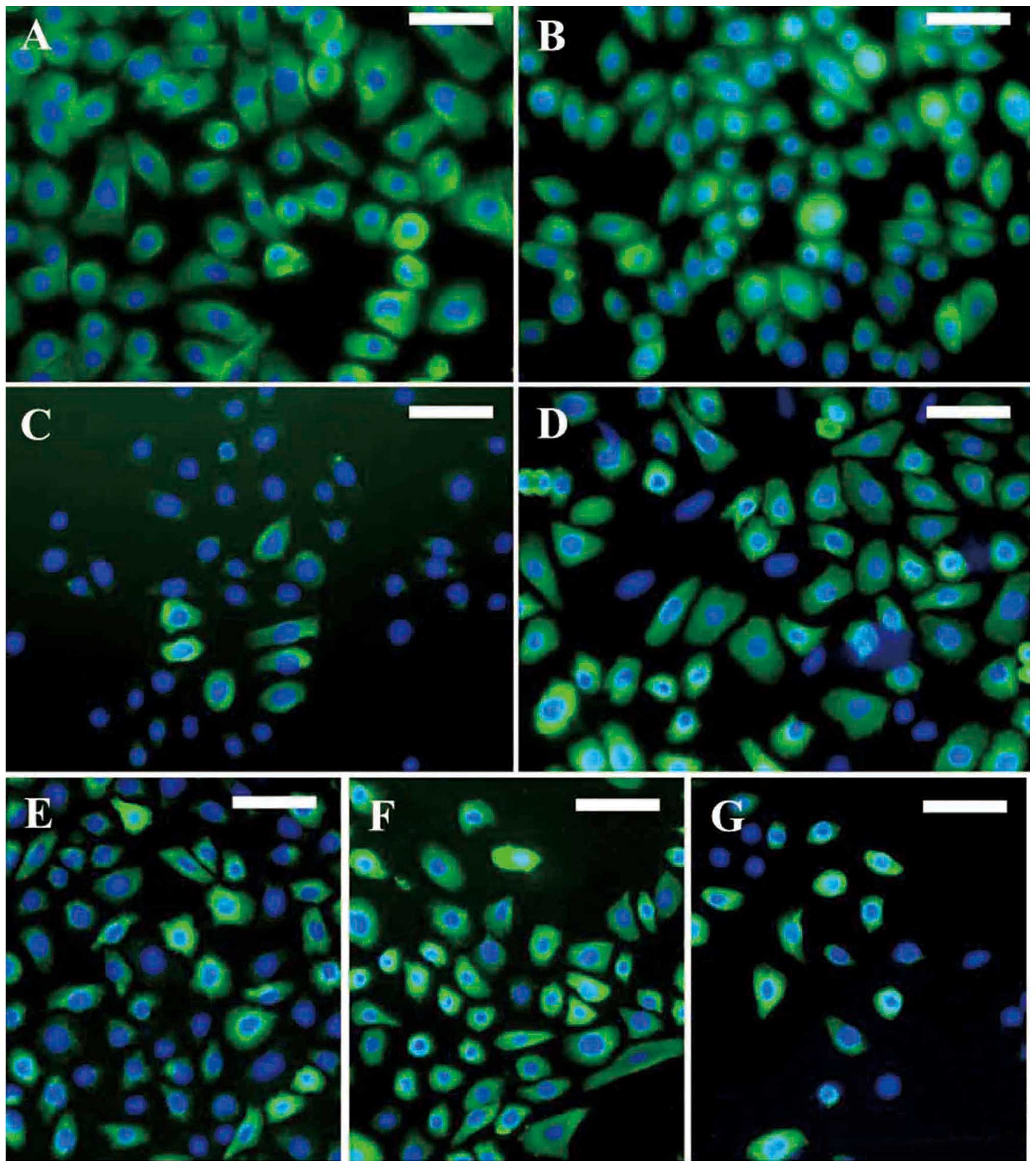
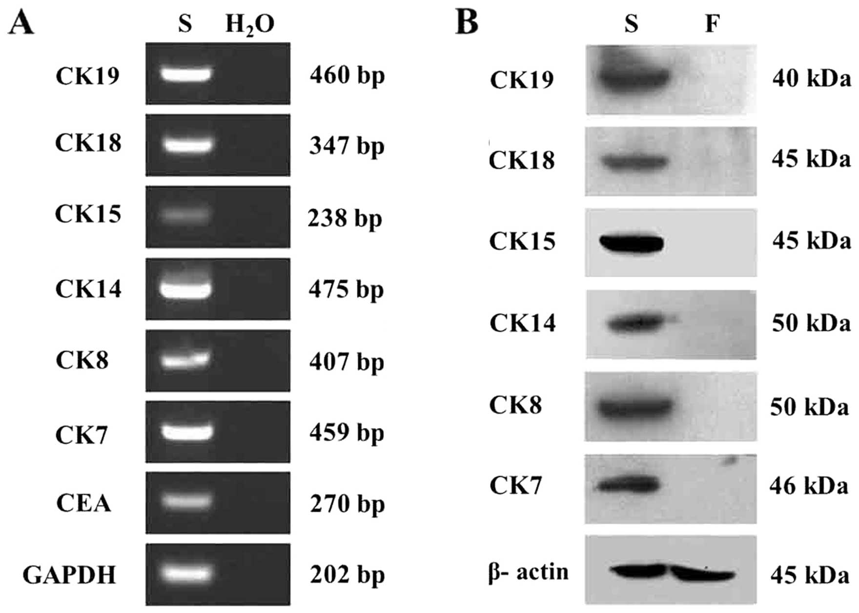
Sweat gland epithelial cells (SGECs) have been identified as essential for the regeneration of sweat glands and for the construction of skin substitutes containing skin appendages. Consequently, the isolation, culture and phenotypic characterization of SGECs are of paramount importance. In the present study study, human sweat glands were isolated by pipetting under a phase contrast microscope following digestion with collagenase type Ⅰ. Subsequently, a microscopic organ culture technique was used for the primary culture of human SGECs, and the culture conditions were modified in order to achieve optimal cell growth status. Primary SGECs were identified based on their expression of markers specific for sweat glands, including carcinoembryonic antigen (CEA), CK7, CK8, CK14, CK15, CK18 and CK19. We explored the possible presence of stem cells in human sweat glands by detecting their expression of leucine‑rich repeat-containing G protein‑coupled receptor 5 (LGR5). Primary SGECs achieved a good growth state when cultured under serum‑free conditions. After one passage, the cells cultured in keratinocyte serum-free medium with 1% fetal bovine serum (FBS) still showed a prominent proliferative activity. Phenotypic analysis by immunofluorescence microscopy, reverse transcription-polymerase chain reaction (RT‑PCR), and western blot analysis demonstrated the expression of sweat gland‑specific markers, including CEA, CK7, CK8, CK14, CK15, CK18 and CK19. In addition, RT‑PCR and immunochemistry detected the expression of LGR5. In comparison with traditional serum‑containing conditions, serum‑free culture provides the preferred culture conditions for human SGECs. LGR5 is a novel marker that identifies human sweat gland‑derived stem cells.
View Figures |
Figure 1
|
|
Figure 2
|
|
Figure 3
|
|
Figure 4
|
|
Figure 5
|
|
Figure 6
|
View References
1
|
Bar-Meir E, Mendes D and Winkler E: Skin
substitutes. Isr Med Assoc J. 8:188–191. 2006.
|
2
|
Norbury WB, Jeschke MG and Herndon DN:
Tissue engineered fetal skin constructs for pediatric burns. Crit
Care. 9:533–534. 2005. View
Article : Google Scholar : PubMed/NCBI
|
3
|
Shibasaki M and Crandall CG: Mechanisms
and controllers of eccrine sweating in humans. Front Biosci (Schol
Ed). 2:685–696. 2010. View
Article : Google Scholar : PubMed/NCBI
|
4
|
Lei X, Liu B, Wu J, Lu Y and Yang Y:
Matrigel-induced tubular morphogenesis of human eccrine sweat gland
epithelial cells. Anat Rec (Hoboken). 294:1525–1531. 2011.
View Article : Google Scholar : PubMed/NCBI
|
5
|
Sheng Z, Fu X, Cai S, Lei Y, Sun T, Bai X
and Chen M: Regeneration of functional sweat gland-like structures
by transplanted differentiated bone marrow mesenchymal stem cells.
Wound Repair Regen. 17:427–435. 2009. View Article : Google Scholar : PubMed/NCBI
|
6
|
Huang S, Xu Y, Wu C, Sha D and Fu X: In
vitro constitution and in vivo implantation of engineered skin
constructs with sweat glands. Biomaterials. 31:5520–5525. 2010.
View Article : Google Scholar : PubMed/NCBI
|
7
|
Biedermann T, Pontiggia L,
Böttcher-Haberzeth S, et al: Human eccrine sweat gland cells can
reconstitute a stratified epidermis. J Invest Dermatol.
130:1996–2009. 2010. View Article : Google Scholar : PubMed/NCBI
|
8
|
Buchanan JA, Yeger H, Tabcharani JA, et
al: Transformed sweat gland and nasal epithelial cell lines from
control and cystic fibrosis individuals. J Cell Sci. 95:109–123.
1990.PubMed/NCBI
|
9
|
Xia L: Biological characters of human
eccrine sweat gland epithelial cells and three-dimensional
reconstruction of eccrine sweat glands in vitro. Third Military
Medical University; Chong Qing, China: 2006
|
10
|
Li J, Fu X and Sheng Z: Study on the
relationship between epidermal stem cells and the developing
process of sweat gland in human fetal skin. Zhonghua Shao Shang Za
Zhi. 18:369–371. 2002.(In Chinese).
|
11
|
Li HH, Zhou G, Fu XB and Zhang L: Antigen
expression of human eccrine sweat glands. J Cutan Pathol.
36:318–324. 2009. View Article : Google Scholar : PubMed/NCBI
|
12
|
Hammarström S: The carcinoembryonic
antigen (CEA) family: structures, suggested functions and
expression in normal and malignant tissues. Semin Cancer Biol.
9:67–81. 1999.PubMed/NCBI
|
13
|
Ito M, Liu Y, Yang Z, Nguyen J, Liang F,
Morris RJ and Cotsarelis G: Stem cells in the hair follicle bulge
contribute to wound repair but not to homeostasis of the epidermis.
Nat Med. 11:1351–1354. 2005. View
Article : Google Scholar : PubMed/NCBI
|
14
|
Langbein L, Rogers MA, Praetzel S, Cribier
B, Peltre B, Gassler N and Schweizer J: Characterization of a novel
human type II epithelial keratin K1b, specifically expressed in
eccrine sweat glands. J Invest Dermatol. 125:428–444. 2005.
View Article : Google Scholar : PubMed/NCBI
|
15
|
Nakamura M and Tokura Y: The Localization
of label-retaining cells in eccrine glands. J Invest Dermatol.
129:2077–2078. 2009. View Article : Google Scholar : PubMed/NCBI
|
16
|
Ohyama M, Terunuma A, Tock CL, et al:
Characterization and isolation of stem cell-enriched huma hair
follicle bulge cells. J Clin Invest. 116:249–260. 2006. View Article : Google Scholar : PubMed/NCBI
|
17
|
Barker N, van Es JH, Kuipers J, et al:
Identification of stem cells in small intestine and colon by marker
gene Lgr5. Nature. 449:1003–1007. 2007. View Article : Google Scholar : PubMed/NCBI
|
18
|
Haegebarth A and Clevers H: Wnt signaling,
lgr5, and stem cells in the intestine and skin. Am J Pathol.
174:715–721. 2009. View Article : Google Scholar : PubMed/NCBI
|