Introduction
Uveal melanoma (UM) is a malignant tumor of the eye
involving the iris, choroid or ciliary body (collectively referred
to as the uvea). It is the most common intraocular tumor, with an
incidence of 5–6 cases per 1,000,000 individuals (1). There is a reported incidence of 4–6
cases per 1,000,000 individuals annually in the US and Europe
(2). UM spreads through the blood
and exhibits a high degree of malignancy.
Angiogenesis is a prognostic factor that has been
extensively investigated in patients with UM (3). It involves the formation of new blood
vessels from the endothelium of the existing vasculature. When a
new tumor reaches a size of 1–2 mm, its further growth requires the
formation of new blood vessels, which may lead to the development
of metastases (4). As one of the
most important biological markers, vascular endothelial growth
factor (VEGF) is an endothelial cell mitogen and permeability
factor that is potently angiogenic in vivo (5). VEGF induces the proliferation,
differentiation and migration of vascular endothelial cells. VEGF
is also required for haematopoiesis in malignant tumors, which
favors primary tumor growth and metastasis (6).
Targeting constitutive VEGF and/or its receptors has
been an attractive approach for cancer therapy. However, the most
widely investigated clinicopathological characteristics associated
with VEGF refer to variables including tumor size, largest tumor
diameter (LTD), depth of invasion, lymph node status and vascular
invasion. Although implicated in the pathogenesis of UM, the
results on the correlation between VEGF and these factors have been
conflicting and inconclusive (7–10).
It has not been elucidated whether the differences
in these studies were due to the limited size of the samples or
genuine heterogeneity. Therefore, in order to gain a full insight
into the clinicopathological characteristics of VEGF expression in
patients with UM, we enrolled data from studies of medical centers.
The clinicopathological significance of our present analysis may
enable a better understanding of the natural history of UM. In
addition, the use of VEGF may be converted from candidate to the
routine clinical setting as a predictor of the outcome of
individual patients.
Materials and methods
Literature search
A search was conducted through PubMed, Embase,
Cochrane Library, CNKI, VIP and Wanfang electronic databases,
without language restrictions. The search was based on the terms
‘melanoma’, ‘vascular endothelial growth factor’ or ‘VEGF’,
‘melanoma’ and ‘neovascularization’. The terms were also modified
according to the different databases. The last query was updated on
January 1, 2013. The references of the retrieved articles were
cross-searched to identify any relevant studies that were
overlooked during the electronic database search.
Inclusion criteria
The inclusion criteria for the primary studies were
as follows: i) the articles included definitively diagnosed UM and
normal eye tissue in humans; ii) all the eye samples with melanoma
were obtained by surgery and immunohistochemical analysis was used
to assess the expression of VEGF in the tumor samples; iii) the UM
patients had not received immune therapy, radiotherapy or
chemotherapy; iv) when multiple studies were published by the same
authors or institutions, the most recent or informative was
selected; and v) studies lacking clinicopathological data for
meta-analysis, review articles without original data and
single-case reports were excluded.
Methodological assessment
Our initial selection for all candidate studies was
based on the careful screening of their abstracts by two
independent reviewers (Meng Yang and Xiaocong Kuang), using a
standardised data collection form, including the following items:
name of first author, year of publication, ethnicity, patient
gender, mean or median age, cell type, LTD, tumor size, scleral
invasion, VEGF assessment method, cut-off value of VEGF positivity
(%), number of readers, blinded reading (the investigator assessing
the slides was blinded to the clinical information) and number of
events in each category of VEGF.
We also screened the references from the relevant
literature, including all the identified studies, without including
additional reviews and editorials. The reference lists of the
retrieved articles were also manually searched. Disagreements were
resolved by consensus between the two readers. In the instance of a
persistent disagreement, the final decision was made by a third
expert investigator (Jianmin Li).
We did not set a predefined minimum number of
patients or a minimum duration of median follow-up for a study to
be included in our meta-analysis. We did not weigh each study by a
quality score, since no such score has received general approval
for use in a meta-analysis, particularly of observational studies,
making the evaluation of its usefulness difficult. Our readers were
not blinded to the studies; however, exclusions were always decided
upon without knowledge of the global result of each study. When
duplicate studies were retrieved, the study involving the highest
number of patients from which data could be extracted (usually the
latest) was included in our meta-analysis, in order to avoid
overlapping between studies.
Statistical analysis
Three categories of stratified models were analyzed.
The first stratified multivariate model was performed in order to
confirm whether VEGF was highly expressed in UM patients compared
with the controls. The second outcome of the meta-analysis was to
assess the clinicopathological characteristics of VEGF expression,
including patient gender, age, cell type, LTD, tumor size and
scleral invasion.
According to the clinical characteristics, the
following elements were combined: high and moderate VEGF
expression; poor and no VEGF expression; mixed-cell and
epithelioid-cell type tumors; tumors >15 mm in LTD; tumors
<15 mm in LTD; small and medium tumor size; patients aged >50
years; and patients aged <50 years.
All statistical analyses were performed using RevMan
statistical analysis software system, version 5.2. A two-tailed
P<0.05 for the summary effect was considered to indicate a
statistically significant difference. The heterogeneity of all the
included studies was assessed by a statistical value I2.
When I2 was <50%, the studies with an acceptable
heterogeneity were considered and the fixed-effects model with the
Mantel-Haenszel method was used; otherwise, a random-effects model
with the DerSimonian and Laird method was adopted. The combined
odds ratio (OR) was initially estimated using forest plots
graphically. For each trial, the OR was estimated by a method
depending on the data provided in the publication. The simplest
method involved the direct retrieval of OR and its 95% confidence
interval (CI) from the original article. If not available, we
assessed the total numbers of events and the numbers of patients at
risk in each group to estimate the OR.
The assessment of publication bias for each of the
pooled study groups was performed mainly by the Egger's linear
regression test. As a supplementary approach, the Begg's rank
correlation was also applied to assess the potential publication
bias. P<0.05 indicated that there was no publication bias in the
study.
Results
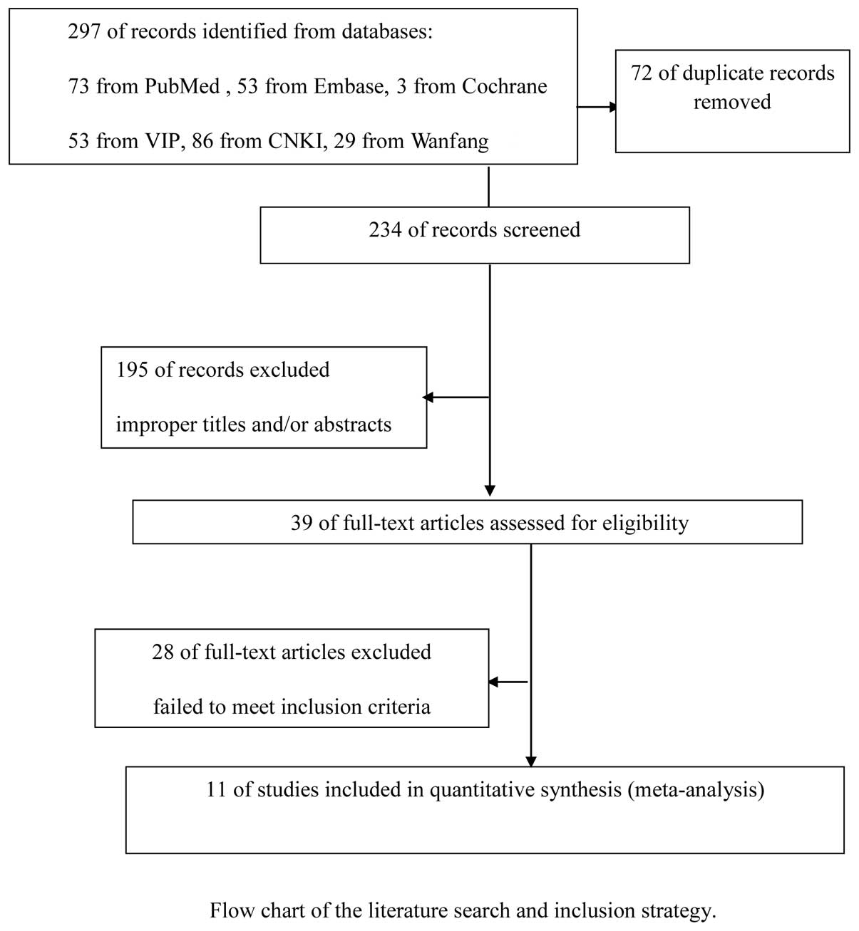
Study selection
A total of 297 references were retrieved for initial
reviewing using search strategies as previously described. A total
of 195 citations were excluded from the analysis after the first
screening based on the abstracts or titles. Following exclusion of
the articles that were out of the scope of our meta-analysis, 39
potential studies were identified for detailed evaluation. Upon
further review, a further 28 studies were eliminated due to the
following reasons: 9 studies overlapped with others, 7 studies
measured VEGF with methods other than immunohistochemistry and 12
studies lacked informative clinical data. Finally, 11 studies on
the VEGF expression were included. The selection process for the
studies included in this meta-analysis is summarized in Fig. 1. and the main characteristics of
the eligible studies are summarized in Table I.
 | Table I.Main characteristics of the 11 studies
included in the final meta-analysis. |
Table I.
Main characteristics of the 11 studies
included in the final meta-analysis.
| First author
(publication year) | Language | Population | Study from
PubMed | No. of patients
(M/F) | Median age
(years) | VEGF detection
method | Cut-off for VEGF
positivity (%) | Blinded
readingb | No. of
readersa | OR or HR
estimatec | Resultsd | Refs. |
|---|
| Sheidow et al
(2000) | English | Canada | Yes | 25/22 | 62.1 | Antibody | 5 | Yes | 2 | Data
extrapolated | Positive | 13 |
| Clarijs et al
(2001) | English | Netherlands | Yes | -/- | - | Antibody | - | - | - | Data
extrapolated | Negative | 20 |
| Ugurel et al
(2001) | English | Germany | Yes | 63/62 | - | Antibody | 363.8 pg/ml | - | - | Reported in text | Positive | 19 |
| Boyd et al
(2002) | English | Netherlands | Yes | 14/16 | 63 | Antibody | - | Yes | 1 | Data
extrapolated | Negative | 21 |
| Wang et al
(2003) | Chinese | China | No | 33/32 | 46.1 | Antibody | 5 | - | 2 | Data
extrapolated | Positive | 22 |
| Sahin et al
(2007) | English | Turkey | Yes | 10/8 | 39.5 | Antibody | 25 | Yes | 2 | Data
extrapolated | Positive | 23 |
| Zhao et al
(2009) | Chinese | China | No | 32/26 | 43 | Antibody | 5 | Yes | 2 | Data
extrapolated | Positive | 24 |
| Franco et al
(2010) | English | Italy | Yes | 23/30 | 61.8 | Antibody | - | - | 2 | Data
extrapolated | Positive | 10 |
| Xu et al
(2011) | English | China | Yes | 29/21 | 45.8 | Antibody | 5 | Yes | 2 | Data
extrapolated | Positive | 25 |
| Pang et al
(2012) | Chinese | China | No | 31/18 | 43 | Antibody | - | - | 2 | Data
extrapolated | Positive | 26 |
| Stitt et al
(1998) | English | UK | Yes | 15/10 | 59.7 | Antibody | - | - | - | Data
extrapolated | Positive | 12 |
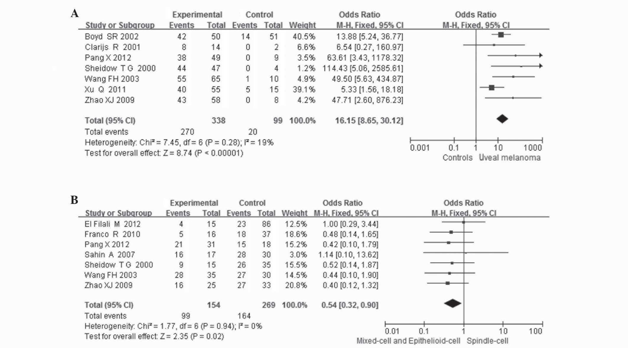
Correlation of VEGF expression between
UM and controls
The combined results from all the studies
demonstrated that the VEGF expression in patients with UM was
significantly higher compared to that in controls in 7 studies (338
patients and 99 controls; OR=16.15, 95%CI: 8.65–30.12,
P<0.00001). There was no statistical heterogeneity among the
studies [I2=19%, degree of freedom (df)=6, P=0.28;
Fig. 2A].
Correlation between VEGF expression and
clinicopathological characteristics
Gender
The VEGF expression in UM according to patient
gender was compared in 5 studies. VEGF expression was observed in
67 of the 102 male patients (66%) and in 62 of the 90 female
patients (69%). There was no statistical heterogeneity among the
studies (I2=0%, df=4, P=0.66). Thus, the fixed-effects
model was used for statistical analysis. No association was
observed between patient gender and VEGF expression (OR=0.69, 95%
CI: 0.35–1.35, P=0.27).
Age
VEGF expression in UM was investigated according to
patient age. The patients were divided into two groups aged >50
and <50 years. VEGF expression was observed in 116 of the 150
(77%) patients in the younger group and 100 of the 165 (61%)
patients in the older group. No statistical heterogeneity was
detected among the studies (I2=13%, df=6, P=0.33). Thus,
the fixed-effects model was used. VEGF expression was significantly
higher among patients aged <50 years (OR=2.08, 95% CI:
1.19–3.62, P=0.01).
Cell type
VEGF expression in UM according to cell type was
compared in 7 studies. VEGF expression was observed in 99 of the
154 (64%) mixed- and epithelioid-cell type and in 164 of the 269
(61%) spindle-cell type tumors. The VEGF expression was lower in
the spindle-cell type tumors compared to the other types (OR=0.54,
95% CI: 0.32–0.90, P=0.02). No statistical heterogeneity was
detected among the studies (I2=0%, df =6, P=0.94;
Fig. 2B).
LTD
The VEGF expression in UM according to LTD was
compared in 5 studies. VEGF expression was observed in 122 of the
182 (67%) tumors with an LTD of >15 mm and in 69 of the 86 (80%)
tumors with an LTD of <15 mm. There was statistical
heterogeneity among the studies (I2=51%, df=4, P=0.09).
Thus, the random-effects model was used for statistical analysis.
No association was observed between LTD and VEGF expression
(OR=0.43, 95% CI: 0.15–1.24, P=0.12).
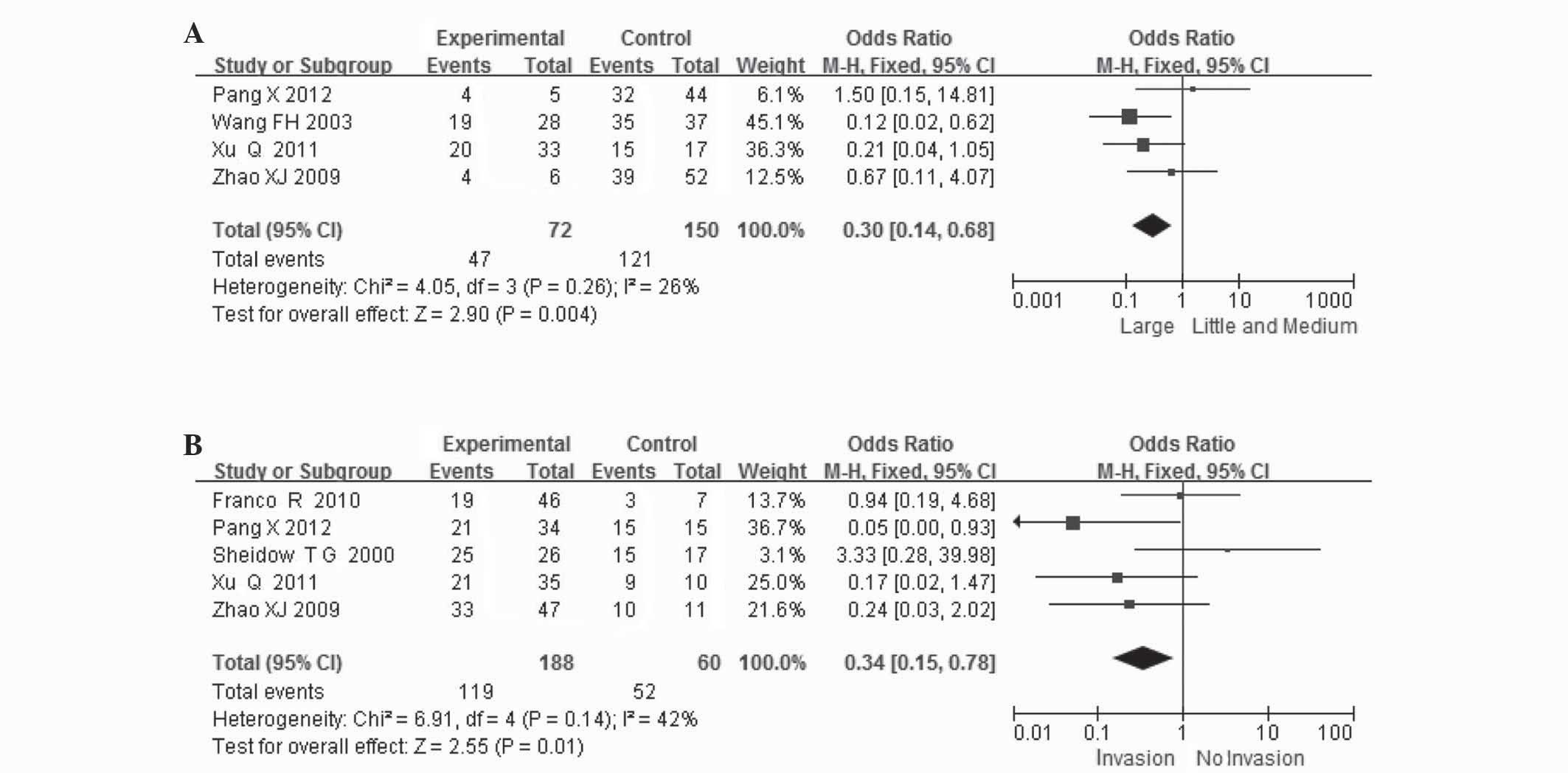
Tumor size
VEGF expression in UM according to tumor size was
compared in 4 studies. VEGF expression was observed in 47 of the 72
(65%) small- and medium-sized and in 121 of the 150 (81%)
large-sized tumors. VEGF expression was significantly higher in
large-sized tumors (OR=0.30, 95% CI: 0.14–0.68, P=0.004). There was
no statistical heterogeneity among the studies (I2=26%,
df=3, P=0.004; Fig. 3A).
Scleral invasion
VEGF expression in UM according to scleral invasion
was compared in 5 studies. VEGF expression was observed in 119 of
the 188 (63%) patients without and in 52 of the 60 (87%) patients
with scleral invasion. VEGF expression was significantly higher in
patients who exhibited scleral invasion (OR=0.34, 95% CI:
0.15–0.78, P=0.01). There was no obvious statistical heterogeneity
among the studies (I2=42%, df=4, P=0.14; Fig. 3B).
Sensitivity analysis and publication
bias
The influence analysis revealed that no individual
study significantly affected the pooled ORs and CIs. When each
study was sequentially removed and the meta-analysis was repeated
with the remaining studies, the pooled OR remained essentially the
same. However, the studies including analyses of VEGF expression
according to patient gender, age, cell type, LTD, tumor size and
scleral invasion, were <9. The Egger's linear regression and
Begg's tests were not used to investigate publication bias.
Discussion
Since the VEGF was identified, there has been an
increasing number of studies on the association between VEGF and
cancer research. It was demonstrated that VEGF expression is
strongly positive in a variety of human malignant tumors in the
mRNA and/or the protein level (11). Pigment and blood vessels are
abundant in uveal tissue. Tumor cells are mainly transferred
through the blood; therefore, angiogenesis is crucial for the
development of the tumor. However, currently available studies on
VEGF expression in UM are sparse and the results obtained are
inconsistent. Kvanta et al (5) reported that VEGF mRNA and protein
expression were strongly positive in retinoblastoma, although there
was no VEGF expression in UM. Stitt et al (12) investigated the expression of VEGF
mRNA, VEGF protein and its receptor in retinoblastoma or UM and
reported a strongly positive VEGF expression in the tumor; the VEGF
expression rate was higher compared to that in the uninvolved
normal retina, choroid and iris. Sheidow et al (13) reported that 37% (16/43) of the
patients with choroidal melanoma developed distant metastases
within the 10-year follow-up, but did not confirm the association
of VEGF expression with distant metastasis, although the expression
of VEGF in the choroidal melanoma cells was strongly positive (94%
positivity rate).
To the best of our knowledge, this is the first
comprehensive and detailed meta-analysis assessing the association
between the expression of VEGF in UM and clinicopathological
characteristics. The stratification of the baseline patient
characteristics, including gender, age, cell type, LTD, tumor size
and scleral invasion, our results demonstrated that VEGF expression
is of clinicopathological value in UM, which may increase the
predictive accuracy of prognosis in UM patients.
Our results demonstrated that high VEGF expression,
as detected by immunohistochemistry, was confirmed in patients with
UM according to evidence-based medicine. Notably, in the subgroup
meta-analysis, we also observed that VEGF overexpression was
correlated with a patient age of <50 years (P=0.008), which may
explain its prognostic effect to some extent. Similar findings were
also reported by other studies on age (14). However, further studies are
required to assess the association of VEGF expression with age. We
also observed that VEGF expression was significantly higher in
mixed-cell and epithelioid-cell tumors and lower in spindle-cell
tumors.
The association between tumor size and VEGF
expression was not clearly determined. Our meta-analysis indicated
that the VEGF expression was increased in large-sized tumors and
decreased in small- and medium-sized tumors. We also demonstrated
that VEGF expression was significantly higher in patients with
scleral invasion. A collaborative ocular melanoma study in the USA
investigated 1,091 enucleation specimens and reported a scleral
invasion rate of 55% (15). The
data mentioned previously questions the feasibility and safety of
the implementation of local tumor resection and may explain the
high recurrence rate following tumor resection in patients with UM
(16). Those findings may also
explain the increased formation of tumor microvasculature with the
increasing degree and local infiltration of UM.
High VEGF expression induces the formation of a rich
vascular network and nutritious environment, which is an active
process that requires degradation of the extracellular matrix and
increase in vascular permeability of blood and lymphatic vessels,
favoring the progression of tumor cells into the blood and
lymphatic vascular space (17).
This may offer an explanation for the observed strong statistical
association of VEGF overexpression with tumor invasion and
metastasis (18).
The present study had several limitations. Firstly,
although we did not detect significant publication bias among the
studies, apart for erythrocyte sedimentation rate (ESR), it is
uncertain whether the cases are comparably representative.
Furthermore, the studies were observational and, therefore, more
prone to biases compared with prospective randomized controlled
studies. We detected publication bias for ESR (P=0.2) and there may
be missing information which may reflect a negative or a more
conservative association of ESR with DR4. More samples are required
to validate the reliability of our conclusions.
In conclusion, our meta-analysis demonstrated that
VEGF may be a marker that may enable earlier identification of
high-risk patients and guide clinical decision-making regarding
therapy and outcome. The evaluation of serum levels of VEGF
expression may therefore play an important role in selecting
melanoma patients for antiangiogenic therapy (19).
However, our conclusion should be interpreted with
caution, since this analysis would be ideally performed on series
of patient data. Further investigation into this subset of patients
from other studies should assess the generalization of results
prior to the implementation of VEGF in the routine clinical
management of UM patients. Future prospective studies investigating
the association of VEGF expression with survival or response to
antiangiogenic therapy are required. The assessment of these
angiogenic markers may be better standardised in future
studies.
Acknowledgements
This study was supported by the Funds
for Guangxi Zhuang Autonomous Region Science and Technology Hall
(grant no. 1140003B-86).
References
|
1.
|
Dobner BC, Riechardt AI, Joussen AM,
Englert S and Bechrakis NE: Expression of haematogenous and
lymphogenous chemokine receptors and their ligands on uveal
melanoma in association with liver metastasis. Acta Ophthalmol.
90:e638–e644. 2012. View Article : Google Scholar : PubMed/NCBI
|
|
2.
|
Petousis V and Finger PT: Current methods
for the diagnosis and treatment of choroidal melanoma. US
Ophthalmic Rev. 5:62–69. 2012.
|
|
3.
|
Woodman SE: Metastatic uveal melanoma:
biology and emerging treatments. Cancer J. 18:148–152. 2012.
View Article : Google Scholar : PubMed/NCBI
|
|
4.
|
Barak V, Pe'er J, Kalickman I and Frenkel
S: VEGF as a biomarker for metastatic uveal melanoma in humans.
Curr Eye Res. 36:386–390. 2011. View Article : Google Scholar : PubMed/NCBI
|
|
5.
|
Kvanta A, Steen B and Seregard S:
Expression of vascular endothelial growth factor (VEGF) in
retinoblastoma but not in posterior uveal melanoma. Exp Eye Res.
63:511–518. 1996. View Article : Google Scholar : PubMed/NCBI
|
|
6.
|
Musumeci F, Radi M, Brullo C and Schenone
S: Vascular endothelial growth factor (VEGF) receptors: drugs and
new inhibitors. J Med Chem. 55:10797–10822. 2012. View Article : Google Scholar : PubMed/NCBI
|
|
7.
|
Erdei E and Torres SM: A new understanding
in the epidemiology of melanoma. Expert Rev Anticancer Ther.
10:1811–1823. 2010. View Article : Google Scholar
|
|
8.
|
El Filali M, van der Velden PA, Luyten GP
and Jager MJ: Anti-angiogenic therapy in uveal melanoma. Dev
Opthalmol. 49:117–136. 2012.PubMed/NCBI
|
|
9.
|
Spagnolo F, Caltabiano G and Queirolo P:
Uveal melanoma. Cancer Treat Rev. 38:549–553. 2012. View Article : Google Scholar
|
|
10.
|
Franco R, Botti G, Mascolo M, et al:
CXCR4-CXCL12 and VEGF correlate to uveal melanoma progression.
Front Biosci (Elite Ed). 2:13–21. 2010. View
Article : Google Scholar : PubMed/NCBI
|
|
11.
|
Berse B, Brown LF, Van De Water L, Dvorak
HF and Senger DR: Vascular permeability factor (vascular
endothelial growth factor) gene is expressed differentially in
normal tissues, macrophages, and tumors. Mol Biol Cell. 3:211–220.
1992. View Article : Google Scholar
|
|
12.
|
Stitt AW, Simpson DA, Boocock C, Gardiner
TA, Murphy GM and Archer DB: Expression of vascular endothelial
growth factor (VEGF) and its receptors is regulated in eyes with
intra-ocular tumours. J Pathol. 186:306–312. 1998. View Article : Google Scholar : PubMed/NCBI
|
|
13.
|
Sheidow TG, Hooper PL, Crukley C, Young J
and Heathcote JG: Expression of vascular endothelial growth factor
in uveal melanoma and its correlation with metastasis. Br J
Ophthalmol. 84:750–756. 2000. View Article : Google Scholar : PubMed/NCBI
|
|
14.
|
Perrone G, Santini D, Vincenzi B, Zagami
M, La Cesa A, Bianchi A, Altomare V, Primavera A, Battista C,
Vetrani A, Tonini G and Rabitti C: COX-2 expression in DCIS:
correlation with VEGF, HER-2/neu, prognostic molecular markers and
clinicopathological features. Histopathology. 46:561–568. 2005.
View Article : Google Scholar : PubMed/NCBI
|
|
15.
|
Schori H, Kipnis J, Yoles E, WoldeMussie
E, Ruiz G, Wheeler LA and Schwartz M: Vaccination for protection of
retinal ganglion cells against death from glutamate cytotoxicity
and ocular hypertension: implications for glaucoma. Proc Natl Acad
Sci USA. 98:3398–3403. 2001. View Article : Google Scholar : PubMed/NCBI
|
|
16.
|
Damato BE, Paul J and Foulds WS:
Predictive factors of visual outcome after local resection of
choroidal melanoma. Br J Ophthalmol. 77:616–623. 1993. View Article : Google Scholar : PubMed/NCBI
|
|
17.
|
Schwarz MA and Cleaver OB: Development of
the pulmonary endothelium in development of the pulmonary
circulation: vasculogenesis and angiogenesis. The Pulmonary
Endothelium: Function in Health and Disease. Voelkel NF and Rounds
S: 1st edition. Wiley-Blackwell; Chichester: pp. 3–24. 2009
|
|
18.
|
Ding GX, Feng CC, Song NH, et al:
Paraneoplastic symptoms: Cachexia, polycythemia, and hypercalcemia
are, respectively, related to vascular endothelial growth factor
(VEGF) expression in renal clear cell carcinoma. Urol Oncol. Apr
23–2012.(Epub ahead of print).
|
|
19.
|
Ugurel S, Rappl G, Tilgen W and Reinhold
U: Increased serum concentration of angiogenic factors in malignant
melanoma patients correlates with tumor progression and survival. J
Clin Oncol. 19:577–583. 2001.PubMed/NCBI
|
|
20.
|
Clarijs R, Schalkwijk L, Ruiter DJ and de
Waal RM: Lack of lymphangiogenesis despite coexpression of VEGF-C
and its receptor Flt-4 in uveal melanoma. Invest Ophthalmol Vis
Sci. 42:1422–1428. 2001.PubMed/NCBI
|
|
21.
|
Boyd SR, Tan DS, de Souza L, et al: Uveal
melanomas express vascular endothelial growth factor and basic
fibroblast growth factor and support endothelial cell growth. Br J
Ophthalmol. 86:440–447. 2002. View Article : Google Scholar : PubMed/NCBI
|
|
22.
|
Wang FH, Li B, Sun XL, et al: Correlation
of tumor angiogenesis with clinicopathologic prognostic parameters
in choroidal melanoma. Chin J Ophthalmol. 39:68–72. 2003.(In
Chinese).
|
|
23.
|
Sahin A, Kiratli H, Tezel GG, Soylemezoglu
F and Bilgic S: Expression of vascular endothelial growth factor A,
matrix metalloproteinase 9 and extravascular matrix patterns in
iris and ciliary body melanomas. Ophthalmic Res. 39:40–44. 2007.
View Article : Google Scholar : PubMed/NCBI
|
|
24.
|
Zhao XJ, Li B, Li LQ, Gao F and Sun XL:
Relationship of survivin and VEGF with angiogenesis in choroidal
melanoma. Chin Ophthal Res. 27:770–773. 2009.(In Chinese).
|
|
25.
|
Xu Q, Zhao GQ, Zhao J, et al: Expression
and significance of factors related to angiogenesis in choroidal
melanoma. Int J Ophthalmol. 4:49–54. 2011.PubMed/NCBI
|
|
26.
|
Pang X, Yunhui Q and Yuewe M: Expressions
of A disintegrin and metalloproteinase 9 and vascular endothelial
growth factor in ocular malignant melanomas. Rec Adv Ophthalmol.
32:620–623. 2012.(In Chinese).
|