Introduction
Primary intraosseous squamous cell carcinoma
(PIOSCC) is a central jaw carcinoma derived from odontogenic
epithelial remnants. PIOSCC is defined as a SCC arising within the
jaw, and it has no initial connection with the oral mucosa. This
tumor was first described by Loos (1)
in 1913 as a central epidermoid carcinoma of the jaw. Willis
(2) renamed the tumor as
intraalveolar epidermoid carcinoma in 1948. In 1972, Pindborg et
al (3) suggested the term primary
intraosseous carcinoma (PIOC) and classified the lesion as an
odontogenic carcinoma. Subsequently, the classification of
odontogenic carcinomas was modified in several studies (4–6). In 2005,
Eversole et al (7) introduced
the term ‘primary intraosseous SCC’ (PIOSCC) and further
categorized this entity into 3 types: Type 1 for solid tumors, type
2 for carcinomas arising from odontogenic cysts and type 3 for
carcinomas associated with odontogenic tumors.
In PIOSCC type 2, an SCC results from malignant
transformation of an odontogenic cyst. To establish the presence of
such malignant transformation, certain diagnostic criteria must be
fulfilled (8). The estimated
incidence of PIOSCC type 2 has been shown to range from 0.1 to 1.8%
of all oral cancers (9–12). Bodner et al analyzed 116
reported cases of PIOSCC arising in an odontogenic cyst and found
that the tumors predominantly affected the mandible, whereas the
maxilla was affected in 21% of the cases (13).
We herein report a case of PIOSCC derived from a
maxillary odontogenic cyst. We have also reviewed the literature
for cases of PIOSCC arising from maxillary cysts, including
keratocystic odontogenic tumors, with respect to the diagnosis,
prognosis and treatment of these tumors. Written informed consent
was obtained from the patient for the publication of his case
details.
Case report
In August, 2010, a 37-year-old Japanese man visited
the Department of Oral Surgery in another hospital, with a 4-month
history of a painful swelling in the left maxillary gingiva.
Following clinical examination, the patient was diagnosed with
apical periodontitis and antibiotics were prescribed; however, no
improvement was noted. In October, 2010, the patient presented with
a fistula in the left maxillary gingiva. A computed tomography (CT)
scan revealed an area of bone resorption in the lateral nasal wall.
The histopathological examination of the biopsy specimen indicated
suspected SCC. The patient was then referred to the Dental Hospital
of Tokyo Medical and Dental University (Tokyo, Japan) for extensive
examination and treatment.
Extraoral examination revealed a swelling without
paresthesia in the left buccal region. Intraoral examination
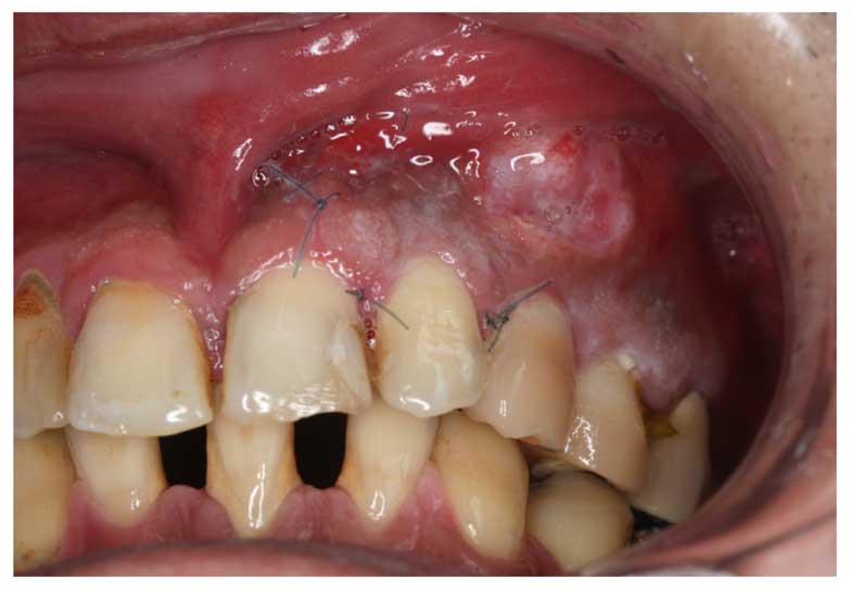
revealed a painful ulcer in the maxillary buccal gingiva of the
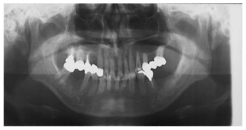
left canine, caused by the previous biopsy (Fig. 1). A panoramic radiograph revealed no
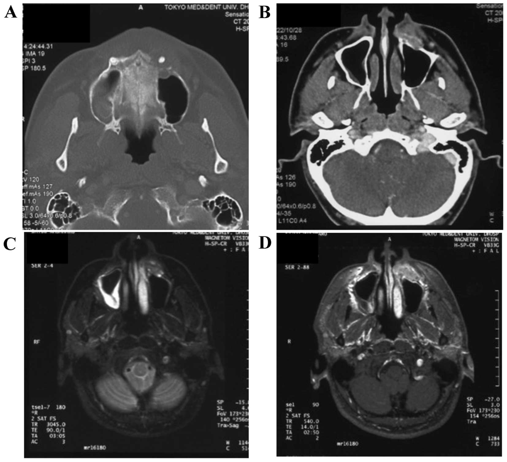
obvious cyst-like radiolucent lesion (Fig. 2); however, a contrast-enhanced CT
revealed a soft tissue mass (30×16×22 mm3) with
irregular borders and heterogeneous density in the left anterior
maxilla, which had invaded and damaged the left lateral nasal wall
and the left bottom edge of the left maxillary sinus (Fig. 3A and B). Contrast-enhanced magnetic
resonance imaging (MRI) revealed a high-density lesion on
T2-weighted images, indicating expansion of the tumor to the base
of the left nasal ala (Fig. 3C and
D). The previous biopsy specimen from the referring hospital
was reevaluated and diagnosed as suspected odontogenic
carcinoma.
Based on this diagnosis of odontogenic carcinoma,
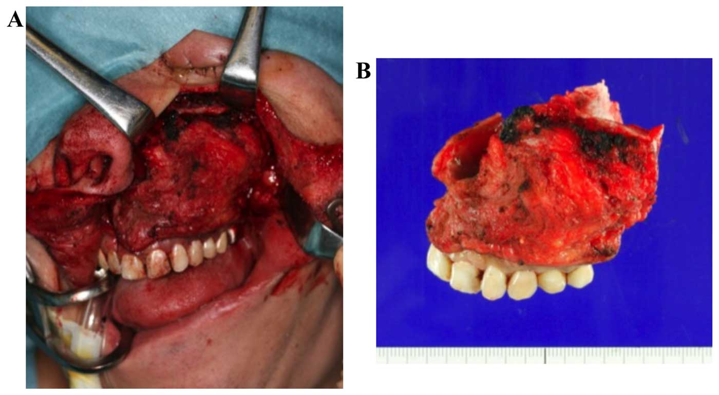
surgical treatment was scheduled. In November, 2010, the patient
underwent partial maxillectomy through a Weber-Ferguson incision.
Due to suspected tumor infiltration of the soft tissues at the base
of the left nasal ala, part of the skin was simultaneously
resected. Following resection, the raw surface of the buccal soft
tissues was covered using a full-thickness skin graft from the
groin. An intraoral incision was made from the right maxillary
central incisor area to the left maxillary first molar area. The
upward incision line was set under 5 mm from the infraorbital
margin (Fig. 4). The postoperative
course was uneventful, and a maxillary prosthesis was placed. A
follow-up fluorodeoxyglucose-positron emission tomography (FDG-PET)
scan revealed no recurrence, metastasis, or carcinoma in other
regions. The patient has been followed up for 53 months, with no
recurrence of the disease.
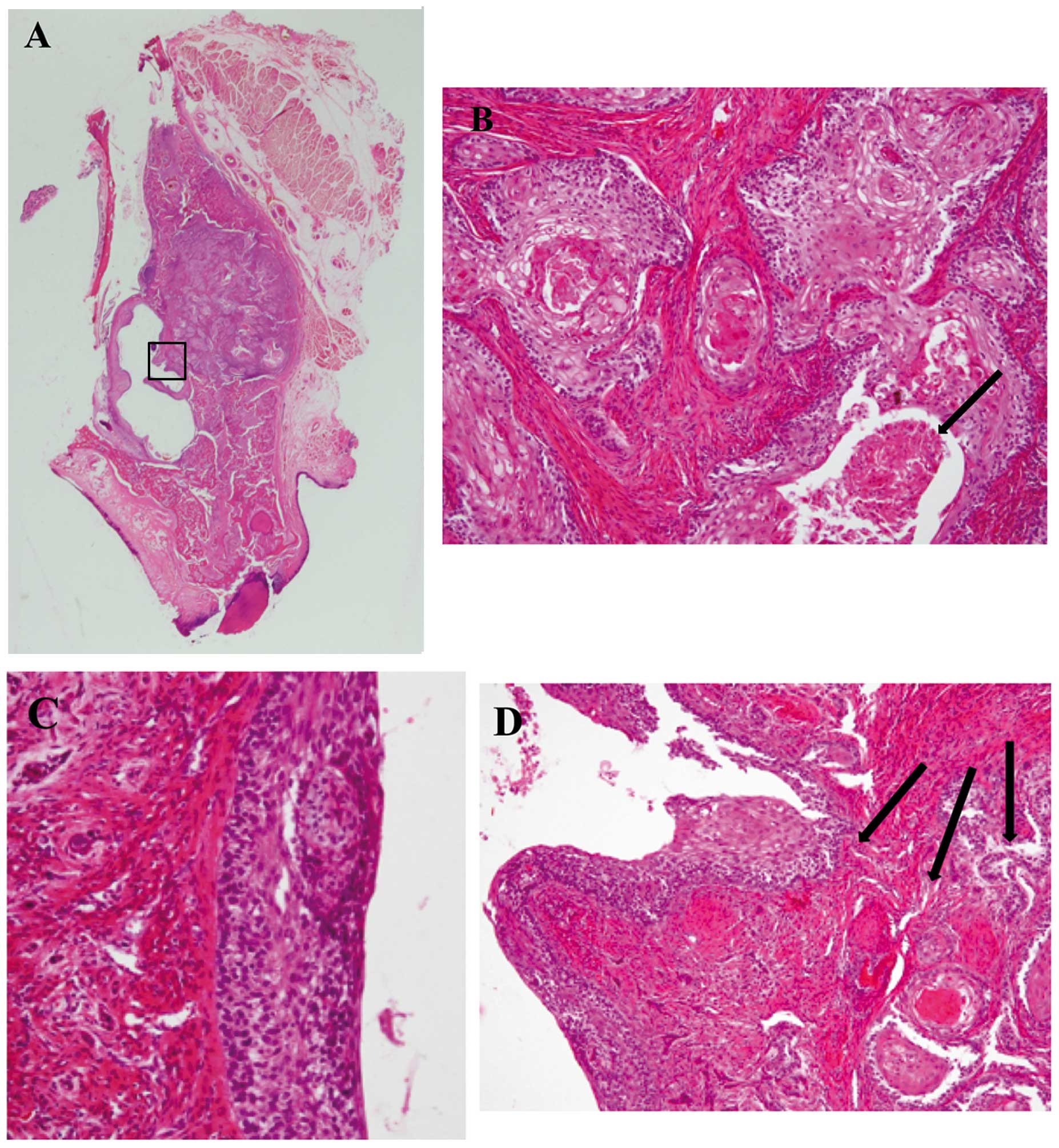
The histological examination revealed a centrally
located tumor within the maxillary bone, without any connection to
the oral or maxillary sinus mucosa, consisting of solid and cystic
components (Fig. 5A). The solid
components exhibited islands of neoplastic squamous epithelium with
keratinization and central necrosis of the tumor nests (Fig. 5B). By contrast, in the cystic
components, the lining epithelium consisted of parakeratinized
squamous epithelium. However, palisaded basal cells, typically seen
in keratocystic odontogenic tumors, were not identified in the
lining epithelium. Parts of the lining epithelium exhibited
moderate to severe dysplastic changes (Fig. 5C), with focal areas of transition to
invasive keratinized SCC nests (Fig.
5D). Based on the clinical data and histopathological findings,
this lesion was diagnosed as a PIOSCC derived from an odontogenic
cyst.
Discussion
PIOSCC is a rare central jaw carcinoma derived from
odontogenic epithelial remnants. According to the 2005 World Health
Organisation Classification of Tumours, the subcategories of PIOSCC
include a solid tumor that invades marrow spaces and induces
osseous resorption (type 1), an SCC arising from the lining of an
odontogenic cyst (type 2) and an SCC in association with other
benign epithelial odontogenic tumors (type 3) (7). The present case was classified as type
2, as it fulfilled the criteria detailed below.
In 1975, Gardner (8)
proposed the following criteria for the diagnosis of SCC arising in
an odontogenic cyst: i) It should be confirmed histologically that
the epithelial lining of the cyst has undergone malignant
transformation to SCC; ii) clinical examinations should reveal no
SCC of the gingiva or oral mucosa and the reported neoplasms should
be centrally located within the bone tissue of the jaws; and iii)
no primary neoplasm at distant sites should be detected.
A fourth criterion was later added by Waldron and
Mustoe: The possibility that the lesion in question represents a
metastasis from a distant primary tumor must be ruled out by
physical and radiological examinations and the subsequent clinical
course (6).
With regard to the present case, the microscopic
examination revealed the presence of a cyst consisting of both
normal stratified squamous epithelium and SCC. Histopathologically,
the resected specimen exhibited an area of transition from normal
cystic epithelium to invasive SCC, along with normal oral mucosa.
This is a noteworthy finding, as in previous studies the carcinoma
destroyed the odontogenic cyst, rendering it difficult to determine
the actual site of malignant transformation (14) Moreover, follow-up FDG-PET scans showed
no carcinoma in any other areas. Therefore, we considered the tumor
in our patient to fulfil all of Gardner's criteria; accordingly,
PIOSCC arising from an odontogenic cyst (type 2) was diagnosed.
In retrospect, it was difficult to diagnose this
lesion as a malignant tumor based on the intraoral examination and
the panoramic radiograph prior to treatment. CT and MRI
examinations showed malignant findings but did not reveal the
presence of the cyst. These factors suggest the significance of a
histopathological examination and confirmation of the diagnosis
prior to initial treatment.
Bodner et al (13) analyzed 116 reported cases of PIOSCC
arising in an odontogenic cyst and found that the majority of the
cases exhibited mandibular involvement, whereas the maxilla was
affected in 21% of the cases. For the present report, we searched
for cases of PIOSCC derived from a maxillary odontogenic cyst. A
review of 29 cases, including the present case, is presented in
Table I (9–12, 15–37). In
the 29 cases, the patient age ranged from 14 to 79 years, with a
mean age of 47.0 years. The male:female ratio was 2.5:1. The most
common clinical symptom was swelling (72.7%, 16/22) with or without
pain. The most common initial treatment approach was enucleation or
local resection (48.0%, 12/25) under the diagnosis of odontogenic
cyst, and none of these 12 patients had undergone a preliminary
biopsy. In the majority of the patients, additional operations,
such as maxillectomy, were performed. However, 8 cases (40.0%,
8/20) were histologically confirmed as malignant tumors by biopsy
prior to initial treatment. Postoperative treatment was
administered to 9 patients (radiation therapy, n=8; and
chemoradiation, n=1).
 | Table I.Review of published reports on primary
intraosseous squamous cell carcinoma arising from a maxillary
odontogenic cyst. |
Table I.
Review of published reports on primary
intraosseous squamous cell carcinoma arising from a maxillary
odontogenic cyst.
| Case | Author (Refs.) | Year | Gender | Age (years) | Symptom | Location | Diagnosis of
biopsy | Initial
treatment | Type of lining
epithelium | Follow-up
(months) |
|---|
| 1 | Axhausen (15) | 1938 | F | 14 | Unknown | A | Unknown | Unknown | Unknown | Unknown |
| 2 | Mann (16) | 1944 | M | 26 | Swelling | A | Unknown | Unknown | Dentigerous cyst | Unknown |
| 3 | Frankl(17) | 1949 | F | 38 | Pain | A | Unknown | Partial
maxillectomy | Unknown | 5 |
| 4 | Martensson(18) | 1955 | M | 49 | Painless
swelling | Unknown | Not performed | Enucleation | Unknown | 24 |
| 5 | Kodel(19) | 1961 | M | 58 | Pain | A | Unknown | Subtotal
maxillectomy | Unknown | Unknown |
| 6 | Williams(20) | 1963 | M | 59 | Pain | A | SCC | Partial
maxillectomy | Unknown | 4 |
| 7 | Lee and Loke(21) | 1967 | M | 57 | Painless
swelling | A | Not performed | Partial
maxillectomy | Primordial
cyst | 36 |
| 8 | Bannerjee(22) | 1967 | M | 37 | Swelling | A | Not performed | Enucleation | Unknown | 10 |
| 9 | Hampl (23) | 1973 | M | 38 | None | P | Not performed | Extraction and
curettage | Unknown | Unknown |
| 10 | Areen (24) | 1981 | M | 60 | Painful
swelling | A | Unknown | Local
resection | OKC | 19 |
| 11 |
Nithiananda(25) | 1983 | M | 59 | Painless
swelling | A | SCC | Maxillectomy | Odontogenic
cyst | 36 |
| 12 | Pearcey (26) | 1985 | F | 79 | Swelling | Unknown | Not performed | Enucleation | Unknown | 31 |
| 13 | Van Der Waal
(11) | 1985 | M | 45 | Painful
swelling | A | Not performed | Enucleation | Unknown | 24 |
| 14 | Van Der Waal
(11) | 1985 | M | 70 | Unknown | P | SCC |
Hemimaxillectomy | Unknown | 12 |
| 15 | Kreidler (10) | 1985 | Unknown | Unknown | Unknown | A | Unknown | Partial
maxillectomy | Unknown | Unknown |
| 16 | Otten (9) | 1985 | F | 32 | Unknown | P | Unknown | Unknown | Unknown | Unknown |
| 17 | Siar and
Ng(27) | 1987 | M | 40 | Sinus swelling | P | Not performed | Enucleation | OKC | Unknown |
| 18 | Stoelinga (12) | 1988 | F | 72 | Unknown | A | Unknown | Enucleation | OKC | 1 |
| 19 | Berenholtz
(28) | 1988 | F | 79 | Ill-fitting
dentures | A | Not performed | Enucleation | Dentigerous
cyst | 30 |
| 20 | Müller (29) | 1991 | M | 56 | Swelling | A | Not performed | Total removal | Unknown | Unknown |
| 21 | Berens (30) | 2000 | M | 40 | Unknown | A | Not performed | Enucleation | Unknown | 12 |
| 22 | Makowski (31) | 2001 | M | 34 | Painful
swelling | P | Not performed | Enucleation | OKC | 42 |
| 23 | Saito (32) | 2002 | M | 58 | Painful
swelling | P | SCC | Unknown | Unknown | Unknown |
| 24 | Chaisuparat
(33) | 2006 | F | 18 | Swelling | A | Unknown | Partial
maxillectomy | OKC | 44 |
| 25 | Mohyuddin (34) | 2011 | M | 40 | Unknown | A | SCC | Partial
maxillectomy | Unknown | 12 |
| 26 | Maria (35) | 2011 | M | 54 | Painless
swelling | P | SCC developing in
an OKC | Partial
maxillectomy | OKC | 24 |
| 27 | Uchida (36) | 2013 | M | 75 | Feeling of
incorrect placement | A | Not performed | Extirpation | Dentigerous
cyst | 18 |
| 28 | Jain (37) | 2013 | F | 38 | Swelling | A | PIOSCC arising from
a radicular | Partial
maxillectomy, ND | Radicular cyst | Unknown |
| 29 | Morita (present
case) | 2015 | M | 37 | Painful
swelling | P | Odontogenic
carcinoma | Partial
maxillectomy | Unclassified | 53 |
Neck metastases from PIOSCC derived from maxillary
odontogenic cysts are rare, with only 4 cases (19.0%, 4/21)
reported to date. Nomura et al (38) reported that the probability of lymph
node metastasis was 4.4% (5/113) in PIOSCC type 2. Thus, neck
dissection should be performed only when required.
Overall, 10 patients were followed up for >2
years and 3 patients succumbed (2 deaths were caused by cancer and
1 was due to another disease). In the 2 cancer deaths, a
preliminary biopsy had not been performed. The 2-year survival rate
was 83.3% (10/12). Chantravekin et al (39) reported that the 2-year survival rate
was 60.0%, while Bodner et al (13) reported that the 2- and 5-year survival
rates were 62 and 38%, respectively. There were significant
differences in survival rate between the present study and previous
reports. An underlying reason may be that the previous reports also
included other types of odontogenic carcinoma, since the definition
of PIOSCC has been modified several times.
Various odontogenic cysts have been reported to be
associated with PIOSCC. Certain types of odontogenic cysts, such as
residual cysts, dentigerous cysts and keratocystic odontogenic
tumors, tend to undergo malignant transformation (13,39,40). As
described in Table I, although
odontogenic keratocysts, which include orthokeratinized and
parakeratinized variants, exhibited the highest incidence among
odontogenic cysts, several reports have previously revealed the
exact types of odontogenic cysts associated with malignant tumors.
The present case exhibited parakeratosis of the lining epithelium
of the cyst, while palisaded basal cells were not identified in the
lining epithelium and the epithelial layer was too thin for
accurately diagnosing a keratocystic odontogenic tumor. Therefore,
the exact type of odontogenic cyst was difficult to diagnose in the
present case.
In conclusion, this report described a case of
PIOSCC derived from a maxillary odontogenic cyst, along with a
review of 29 cases of PIOSCC with maxillary involvement, focusing
on the clinical and histopathological findings. Due to the
limitation of the small number of cases, the treatment approach for
odontogenic carcinomas, including PIOSCC, has yet to be
standardized. Therefore, careful documentation and follow-up are
recommended for each case of type 2 PIOSCC.
References
|
1
|
Loos D: Central epidermoid carcinoma of
the jaws. Dtsch Monatsschr Zahnheilk. 31:3081913.
|
|
2
|
Willis RA: Pathology of Tumors (1st).
London: Butterworth & Co. 310–316. 1948.
|
|
3
|
Pindborg JJ, Kramer IRH and Torloni H:
Histological Typing of Odontogenic Tumors. Jaw Cysts and Allied
Lesions (1st). (Geneva). World Health Organization. 35–36.
1972.
|
|
4
|
Elzay RP: Primary intraosseous carcinoma
of the jaws. Review and update of odontogenic carcinomas. Oral Surg
Oral Med Oral Pathol. 54:299–303. 1982. View Article : Google Scholar : PubMed/NCBI
|
|
5
|
Slootweg PJ and Muller H: Malignant
ameloblastoma or ameloblastic carcinoma. Oral Surg Oral Med Oral
Pathol. 57:168–176. 1984. View Article : Google Scholar : PubMed/NCBI
|
|
6
|
Waldron CA and Mustoe TA: Primary
intraosseous carcinoma of mandible with probable origin in an
odontogenic cyst. Oral Surg Oral Med Oral Pathol. 67:716–724. 1989.
View Article : Google Scholar : PubMed/NCBI
|
|
7
|
Eversole LR, Siar CH and van der Waal I:
Primary intraosseous squamous cell carcinomas In: World Health
Organization Classification of Tumors. Pathology and Genetics Head
and Neck Tumors. Barnes L, Evson JW, Reichart P and Sidransky D:
(Lyon). IACR Press. 290–291. 2005.
|
|
8
|
Gardner AF: A survey of odontogenic cysts
and their relationship to squamous cell carcinoma. Dent J.
41:161–167. 1975.PubMed/NCBI
|
|
9
|
Otten JE, Joos U and Schilli W:
Carcinogenesis in the apex of the cyst-forming odontogenic
epithelium. Dtsch Zahnhartl Z. 40:544–547. 1985.(In German).
|
|
10
|
Kreidler J, Haas S and Kamp W:
Carcinogenesis in jaw cyst. 2 case reports. Dtsch Zahnhartl Z.
40:548–550. 1985.(In German).
|
|
11
|
van der Waal I, Rauhamaa R, van der Kwast
WA and Snow GB: Squamous cell carcinoma arising in the lining of
odontogenic cysts. Report of 5 cases. Int J Oral Surg. 14:146–152.
1985. View Article : Google Scholar : PubMed/NCBI
|
|
12
|
Stoelinga PJ and Bronkhorst FB: The
incidence, multiple presentation and recurrence of aggressive cysts
of the jaws. J Craniomaxfac Surg. 16:184–195. 1988. View Article : Google Scholar
|
|
13
|
Bodner L, Manor E, Shear M and van der
Waal I: Primary intraosseous squamous cell carcinoma arising in an
odontogenic cyst: A clinicopathologic analysis of 116 reported
cases. J Oral Pathol Med. 40:733–738. 2011. View Article : Google Scholar : PubMed/NCBI
|
|
14
|
Gardner AF: The odontogenic cyst as a
potential carcinoma: A clinicopathological appraisal. J Am Dent
Assoc. 78:746–755. 1969. View Article : Google Scholar : PubMed/NCBI
|
|
15
|
Axhausen G: Die Kiefergeschwülste.
Berichte vom IX Internationalen Zahnärztekongress der FDI (Wien).
1936.
|
|
16
|
Mann JB, Ash JE and Bernier JL: Atlas of
dental and oral pathology (3rd). Chicago: American Dental
Association. 1944.
|
|
17
|
Frankl Z and Wiesner J: Cancer starting
from a cyst. Dent Item Interest. 71:5641949.
|
|
18
|
Martensson G: Cyst and carcinoma of the
jaws. Oral Surg Oral Med Oral Pathol. 8:673–681. 1955. View Article : Google Scholar : PubMed/NCBI
|
|
19
|
Kodel G: Zur Malignen Umwandlung
odontogener Kieferzysten. Dtsch Zahn Mund Kieferhk. 36:891961.
|
|
20
|
Williams IE and Newman CW: Squamous cell
carcinoma associated with a dentigerous cyst of the maxilla. Review
and report of a case. Oral Surg Oral Med Oral Pathol. 16:1012–1016.
1963. View Article : Google Scholar : PubMed/NCBI
|
|
21
|
Lee KW and Loke SJ: Squamous cell
carcinoma arising in a dentigerous cyst. Cancer. 20:2241–2244.
1967. View Article : Google Scholar : PubMed/NCBI
|
|
22
|
Bannerjee SC: Squamous cell carcinoma in a
maxillary cyst. Oral Surg Oral Med Oral Pathol. 23:193–200. 1967.
View Article : Google Scholar : PubMed/NCBI
|
|
23
|
Hampl PF and Harrigan WF: Squamous cell
carcinoma possibly arising from an odontogenic cyst: Report of a
case. J Oral Surg. 31:359–362. 1973.PubMed/NCBI
|
|
24
|
Areen RG, McClatchey KD and Baker HL:
Squamous cell carcinoma developing in an odontogenic keratocyst.
Report of a case. Arch Otolaryngol. 107:568–569. 1981. View Article : Google Scholar : PubMed/NCBI
|
|
25
|
Nithiananda S: Squamous cell carcinoma
arising in the lining of odontogenic cyst. Br J Oral Surg.
21:56–62. 1983. View Article : Google Scholar : PubMed/NCBI
|
|
26
|
Pearcey RG: Squamous cell carcinoma
arising in dental cyst. Clin Radiol. 36:387–388. 1985. View Article : Google Scholar : PubMed/NCBI
|
|
27
|
Siar CH and Ng KH: Squamous cell carcinoma
in an orthokeratinised odontogenic keratocyst. Int J Oral
Maxillofac Surg. 16:95–98. 1987. View Article : Google Scholar : PubMed/NCBI
|
|
28
|
Berenholz L, Gottlieb RD, Cho SY and Lowry
LD: Squamous cell carcinoma arising in a dentigerous cyst. Ear Nose
Throat. 67:764–766, 768. 1988.
|
|
29
|
Müller S and Waldron CA: Primary
intraosseous squamous carcinoma. Report of two cases. Int J Oral
Maxillofac Surg. 20:362–365. 1991. View Article : Google Scholar : PubMed/NCBI
|
|
30
|
Berens A, Kramer FJ, Kuettner C, Eckardt A
and Kreft A: Growth of a squamous epithelial carcinoma in an
odontogenic cyst. Mund Kiefer Gesichtschir. 4:330–334. 2000.(in
German). View Article : Google Scholar : PubMed/NCBI
|
|
31
|
Makowski G, McGuff S and Van Sickels JE:
Squamous cell carcinoma in a maxillary odontogenic cyst. J Oral
Maxillofac Surg. 59:76–80. 2001. View Article : Google Scholar : PubMed/NCBI
|
|
32
|
Saito T, Okada H, Akimoto Y and Yamamoto
H: Primary intraosseous carcinoma arising from an odontogenic cyst:
A case report and review of the Japanese cases. J Oral Sci.
44:49–53. 2002. View Article : Google Scholar : PubMed/NCBI
|
|
33
|
Chaisuparat R, Coletti D, Kolokythas A,
Ord RA and Nikitakis NG: Primary intraosseous odontogenic carcinoma
arising in an odontogenic cyst or de novo: A clinicopathologic
study of six new cases. Oral Surg Oral Med Oral Pathol Oral Radiol
Endodod. 101:194–200. 2006. View Article : Google Scholar
|
|
34
|
Mohyuddin N and Yao M: Primary
intraosseous carcinoma of the anterior maxilla: An unusual case and
review of the literature. Ear Nose Throat J. 90:E35–E37.
2011.PubMed/NCBI
|
|
35
|
Maria A, Sharma Y and Chhabria A: Squamous
cell carcinoma in a maxillary odontogenic keratocyst: A rare
entity. Natl J Maxillofac Surg. 2:214–218. 2011. View Article : Google Scholar : PubMed/NCBI
|
|
36
|
Uchida K, Ochiai T, Sinohara A, Miki M,
Muto A, Yoshinari N, Hasegawa H and Taguchi A: Primary intraosseous
odontogenic carcinoma arising from a dentigerous cyst. J Hard
Tissue Biol. 22:375–382. 2013. View Article : Google Scholar
|
|
37
|
Jain M, Mittal S and Gupta DK: Primary
intraosseous squamous cell carcinoma arising in odontogenic cysts:
An insight in pathogenesis. J Oral Maxillofac Surg. 71:e7–e14.
2013. View Article : Google Scholar : PubMed/NCBI
|
|
38
|
Nomura T, Monobe H, Tamaruya N, Kishishita
S, Saito K, Miyamoto R and Nakao K: Primary intraosseous squamous
cell carcinoma of the jaw: Two new cases and review of the
literature. Eur Arch Otorhinolaryngol. 270:375–379. 2013.
View Article : Google Scholar : PubMed/NCBI
|
|
39
|
Chantravekin Y, Rungsiyanont S, Tang P,
Tungpisityotin M and Swasdison S: Primary intraosseous squamous
cell carcinoma derived from odontogenic cyst: Case report and
review of 56 cases. Asian J Oral Maxillofac Surg. 20:215–220. 2008.
View Article : Google Scholar
|
|
40
|
Eversole LR, Sabes WR and Rovin S:
Aggressive growth and neoplastic potential of odontogenic cysts:
With special reference to central epidermoid and mucoepidermoid
carcinomas. Cancer. 35:270–282. 1975. View Article : Google Scholar : PubMed/NCBI
|


