Introduction
Prostate cancer is the most common solid malignancy
in men in the Western world (1).
In Taiwan, over the decade from 2010 to 2019, prostate cancer had
the highest 10-year percent change rate of all types of cancer
among men (2,3). Similar to other Asian populations,
Taiwanese patients exhibit distinct characteristics compared with
those in Western countries; these differences include variations in
stage distribution, attitudes toward therapy modality and response
to androgen deprivation therapy (4-6).
Prostate cancer has a complex pathogenesis; while genetic factors,
aging and ethnicity are non-modifiable risk factors, environmental
factors, including infectious agents, dietary carcinogens or other
environmental disruption compounds, such as phthalates (7), are modifiable. According to the
cancer registry in Taiwan, the annual incidence of new prostate
cancer diagnoses is rising, along with an increase in cancer
treatment-related mortality and morbidities (8). Accumulating evidence has indicated
that oxidative stress is associated with prostate cancer,
suggesting that antioxidants may have the ability to protect men
from this disease (9,10). Therefore, it is mandatory to
investigate the relationship between environmental exposure-related
oxidative stress and prostate cancer pathogenesis.
Oxidative stress can lead to DNA damage by causing
modifications to the DNA bases. Guanine, one of the DNA bases, is
particularly susceptible to oxidation, forming
8-hydroxydeoxyguanosine (8-OHdG). This modified DNA base is then
released into the bloodstream and excreted in the urine. Notably,
urinary 8-OHdG does not solely originate in the prostate, it
reflects systemic oxidative stress, indicating the extent of DNA
damage occurring throughout the body. Urinary 8-OHdG serves as a
reliable and non-invasive biomarker of oxidative stress, and higher
levels of urinary 8-OHdG indicate increased oxidative stress and
potential DNA damage within cells (11). Elevated levels of urinary 8-OHdG
have been associated with various pathogeneses linked to oxidative
stress, including aging, metabolic diseases (12), cardiovascular diseases (13), neurodegenerative disorders
(14) and cancer (15). In a previous immunohistochemical
study, 8-OHdG was detected more in prostate cancer tissues than in
benign prostate tissues (16). In
addition, our previous study demonstrated that prostate volume was
significantly positively associated with urinary 8-OHdG and serum
inducible nitric oxide synthase (iNOS) (17). Furthermore, not only prostate
enlargement, but also serum prostate-specific antigen (PSA) levels,
were shown to be positively correlated with urinary 8-OHdG levels
in a cohort of 207 men with a mean age of 62.5 years (17). Therefore, the present study aimed
to investigate the relationship between urinary 8-OHdG levels and
prostate carcinogenesis, in order to investigate the clinical
relevance of urinary 8-OHdG levels in patients with prostate cancer
at initial diagnosis.
Materials and methods
Patient population and study
samples
The present single institution study obtained the
approval and institutional oversight of the Institutional Review
Board (IRB) for the Protection of Human Subjects at National Cheng
Kung University Hospital (NCKUH; Tainan, Taiwan; IRB no.
A-ER-101-181) on November 21, 2012. Patients were recruited from
NCKUH between November 2012 and October 31, 2020. At the time of
specimen collection, all patients provided written informed consent
for study participation, subsequent retrospective review and
publication. The patients who met the National Comprehensive Cancer
Network criteria of prostate biopsy for prostate cancer diagnosis
were invited to participate in this cohort study, and their data
were collected into a transrectal ultrasound of prostate (TRUS)
biopsy database created for this research (18). The indications for prostate biopsy
were men aged >40 years old that exhibited any of the following
criteria: i) Abnormal digital rectal examination with or without
PSA elevation; ii) PSA >3.0 ng/ml in those with a suspicious
family history of prostate cancer or a progressive increase in PSA
in three consecutive measurements; iii) PSA >4.0 ng/ml. Patients
with Foley catheterization, suprapubic cystostomy or an active
urinary tract infection were excluded from the current study.
Self-voided mid-stream urine was collected immediately before
receiving a prostate biopsy, which was performed at an outpatient
clinic of NCKUH as scheduled in the morning. All patients with
newly diagnosed prostate cancer received subsequent staging and
treatment according to the prostate cancer treatment guidelines at
NCKUH and the d'Amico prostate cancer risk stratification (19). Those with a non-malignant biopsy
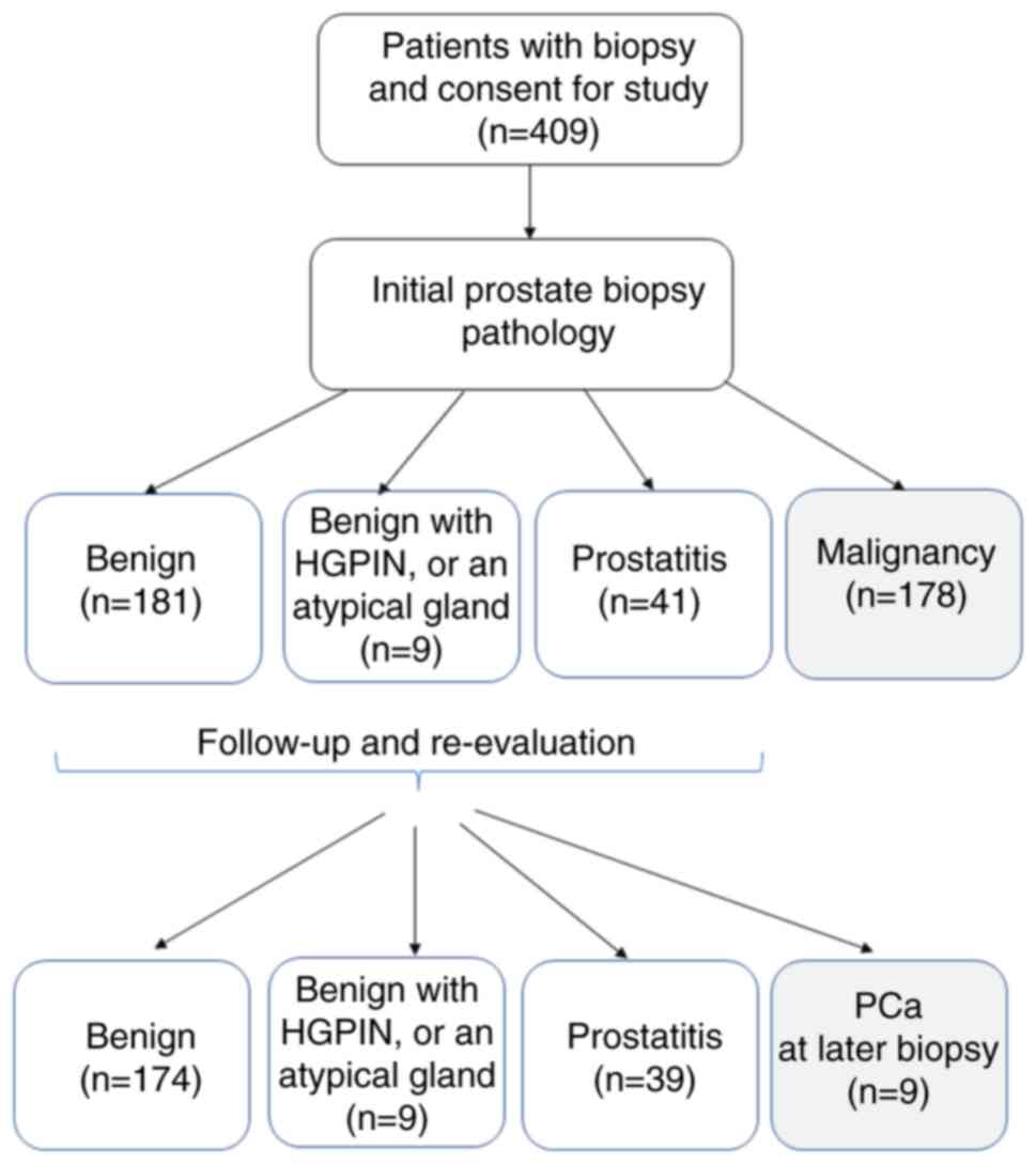
were kept on for follow-up studies (18). A Consolidated Standards of
Reporting Trials flowchart is shown in Fig. 1, which is a standardized diagram
utilized to illustrate participant flow (20). The study was performed after
obtaining the participants' informed consent in compliance with The
Declaration of Helsinki.
Urinary 8-OHdG and N-terminal
telopeptide (NTx) ELISA
Once the urine specimen was collected, it was
centrifuged at room temperature for 10 min at 600 x g, and
separated into two portions, supernatants and cell pellets. The
cell pellet was collected after harvesting 1-2 ml of the
supernatant without disturbing the pellet. Both portions were
separately stored at -80˚C. The supernatant without cells, cell
debris or other particulates (such as crystals) was investigated
using a urinary 8-OHdG ELISA according to a previously described
method (17). The results were
normalized to urine creatinine levels. Urinary 8-OHdG (oxidatively
damaged DNA adducts) levels were measured using a highly sensitive
8-OHdG Check ELISA kit (cat. no. KOG-200SE; Japan Institute for the
Control of Aging); the sensitivity of this kit was reported as
0.125 ng/ml. Urinary NTx levels were measured with the human NTx I
(Cross Linked N-telopeptide of type I collagen) ELISA kit (cat. no.
E-EL-H0836; Elabscience; Elabscience Bionovation Inc.) according to
the manufacturer's protocol; this kit recognizes human NTx Ⅰ in
studied samples, and the sensitivity was reported as 1.88 ng/ml and
the detection range as 3.13-200 ng/ml. There is no significant
interference or cross-reactivity between human NTx Ⅰ and its
analogs (standards or controls), and the coefficient of variation
is <10%, according to the manufacturer's manual; thus indicating
that this assay is highly specific and accurate.
Statistical analysis
The normal distribution of the data was assessed
using descriptive statistics and frequency distribution, followed
by the Shapiro-Wilk normality test. The comparison of urinary
8-OHdG levels among patients with benign prostatitis and
malignancy, or localized, locally advanced and de novo
metastatic diseases, was calculated using one-way ANOVA and
Spearman's rank correlation coefficient. If the ANOVA test was
significant, a post-hoc analysis with Tukey's multiple comparisons
test was performed to compare the differences between each group.
Unpaired Student's t-test was used to compare the differences
between initial and later diagnosis of prostate cancer.
χ2 test or Fisher's exact test (when the expected count
was ≤5 in ≥20% of cells) was used to assess contingency tables. The
association between urinary 8-OHdG and NTx levels was determined
using a linear regression analysis. The association of urinary
8-OHdG/creatinine ratio with age, PSA, prostate volume and PSA
density (PSAD; PSA level divided by prostate volume) was also
assessed using linear regression analysis. Statistical analysis was
performed using SPSS version 12.0 (SPSS, Inc.) and GraphPad Prism
6.00 for Windows (Dotmatics) software packages. P<0.05
(two-sided) was considered to indicate a statistically significant
difference.
Results
Patient characteristics
From November 2012 to October 31, 2020, 409 patients
were enrolled in the current study at an outpatient clinic before
prostate biopsy, including 190 patients with a benign biopsy
(including an atypical gland and prostate intraepithelial
neoplasm), 41 patients with prostatitis and 178 with malignancy.
The median age of the patients was 68 years (range, 43-91 years).
As compared with non-malignant (benign or prostatitis) patients,
those with a malignant histology were older (P<0.0001), and had
higher PSA values (P=0.001), a lower prostate volume (P<0.0001)
and higher PSAD (P=0.010) (Table
I).
 | Table IBasic characteristics of the studied
cohort at initial biopsy. |
Table I
Basic characteristics of the studied
cohort at initial biopsy.
| Parameter | Entire cohort | Benign (PIN,
AG) | Prostatitis | Malignancy |
P-valuea |
P-valueb,
Malignancy vs. Benign/Malignancy vs. Prostatitis |
|---|
| Number of
patients | 409 | 190 | 41 | 178 | - | - |
| Age, years | | | | | <0.0001 |
<0.0001/0.0002 |
|
Median
(range) | 68 (43-91) | 66 (43-86) | 66 (50-80) | 71 (53-91) | | |
|
25-75%
percentile | 62, 74 | 61, 72 | 60, 72 | 66, 76 | | |
| PSA, ng/ml | | | | | 0.001 | 0.0013/0.0868 |
|
Geometric
mean | 13.91 | 7.7 | 8.9 | 28.8 | | |
|
95% CI | 12.2-15.8 | 7.1-8.5 | 7.2-10.1 | 22.7-36.6 | | |
|
Range | 0.6-8332 | 0.6-41.9 | 1.4-49.7 | 1.8-8332 | | |
| Prostate volume,
ml | | | | | <0.0001 |
<0.0001/0.0234 |
|
Mean ±
SD | 49.8±35.4 | 55.0±27.1 | 54.5±26.5 | 43.1±21.5 | | |
| PSAD, ng/ml/ml | | | | | 0.010 | 0.0107/0.1944 |
|
Geometric
mean | 0.32 | 0.16 | 0.18 | 0.75 | | |
|
95% CI | 0.28-0.36 | 0.14-0.17 | 0.15-0.22 | 0.59-0.94 | | |
|
Range | 0.03-307 | 0.03-1.55 | 0.06-0.97 | 0.06-307 | | |
| Urinary creatinine,
mg/dl | | | | | 0.680 | |
|
Geometric
mean | 73.5 | 73.0 | 74.3 | 74.0 | | |
|
95% CI | 68.6-78.9 | 66.1-80.6 | 57.9-95.4 | 66.3-82.5 | | |
| 8-OHdG, ng/ml | | | | | 0.144 | |
|
Geometric
mean | 3.3 | 3.0 | 3.2 | 3.6 | | |
|
95% CI | 2.9-3.7 | 2.6-3.6 | 2.2-4.6 | 3.0-4.4 | | |
| 8-OHdG/creatinine,
x10-6 | | | | | 0.377 | |
|
Geometric
mean | 4.5 | 4.17 | 4.29 | 4.77 | | |
|
95% CI | 4.1-4.9 | 3.72-4.67 | 3.39-5.43 | 4.26-5.34 | | |
|
8-OHdG/creatinine/prostate volume,
x10-6/ml | | | | | 0.004 | 0.0065/0.070 |
|
Median | 0.10 | 0.09 | 0.08 | 0.12 | | |
|
95% CI | 0.03-0.31 | 0.03-0.27 | 0.03-0.25 | 0.04-0.38 | | |
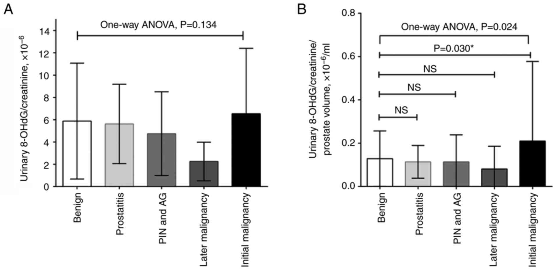
Urinary 8-OHdG levels and
normalization with urine creatinine level and prostate volume
At the time of prostate biopsy, urinary 8-OHdG
levels in patients with prostate cancer were higher than those
without malignancy (3.6 vs. 3.2 or 3.0 ng/ml; P=0.144; Table I); however, this was not
significant. During correction for urinary creatinine levels,
malignant patients had higher ratios of urinary 8-OHdG/creatinine
than the non-malignant patients (4.77x10⁻⁶ vs.
4.29x10⁻⁶ or 4.17x10⁻⁶; P=0.377; Table I); however, this was also not
significant. Since prostate enlargement can be induced by oxidative
stress, and there was a borderline association between prostate
volume and 8-OHdG/creatinine ratio in the current study (Table SI), the present study further
corrected the ratio with prostate volume (17). The results demonstrated that
patients with malignancies exhibited a significantly higher ratio
of urinary 8-OHdG/creatinine per prostate volume than the other
subgroups (0.12x10⁻⁶/ml vs. 0.08x10⁻⁶/ml or
0.09x10⁻⁶/ml; P=0.004; Table I). A post-hoc analysis with Tukey's
multiple comparisons test showed that the initial malignancy group
exhibited a higher urine 8-OHdG/creatinine ratio per prostate
volume than the benign group (P=0.030; Fig. 2).
During follow-up, another nine patients were
diagnosed with prostate cancer, including two from the prostatitis
subgroup and seven from the initial benign subgroup (Table SII). Among the four subgroups with
an initially benign biopsy, there were no significant differences
in age, PSA, prostate volume, urinary 8-OHdG/creatinine ratio or
urinary 8-OHdG/creatinine ratio per prostate volume (P>0.05);
however, the later malignancy group exhibited highest PSAD levels
within all the groups (P=0.027).
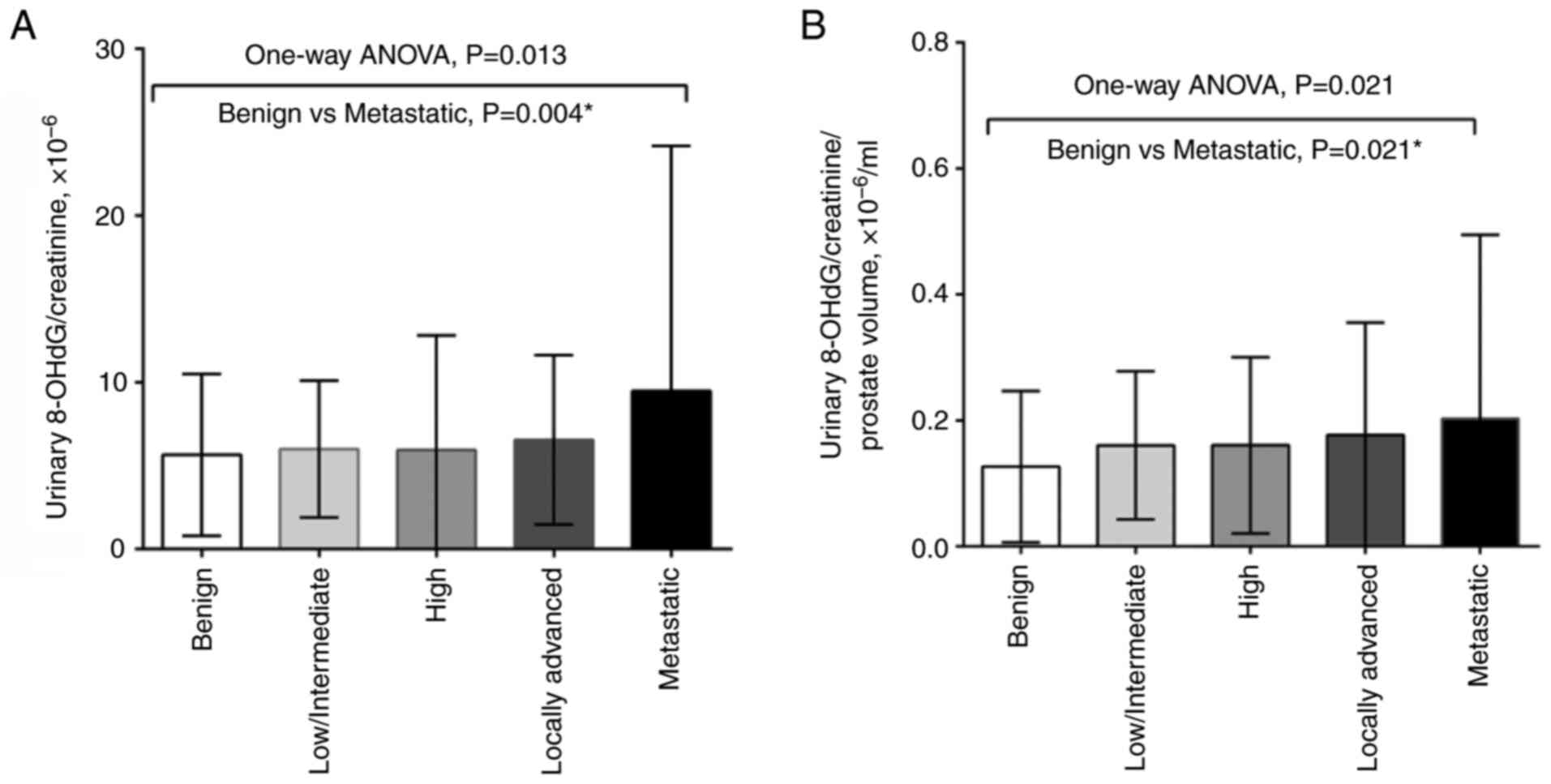
Prostate cancer risk stratification
and urinary 8-OHdG/creatinine ratio
During the study period, a total of 187 patients
were diagnosed with prostate cancer, including 178 at the initial
biopsy and nine at a later repeat biopsy. As compared with patients
with prostate cancer diagnosed at the initial biopsy, those with
later malignancy were younger and had lower urinary
8-OHdG/creatinine ratios (P=0.010 and P=0.031, respectively). Due
to the small number of patients diagnosed with a later malignancy,
there were no statistical differences in Gleason grading, clinical
TNM stage (21) and cancer
recurrent risk when comparing with patients initially diagnosed
with prostate cancer (Fisher's exact test; P=0.578, P=0.404, and
P=0.183, respectively) (Table
II). A total of 50 patients received subsequent radical
prostatectomy, and there was no difference between prostate cancer
at initial diagnosis and later diagnosis in terms of pathological
staging or grade (Table
SIII).
| Table IIClinicopathological characteristics
of patients with PCa according to the time of diagnosis. |
Table II
Clinicopathological characteristics
of patients with PCa according to the time of diagnosis.
| Parameter | Total | Initial PCa | Later PCa | P-value |
|---|
| Number of
patients | 187 | 178 | 9 | |
| Median age, years
(range) | 71 (53-91) | 71 (53-91) | 63 (55-73) | 0.010a |
|
25%-75%
percentile | 65-76 | 66-76 | 61-70 | |
| Mean PSA, ng/ml
(95% CI) | 27.5
(21.9-34.6) | 28.8
(22.7-36.6) | 10.9
(7.0-16.8) | 0.471a |
| Mean ±SD prostate
volume, ml | 43.0±21.6 | 43.1±21.5 | 41.5±24.5 | 0.833a |
| Mean PSAD,
ng/ml2 (95% CI) | 0.72
(0.57-0.89) | 0.75
(0.59-0.94) | 0.31
(0.19-0.50) | 0.560a |
| Mean 8-OHdG/crea.,
x10-6 (95% CI) | 4.6 (4.1-5.3) | 4.8 (4.2-5.6) | 1.7 (0.9-3.2) | 0.031a |
| Median
8-OHdG/crea./PV, x10-6/ml (95% CI) | 0.12
(0.10-0.14) | 0.12
(0.10-0.15) | 0.05
(0.02-0.09) | 0.294a |
| Gleason grade | | | | 0.578b |
|
3+3 | 58 | 53 | 5 | |
|
3+4 | 21 | 20 | 1 | |
|
4+3 | 29 | 28 | 1 | |
|
4+4, 3+5,
5+3 | 33 | 32 | 1 | |
|
5+5, 5+4,
4+5 | 46 | 45 | 1 | |
| Clinical TNM
staging | | | | 0.404b |
|
cT1c N0 | 53 | 48 | 5 | |
|
cT2a,b,c
N0 | 37 | 35 | 2 | |
|
cT3aN0 | 20 | 19 | 1 | |
|
cT3b-4N0 | 27 | 26 | 1 | |
|
Any cT,
cN+ | 11 | 11 | 0 | |
|
Any cT, Any
cN, M+ | 39 | 39 | 0 | |
| D'Amico prostate
cancer risk stratification | | | | 0.183b |
|
Localized
(low risk) | 25 | 24 | 1 | |
|
Localized
(intermediate risk) | 41 | 36 | 5 | |
|
Localized
(high risk) | 44 | 42 | 2 | |
|
Locally
advanced (very high risk) | 26 | 25 | 1 | |
|
Nodal | 10 | 10 | 0 | |
|
Metastatic | 41 | 41 | 0 | |
| Treatment
options | | | | 0.083b |
|
Active
surveillance/observation | 22 | 20 | 2 | |
|
Radical
prostatectomy | 50 | 45 | 5 | |
|
Radiotherapy | 31 | 29 | 2 | |
|
ADT | 51 | 51 | 0 | |
|
Visit other
hospitals or options | 33 | 33 | 0 | |
Significant differences in urinary 8-OHdG/creatinine
ratio were detected between different prostate cancer risk groups
using one-way ANOVA, and the Tukey's post hoc test indicated that
the significant differences were between the benign and metastatic
groups, regardless of whether the urinary 8-OHdG/creatinine ratio
was normalized to prostate volume (P=0.004 and P=0.021,
respectively) (Fig. 3). The
Spearman's rank correlation coefficient revealed that was a
negligible correlation between patient risk group and urinary
8-OHdG levels, with or without prostate volume correction (Fig. S1).
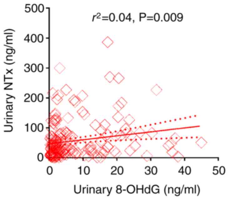
Association of 8-OHdG and NTx levels
in the urine
Urinary NTx levels were measured in a total of 188
studied patients, including 178 patients diagnosed with prostate
cancer at the initial biopsy and 10 random patients with an initial
benign biopsy. The geometric mean value of urinary NTx level was
30.8 ng/ml with a range of 0.5-386 ng/ml (95% CI, 47.5-65.6 ng/ml).
The association between 8-OHdG and NTx levels in the urine was
analyzed, and the results showed a weakly positive association
between both factors in the urine (linear regression,
r2=0.04; P=0.009) (Fig.
4).
Discussion
The present study demonstrated that urinary 8-OHdG
levels with prostate volume correction were elevated in patients
with malignancy at the time of prostate biopsy compared with those
with non-malignant histology, although there was an absence of a
direct association between urinary 8-OHdG and cancer in this
analysis. Significant differences in urinary 8-OHdG/creatinine
ratio were also determined between the different prostate cancer
risk groups. Moreover, urinary 8-OHdG levels were significantly
associated with urinary NTx levels. Taken together, urinary 8-OHdG
levels may reflect the involvement of oxidative stress in the
initiation and progression of prostate carcinogenesis.
Several studies have reported on the association
between oxidative stress and prostate cancer (22-24),
with several studies noting changes in pro-oxidant/antioxidant
balance in clinical prostate cancer samples, rodent models and
prostate cell lines (9,25,26).
In a previous study, malignant prostate samples demonstrated
elevated levels of thiobarbituric acid reactive substances and
reduced levels of glutathione peroxidase and CuZn-superoxide
dismutase (SOD) in comparison with the benign group (27). Upregulated oxidative stress
profiles have also been detected in human prostate cancer and it
has been suggested that antioxidant defense systems might be
impaired (28). Miyake et
al (29) reported that the
urinary 8-OHdG/creatinine ratio in patients with prostate cancer
was significantly elevated as compared with that in age-matched
healthy controls, which indicates the importance of oxidative
stress in the early events of prostate cancer, including cell
proliferation, differentiation, apoptosis and carcinogenesis.
Ohtake et al (16) reported
that 8-OHdG was more highly expressed in prostate cancer tissues in
comparison to benign prostate tissues using immunohistochemical
analysis. In addition, our recent publication identified a positive
association between serum PSA levels (or PSAD) and urinary 8-OHdG
levels in men with a mean age of 62.5 years, which was associated
with prostate enlargement (17).
Since urinary 8-OHdG is not solely of prostate origin, it reflects
systemic oxidative stress, indicating the extent of DNA damage
occurring throughout the body. According to our previous study,
prostate volume was significantly positively associated with
urinary 8-OHdG and serum iNOS levels (17), thus the present study normalized
urinary 8-OHdG levels to prostate volume. The results provide
further evidence that oxidative stress may partly contribute to the
initiation of prostate cancer and prostate enlargement. To the best
of our knowledge, the present study is the first to demonstrate a
significant association between urinary 8-OHdG levels and d'Amico
risk stratification, thereby aiding in predicting patient risk
during initial diagnosis.
Our previous study showed that urinary
di(2-ethylhexyl)phthalate (DEHP) metabolites, which serve as
evidence of phthalate exposure, were associated with urinary 8-OHdG
levels, serum PSA and prostate volume (17). Phthalates are well-known inducers
of oxidative stress associated with several diseases (30,31).
Numerous studies have proposed that exposure to phthalates, both
in vivo and in vitro, leads to oxidative stress in
patients, primarily by decreasing the levels of SOD and glutathione
(17); however, their role in
prostate carcinogenesis remains unclear. Some in vitro
studies have shown that prostate cancer cells exhibit various
inherent responses to oxidative stress, including reactive oxygen
species generation, and activation of matrix metalloproteinase-9
(MMP-9) by causing changes in the MMP-9 structure, which is
involved in the breakdown of extracellular matrix components that
is required for aggressive phenotypes (32). In addition, some phthalates (such
as DEHP, benzyl butyl phthalate and diisobutyl phthalate) can
upregulate cyclin D1 and proliferating cell nuclear antigen,
downregulate P21 and stimulate LNCaP proliferation (33). Phthalates may also promote prostate
cancer progression through the Hedgehog pathway in LNCaP cells
(34). Although there is a lack of
causal mechanistic experiments regarding oxidative stress and
prostate cancer progression, the present results demonstrated
significant differences in urinary 8-OHdG/creatinine ratio between
the different prostate cancer risk groups, and identified a
positive relationship between urinary 8-OHdG levels and NTx levels.
Such evidence provides strong clinical evidence that oxidative
stress serves an important role in the process of prostate cancer
progression. However, Spearman's rank correlation coefficient
showed no significant correlation between patient risk group and
urinary 8-OHdG levels; this lack of correlation indicates the need
for further research to explore potential non-linear relationships
or other influencing factors.
Although these cellular and clinical studies support
that oxidative stress is crucial in the etiology or progression of
prostate cancer, several large clinical trials have failed to
support the ability of supplemental dietary antioxidants, such as
vitamin E, to reduce prostate cancer risk. These studies include
the Prostate, Lung, Colorectal and Ovarian; Alpha-Tocopherol,
Beta-Carotene Prevention; and Selenium and Vitamin E Cancer
Prevention Trial studies (35-37).
However, vitamin E has been reported to exhibit some benefit in a
particular subgroup of patients; smokers supplemented with 50 mg
vitamin E daily exhibited a significantly lower prostate cancer
incidence (32%) and a significantly lower prostate cancer mortality
rate (41%) than those assigned to receive a placebo (38). Despite the absence of clinical
trials supporting the efficacy of supplemental dietary
antioxidants, it may still be worthwhile exploring therapies that
target the antioxidant response.
Notably, 8-OHdG is an oxidized nucleoside of DNA
that is the most frequently studied and detected biomarker of
oxidative DNA damage. Upon DNA repair, 8-OHdG is soon excreted in
the urine, and can be measured using ELISA and HPLC-MS methods.
Evidence has demonstrated that urinary 8-OHdG is a well-known
biomarker of generalized, cellular oxidative stress, and a risk
factor for atherosclerosis, diabetes and cancer (13). By contrast, blood 8-OHdG may
represent systemic oxidative stress from the tissues and
circulating lymphocytes. Few studies have focused on blood 8-OHdG
because several challenges exist for detecting 8-OHdG in the blood;
notably, techniques such as ELISA may not be sensitive enough, as
their detection limits can be higher than the normal range found in
healthy individuals (39,40). Our previous study demonstrated that
urinary 8-OHdG levels were positively associated with prostate
enlargement (17). Therefore, the
urinary 8-OHdG levels in the current study theoretically reflect
oxidative stress both in the prostate and the whole body. The
prostate is one of the most vulnerable organs in the body. While
the absence of a direct association between urinary 8-OHdG and
cancer is noted in the present analysis, it does not preclude its
potential value as an indicator of oxidative stress or DNA damage,
both of which are linked to cancer initiation and progression.
Notably, compared with prostate volume, the normalized urinary
8-OHdG levels were a worse marker for predicting prostate cancer.
However, urinary 8-OHdG level reflects systemic oxidative stress,
which not only promotes prostate enlargement (17), but might induce the initiation and
progression of prostate cancer, as evidenced by the association
between urinary 8-OHdG and NTx levels. Further research into the
biological mechanisms underlying how environmental exposures induce
oxidative stress, as evidenced by urinary 8OH-dG levels, and their
connection to prostate cancer could illuminate their potential
clinical relevance for diagnosing and treating the disease.
There were several limitations in the current study.
First, there are several factors associated with oxidative stress,
such as aging, prostate volume, infection/inflammation, cancer and
environmental exposure. In the current study, it was revealed that
prostate volume and cancer were the two most notable contributing
factors to oxidative stress. In contrast with large-scale clinical
trials, the number of study patients in the current study was
relatively small to explore the effect of other factors. Second,
urinary 8-OHdG levels might be influenced by cancer volume.
Although the impact of tumor percentage was not investigated in the
current study, the results revealed that urinary 8-OHdG levels were
associated with risk stratification, which might partly reflect
tumor burden. A future study may be conducted with more patients
receiving radical prostatectomy. Third, the research objective was
to utilize the existing TRUS biopsy database to observe potential
clinically significant phenomenal; therefore, the required sample
size in the current study was not explicitly calculated, which may
influence the statistical power. Since the present study enrolled
409 patients, it may be considered a reasonable size. The present
data also showed that patient age may be a better marker than the
urinary 8-OHdG/creatinine ratio regarding prostate cancer detection
(P=0.01 vs. P=0.031). Notably, as the present study utilized an
existing TRUS biopsy database, disparities in both population size
and age distribution were present during group comparisons;
therefore, it is difficult to make comparisons to determine which
factor is better. Additionally, some patient information was not
collected and analyzed in the current study, including smoking,
drug history, occupation history, comorbidities and medication
history. These factors may interfere with urinary 8-OHdG levels. In
future studies, increasing participant enrollment with detailed
medical and social history records may help mitigate these
potential biases. Fourth, the present study did not perform
immunohistochemical analysis to clarify whether 8-OHdG was
expressed more highly in prostate cancer tissues than in benign
prostate tissues, and 8-OHdG was not measured in the blood for
comparison with urinary levels. Although both the blood test and
tissue assay for the existence of 8-OHdG might theoretically
reflect the action of oxidative stress on prostate carcinogenesis,
blood 8-OHdG cannot accurately reflect the oxidative stress status
of the prostate or malignant tissue. Our previous study showed that
oxidative status and urinary 8-OHdG levels were positively
correlated with prostate enlargement in patients with lower urinary
tract symptoms that exhibited relatively normal serum PSA levels
(17). Therefore, we focused on
the influence of oxidative stress on prostate carcinogenesis in the
present study. It was hypothesized that the action of phthalate
exposure is not specific for prostate enlargement but also for
prostate cancer. The non-specific effect of phthalate exposure on
the prostate is similar with the non-specific characteristic of the
origin of PSA. Both benign and malignant prostate tissue can
produce and secrete PSA into the lumen of the prostate gland, which
can leak into the blood stream, and benign prostate cells can
produce more PSA mRNA than malignant prostate cancer cells
(41). The present study
demonstrated a substantial association between prostate
volume-normalized urinary 8-OHdG/creatinine ratio and the
aggressiveness of prostate cancer. This finding suggested its
potential as a non-invasive and easily accessible predictor for
assessing the influence of oxidative stress on prostate cancer
aggressiveness at initial diagnosis. Nevertheless, larger studies
are necessary to yield more robust results. At present, we are
dedicated to investigating the influence and molecular mechanism of
phthalate exposure on prostate carcinogenesis in order to validate
and reinforce our findings, thereby improving their clinical
applicability and upholding the highest standards of patient care
through rigorous scientific research.
In conclusion, the present study demonstrated that
patients with prostate cancer had higher urinary levels of the
oxidative stress marker 8-OHdG normalized to prostate volume than
non-malignant patients at the time of prostate biopsy. Furthermore,
urinary 8-OHdG levels normalized to prostate volume were higher in
more advanced disease and were positively associated with urinary
NTx levels. These data highlight the evidence of oxidative stress
in the etiology and progression of prostate cancer; however,
further studies are required to identify the etiology of oxidative
stress in prostate cancer patients.
Supplementary Material
Spearman's rank correlation
coefficient between patient risk groups and urinary 8-OHdG levels
was calculated (A) without or (B) with prostate volume correction.
Risk group 0, benign; risk group 1, low/intermediate risk; risk
group 2, high risk; risk group 3, locally advanced; risk group 4,
nodal/metastatic. 8-OHdG, 8-hydroxydeoxyguanosine; CI, confidence
interval.
Linear regression analysis of urinary
8-hydroxydeoxyguanosine/creatinine ratio (n=405).
Basic characteristics of the studied
patients with an initial benign biopsy.
Pathological characteristics of
patients with PCa according to the time of diagnosis.
Acknowledgements
Not applicable.
Funding
Funding: The present study was supported by Taiwan Ministry of
Science and Technology (grant nos. 111-2314-B-006-108- and
110-2314-B-006-065-MY3), the NCKUH (grant nos. NCKUH-11204018,
NCKUH-11104011, NCKUH-11004022, NCKUH-10902045 and NCKUH-10802035)
and the Ditmanson Medical Foundation Chia-Yi Christian Hospital
(grant no. NCKUCYCH-P-11101-1).
Availability of data and materials
The data generated in the present study may be
requested from the corresponding author.
Authors' contributions
TST, YCJ and YST designed the study. HTT and YST
were primarily responsible for conducting the experiments and
confirm the authenticity of all the raw data. YCH and ISC acquired
and analyzed the data, and wrote the manuscript. LNH and HTT
assisted in data acquisition and interpretation, and edited the
manuscript. All authors read and approved the final version of the
manuscript.
Ethics approval and consent to
participate
The present study obtained the approval and
institutional oversight of the Institutional Review Board (IRB) for
the Protection of Human Subjects at National Cheng Kung University
Hospital (IRB no.: A-ER-101-181). Written informed consent was
obtained from all subjects involved in the study.
Patient consent for publication
Not applicable.
Competing interests
The authors declare that they have no competing
interests.
References
|
1
|
Siegel RL, Miller KD, Wagle NS and Jemal
A: Cancer statistics, 2023. CA Cancer J Clin. 73:17–48.
2023.PubMed/NCBI View Article : Google Scholar
|
|
2
|
Health Promotion Administration (HPA):
2022 Health Promotion Administration Annual Report. HPA, Taipei
City, 2022. https://www.hpa.gov.tw/EngPages/Detail.aspx?nodeid=1070&pid=16384.
Accessed 18 May, 2024.
|
|
3
|
Chiang CJ, Lo WC, Yang YW, You SL, Chen CJ
and Lai MS: Incidence and survival of adult cancer patients in
Taiwan, 2002–2012. J Formos Med Assoc. 115:1076–1088.
2016.PubMed/NCBI View Article : Google Scholar
|
|
4
|
Lin YH, Chen KK and Chiu JH: Use of
Chinese medicine among prostate cancer patients in Taiwan: A
retrospective longitudinal cohort study. Int J Urol. 18:383–386.
2011.PubMed/NCBI View Article : Google Scholar
|
|
5
|
Inamoto T, Azuma H, Hinotsu S, Tsukamoto
T, Oya M, Ogawa O, Kitamura T, Kazuhiro S, Naito S, Namiki M, et
al: Age at diagnosis on prostate cancer survival undergoing
androgen deprivation therapy as primary treatment in daily
practice: Results from Japanese observational cohort. J Cancer Res
Clin Oncol. 140:1197–1204. 2014.PubMed/NCBI View Article : Google Scholar
|
|
6
|
Chen PM, Chen SC, Liu CJ, Hung MH, Tsai
CF, Hu YW, Chen MH, Shen CC, Su TP, Yeh CM, et al: The association
between prostate cancer and mood disorders: A nationwide
population-based study in Taiwan. Int Psychogeriatr. 27:481–490.
2015.PubMed/NCBI View Article : Google Scholar
|
|
7
|
Balistreri CR, Candore G, Lio D and
Carruba G: Prostate cancer: From the pathophysiologic implications
of some genetic risk factors to translation in personalized cancer
treatments. Cancer Gene Ther. 21:2–11. 2014.PubMed/NCBI View Article : Google Scholar
|
|
8
|
Huang WK, Liu CH, Pang ST, Liu JR, Chang
JW, Liaw CC, Hsu CL, Lin YC and See LC: Type of androgen
deprivation therapy and risk of dementia among patients with
prostate cancer in Taiwan. JAMA Netw Open.
3(e2015189)2020.PubMed/NCBI View Article : Google Scholar
|
|
9
|
Shukla S, Srivastava JK, Shankar E, Kanwal
R, Nawab A, Sharma H, Bhaskaran N, Ponsky LE, Fu P, MacLennan GT
and Gupta S: Oxidative stress and antioxidant status in high-risk
prostate cancer subjects. Diagnostics (Basel).
10(126)2020.PubMed/NCBI View Article : Google Scholar
|
|
10
|
Drozdz-Afelt JM, Koim-Puchowska BB and
Kaminski P: Analysis of oxidative stress indicators in Polish
patients with prostate cancer. Environ Sci Pollut Res Int.
29:4632–4640. 2022.PubMed/NCBI View Article : Google Scholar
|
|
11
|
Al-Taie A, Sancar M and Izzettin FV:
Chapter 17-8-Hydroxydeoxyguanosine: A valuable predictor of
oxidative DNA damage in cancer and diabetes mellitus. In: Cancer
(Second Edition). Preedy VR and Patel VB (eds). Academic Press, San
Diego, pp179-187, 2021.
|
|
12
|
Li YS, Song MF, Kasai H and Kawai K:
8-hydroxyguanine in urine and serum as an oxidative stress marker:
Effects of diabetes and aging. J Uoeh. 35:119–127. 2013.PubMed/NCBI View Article : Google Scholar
|
|
13
|
Wu LL, Chiou CC, Chang PY and Wu JT:
Urinary 8-OHdG: A marker of oxidative stress to DNA and a risk
factor for cancer, atherosclerosis and diabetics. Clin Chim Acta.
339:1–9. 2004.PubMed/NCBI View Article : Google Scholar
|
|
14
|
Miyaoka T, Ieda M, Hashioka S, Wake R,
Furuya M, Liaury K, Hayashida M, Tsuchie K, Arauchi R, Araki T, et
al: Analysis of oxidative stress expressed by urinary level of
biopyrrins and 8-hydroxydeoxyguanosine in patients with chronic
schizophrenia. Psychiatry Clin Neurosci. 69:693–698.
2015.PubMed/NCBI View Article : Google Scholar
|
|
15
|
Yano T, Shoji F, Baba H, Koga T, Shiraishi
T, Orita H and Kohno H: Significance of the urinary 8-OHdG level as
an oxidative stress marker in lung cancer patients. Lung Cancer.
63:111–114. 2009.PubMed/NCBI View Article : Google Scholar
|
|
16
|
Ohtake S, Kawahara T, Ishiguro Y,
Takeshima T, Kuroda S, Izumi K, Miyamoto H and Uemura H: Oxidative
stress marker 8-hydroxyguanosine is more highly expressed in
prostate cancer than in benign prostatic hyperplasia. Mol Clin
Oncol. 9:302–304. 2018.PubMed/NCBI View Article : Google Scholar
|
|
17
|
Chang WH, Tsai YS, Wang JY, Chen HL, Yang
WH and Lee CC: Sex hormones and oxidative stress mediated
phthalate-induced effects in prostatic enlargement. Environ Int.
126:184–192. 2019.PubMed/NCBI View Article : Google Scholar
|
|
18
|
Moses KA, Sprenkle PC, Bahler C, Box G,
Carlsson SV, Catalona WJ, Dahl DM, Dall'Era M, Davis JW, Drake BF,
et al: NCCN Guidelines® Insights: Prostate cancer early detection,
version 1.2023. J Natl Compr Canc Netw. 21:236–246. 2023.PubMed/NCBI View Article : Google Scholar
|
|
19
|
D'Amico AV, Whittington R, Malkowicz SB,
Schultz D, Blank K, Broderick GA, Tomaszewski JE, Renshaw AA,
Kaplan I, Beard CJ and Wein A: Biochemical outcome after radical
prostatectomy, external beam radiation therapy, or interstitial
radiation therapy for clinically localized prostate cancer. JAMA.
280:969–974. 1998.PubMed/NCBI View Article : Google Scholar
|
|
20
|
Schulz KF, Altman DG and Moher D: CONSORT
Group. CONSORT 2010 statement: Updated guidelines for reporting
parallel group randomised trials. BMJ. 340(c332)2010.PubMed/NCBI View Article : Google Scholar
|
|
21
|
NCCN Clinical Practice Guidelines in
Oncology. Prostate Cancer (Version 4.2024). National Comprehensive
Cancer Network; 2024. Available at: https://www.nccn.org/guidelines/guidelines-detail?category=1&id=1459.
Accessed 24 July, 2024.
|
|
22
|
Paschos A, Pandya R, Duivenvoorden WC and
Pinthus JH: Oxidative stress in prostate cancer: Changing research
concepts towards a novel paradigm for prevention and therapeutics.
Prostate Cancer Prostatic Dis. 16:217–225. 2013.PubMed/NCBI View Article : Google Scholar
|
|
23
|
Liou GY, C'Lay-Pettis R and Kavuri S:
Involvement of reactive oxygen species in prostate cancer and its
disparity in african descendants. Int J Mol Sci.
25(6665)2024.PubMed/NCBI View Article : Google Scholar
|
|
24
|
Biesiadecki M, Mołoń M, Balawender K,
Kobylińska Z and Galiniak S: Shedding light on the shadows:
Oxidative stress and its pivotal role in prostate cancer
progression. Front Oncol. 14(1393078)2024.PubMed/NCBI View Article : Google Scholar
|
|
25
|
Battisti V, Maders LDK, Bagatini MD, Reetz
LG, Chiesa J, Battisti IE, Gonçalves JF, Duarte MM, Schetinger MR
and Morsch VM: Oxidative stress and antioxidant status in prostate
cancer patients: Relation to Gleason score, treatment and bone
metastasis. Biomed Pharmacother. 65:516–524. 2011.PubMed/NCBI View Article : Google Scholar
|
|
26
|
Rossetto IMU, Santos FR, da Silva HM,
Minatel E, Mesquitta M, Salvador MJ, Montico F and Cagnon VHA:
Tempol effect on oxidative and mitochondrial markers in preclinical
models for prostate cancer. Toxicol Res (Camb).
13(tfae056)2024.PubMed/NCBI View Article : Google Scholar
|
|
27
|
Aydin A, Arsova-Sarafinovska Z, Sayal A,
Eken A, Erdem O, Erten K, Ozgök Y and Dimovski A: Oxidative stress
and antioxidant status in non-metastatic prostate cancer and benign
prostatic hyperplasia. Clin Biochem. 39:176–179. 2006.PubMed/NCBI View Article : Google Scholar
|
|
28
|
Oh B, Figtree G, Costa D, Eade T, Hruby G,
Lim S, Elfiky A, Martine N, Rosenthal D, Clarke S and Back M:
Oxidative stress in prostate cancer patients: A systematic review
of case control studies. Prostate Int. 4:71–87. 2016.PubMed/NCBI View Article : Google Scholar
|
|
29
|
Miyake H, Hara I, Kamidono S and Eto H:
Oxidative DNA damage in patients with prostate cancer and its
response to treatment. J Urol. 171:1533–1536. 2004.PubMed/NCBI View Article : Google Scholar
|
|
30
|
Han Q, Gao X, Wang S, Wei Z, Wang Y, Xu K
and Chen M: Co-exposure to polystyrene microplastics and
di-(2-ethylhexyl) phthalate aggravates allergic asthma through the
TRPA1-p38 MAPK pathway. Toxicol Lett. 384:73–85. 2023.PubMed/NCBI View Article : Google Scholar
|
|
31
|
Zhang Y, Yang Y, Tao Y, Guo X, Cui Y and
Li Z: Phthalates (PAEs) and reproductive toxicity:
Hypothalamic-pituitary-gonadal (HPG) axis aspects. J Hazard Mater.
459(132182)2023.PubMed/NCBI View Article : Google Scholar
|
|
32
|
Kumar B, Koul S, Khandrika L, Meacham RB
and Koul HK: Oxidative stress is inherent in prostate cancer cells
and is required for aggressive phenotype. Cancer Res. 68:1777–1785.
2008.PubMed/NCBI View Article : Google Scholar
|
|
33
|
Zhu M, Huang C, Ma X, Wu R, Zhu W, Li X,
Liang Z, Deng F, Wu J, Geng S, et al: Phthalates promote prostate
cancer cell proliferation through activation of ERK5 and p38.
Environ Toxicol Pharmacol. 63:29–33. 2018.PubMed/NCBI View Article : Google Scholar
|
|
34
|
Yong W, Jiao C, Jianhui W, Yan Z, Qi P,
Xiu W, Zuyue S and Yunhui Z: Mono-2-ethyhexyl phthalate advancing
the progression of prostate cancer through activating the hedgehog
pathway in LNCaP cells. Toxicol In Vitro. 32:86–91. 2016.PubMed/NCBI View Article : Google Scholar
|
|
35
|
Virtamo J, Edwards BK, Virtanen M, Taylor
PR, Malila N, Albanes D, Huttunen JK, Hartman AM, Hietanen P,
Mäenpää H, et al: Effects of supplemental alpha-tocopherol and
beta-carotene on urinary tract cancer: Incidence and mortality in a
controlled trial (Finland). Cancer Causes Control. 11:933–939.
2000.PubMed/NCBI View Article : Google Scholar
|
|
36
|
Lonn E, Bosch J, Yusuf S, Sheridan P,
Pogue J, Arnold JM, Ross C, Arnold A, Sleight P, Probstfield J, et
al: Effects of long-term vitamin E supplementation on
cardiovascular events and cancer: A randomized controlled trial.
JAMA. 293:1338–1347. 2005.PubMed/NCBI View Article : Google Scholar
|
|
37
|
Kirsh VA, Hayes RB, Mayne ST, Chatterjee
N, Subar AF, Dixon LB, Albanes D, Andriole GL, Urban DA and Peters
U: PLCO Trial. Supplemental and dietary vitamin E, beta-carotene,
and vitamin C intakes and prostate cancer risk. J Natl Cancer Inst.
98:245–254. 2006.PubMed/NCBI View Article : Google Scholar
|
|
38
|
Heinonen OP, Albanes D, Virtamo J, Taylor
PR, Huttunen JK, Hartman AM, Haapakoski J, Malila N, Rautalahti M,
Ripatti S, et al: Prostate cancer and supplementation with
alpha-tocopherol and beta-carotene: Incidence and mortality in a
controlled trial. J Natl Cancer Inst. 90:440–446. 1998.PubMed/NCBI View Article : Google Scholar
|
|
39
|
Koide S, Kinoshita Y, Ito N, Kimura J,
Yokoyama K and Karube I: Determination of human serum
8-hydroxy-2'-deoxyguanosine (8-OHdG) by HPLC-ECD combined with
solid phase extraction (SPE). J Chromatogr B Analyt Technol Biomed
Life Sci. 878:2163–2167. 2010.PubMed/NCBI View Article : Google Scholar
|
|
40
|
Korkmaz KS, Butuner BD and Roggenbuck D:
Detection of 8-OHdG as a diagnostic biomarker. J Lab Precis Med.
3(95)2018.
|
|
41
|
Qiu SD, Young CY, Bilhartz DL, Prescott
JL, Farrow GM, He WW and Tindall DJ: In situ hybridization of
prostate-specific antigen mRNA in human prostate. J Urol.
144:1550–1556. 1990.PubMed/NCBI View Article : Google Scholar
|