Introduction
Osteosarcoma (OS) is a highly prevalent malignant
tumor of mesenchymal origin, accounting for 20% of all bone tumors
(1). It occurs mainly in adolescents
and the geriatric population (2).
Etiologically, it is linked to genetic mutations on chromosome 6,
previous exposure to radiotherapy, a high birth weight, repeated
bone trauma and the African race (3). Mortality rates vary according to the
histological grade, with ~3.1 deaths per million affected
individuals annually (4). OS
typically manifests in the long bones of the appendicular skeleton
(5), primarily as localized lesions
in the bone marrow; however, it can also occur in the periosteum,
bone cortex and surrounding soft tissues (6). Histologically, it contains variable
amounts of osteoid and fibrotic material, exhibiting different
cellular predominance of osteoblasts, chondroblasts, or
fibroblasts. It also exhibits various degrees of cellular
degeneration and varying mitotic rates (7). Clinically, it presents with pain,
swelling, limited joint mobility, and, in some cases, pathological
fractures (8). Surgery combined with
adjuvant chemotherapy is regarded as the optimal treatment for this
neoplasm (9). Prompt intervention is
crucial, as the tumor can metastasize to the lungs, requiring
timely treatment to minimize mortality (10). Regrettably, diagnosis can be delayed
in some patients as the symptoms may be mistaken for more common
conditions, as observed when OS occurs during pregnancy. Currently,
the treatment of OS during pregnancy may be controversial due to
potential risks to the fetus (5).
Therefore, the present study describes the case of a
pregnant woman diagnosed with OS in the appendicular skeleton
during the first trimester. She underwent surgical treatment and
adjuvant chemotherapy, exhibiting a positive response to treatment
and successful survival of the gestational product.
Case report
The medical care for the patient discussed in the
present case report was provided at Star Médica Hospital Chihuahua,
Chihuahua, Mexico. The patient's care was administered from August,
2022 to January, 2023, in accordance with the standard protocols
established by the institution and the specific pathology
presented. As of the drafting of this clinical case report, both
the mother and the infant are in good overall health.
A 21-year-old female with no notable medical history
was in the first trimester of a physiological pregnancy, managed
with mineral supplements and multivitamins. Around the 11th week of
gestation, the patient reported swelling and localized pain in the
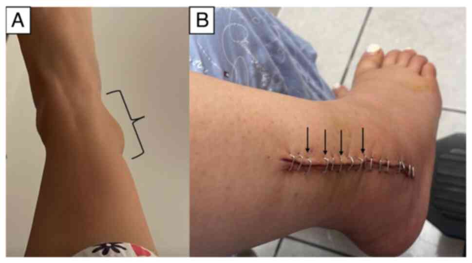
distal third of her right tibia and fibula region (Fig. 1A). Anterior-posterior and lateral
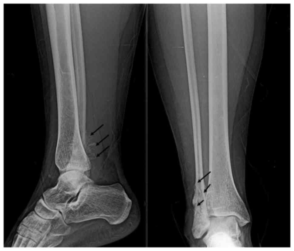
X-rays of the right ankle revealed granular lesions of variable
radiographic density in the region of the right lateral malleolus
(Fig. 2). Given the suspicion of
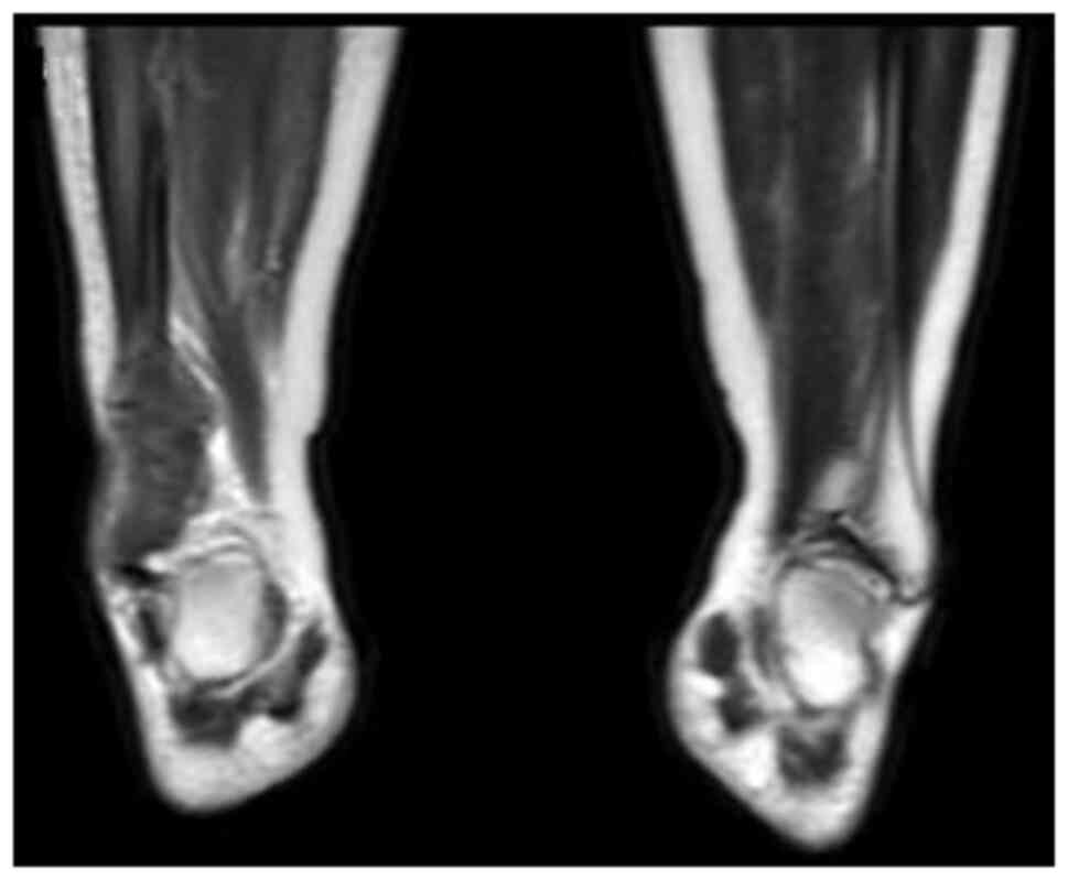
malignancy, contrast-enhanced magnetic resonance imaging (MRI) of
the abdominal-pelvic region, chest, and right ankle was conducted.
The MRI revealed increased volume and signal intensity in the soft
tissues surrounding the lateral malleolus. A non-mineralized bony
matrix lesion was identified in the distal diaphysis of the fibula,
appearing hypointense on the T1 sequence and hyperintense in the
proximal diaphysis, with irregular margins measuring 66x30x20 mm.
The lesion caused cortical destruction and extension into adjacent
soft tissues. Post-contrast imaging revealed avid and heterogeneous
enhancement of the affected region (Fig.
3).
Subsequently, following appropriate preparation, the
patient underwent a biopsy of the affected area. The excised tissue
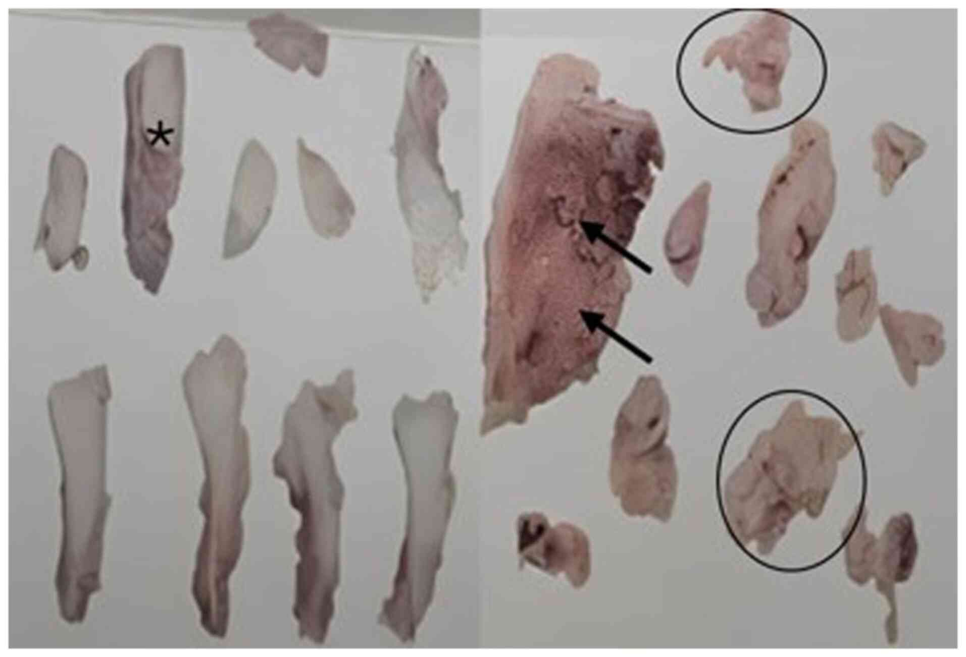
was sent for analysis. The histopathology report described
fragmented tissue measuring 6x4.2x1.2 cm in total, with a white or
pink color, rough or granular texture, soft or semi-solid
consistency, and exhibiting portions of white and hard bone.
Microscopically, features consistent with a malignant mesenchymal
neoplasm affecting bone, periosteum and adjacent fibroadipose
tissue were observed. Abundant osteoid and fibrosis were present.
Clusters or fascicles of spindle, oval, irregular, or polygonal
cells with nuclear enlargement, oval, elongated, or irregular
nuclei, vesicular or hyperchromatic, with one or two prominent
nucleoli, two mitoses in 10 high-power fields, and scant cytoplasm
were observed in the osteoid and fibrous stroma. Multinucleated
giant tumor cells were also noted. The diagnosis of OS involving
bone, periosteum and neighboring fibroadipose tissue, fragmented,
measuring 6x4.2x1.2 cm, was established (Fig. 4). Upon the confirmation of diagnosis,
the patient was closely monitored. At approximately the 15th week
of gestation, she was admitted to the Department of Traumatology
and Orthopedics of Hospital Star Médica Chihuahua (Chihuahua,
Mexico) for the resection of the distal third of the right fibula.
Following the surgical protocol, anesthesia was induced by regional
blockade, and hemostatic control was established. The patient was
positioned in dorsal decubitus, and asepsis and antisepsis
techniques were employed for the lower right limb. Sterile fields
were prepared, and an approach was made to the lateral malleolus.
The skin, subcutaneous tissue and fascia were dissected, and the
area between the long and short fibula and the distal third of the
fibula was approached. A granular-looking tumor measuring ~8x5 cm
was located and excised. Post-surgical cleansing and conventional
wound closure were performed without complications (Fig. 1B). Following recovery, after 1 week
of her admission the patient exhibited good progress and was
admitted for a scheduled outpatient follow-up. Analgesic management
and antibiotic therapy with clindamycin at 300 mg every 6 h for 7
days were prescribed, along with general care of the surgical site
and follow-up at the outpatient clinic. The patient subsequently
continued with an uncomplicated prenatal follow-up. Between 29 and
31 weeks of gestation, adjuvant chemotherapy was initiated with
doxorubicin at 110 mg in 100 cc of 0.9% saline solution and
cisplatin at 140 mg in 1,000 cc of 0.9% saline solution.
Additionally, anti-emetic, anti-inflammatory and anti-edematous
management was provided, along with the administration of 6 mg
pegfilgrastim following chemotherapy to prevent neutropenia.
Following the completion of the chemotherapeutic
regimen, the patient exhibited favorable progress and underwent a
scheduled cesarean section at the beginning of the 37th week of
gestation. The surgery was performed without complications,
yielding a live product with a weight of 2,285 grams, a height of
48 cm, and Apgar scores of 9 at 1 and 5 min. At 1 week postpartum,
the patient underwent three additional sessions of the same
chemotherapeutic regimen. Subsequently, a biopsy was performed to
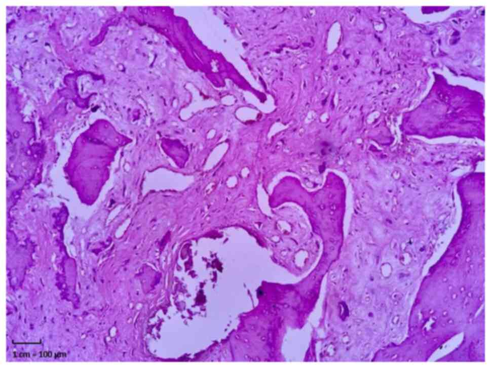
evaluate the antitumor efficacy. A histopathological analysis
revealed multiple bone fragments totaling 8x7 cm, characterized by
a gray-brown, hard, irregular, opaque and trabecular appearance.
The obtained sample was processed using 10% buffered formalin, then
embedded in paraffin and sectioned into 50-µm-thick slices using a
microtome. The sections were placed on slides and stained with
hematoxylin and eosin; these procedures were performed in the
Cytopathology and Oncological Pathology Laboratory,
Histopath®, in accordance with standard guidelines. A
microscopic examination revealed sections of cortical and medullary
bone tissue with distinct intraparenchymal hemorrhage and diffuse
fibrosis, alongside the loss of elements from all three
hematopoietic series (myeloid, erythroid and platelets). Notably,
no residual malignant neoplastic tissue was detected, indicating a
complete pathological response (residual tumor, 0%) (Fig. 5).
A regards the follow-up of the gestational product,
pediatric reports indicate a generally positive outcome, with only
a few unrelated conditions. Notably, a cardiac murmur was diagnosed
at 1 month of age, but was deemed benign. Upon the writing of the
present manuscript, the patient has been asymptomatic for >1
year. Additionally, the infant experienced symptoms of
gastroesophageal reflux, which led to nutritional issues, resulting
in iron-deficiency anemia and proctocolitis with limited weight
gain around the 5th month. All these conditions have been
effectively treated. Currently, all symptoms have resolved, and
there are no pathological findings linking the condition to in
utero exposure to the antineoplastic drug or the maternal OS.
Discussion
The present study describes the case of a young
pregnant woman diagnosed with OS in the distal fibula, who was
successfully treated with surgery and adjuvant chemotherapy. It is
important to highlight that the woman in the present case report
had no evident risk factors for developing OS. Thus, this clearly
demonstrates the unpredictable nature of this neoplasm in young
individuals. Fortunately, long-term follow-up reports for this
woman indicate that both she and the gestational product are in
good health, with no pathologies linked to the primary tumor or
arising from the treatment. Notably, the occurrence of neoplasms
during pregnancy is rare, with an incidence of ~1 case per 1,000
pregnancies (11). A previous review
suggested that the incidence of OS in pregnant women may be on the
rise due to the increasing trend of delaying pregnancy until later
ages (12). However, the age of the
patient in the present study does not align with this trend.
Therefore, it can be hypothesized that the patient in the present
case report likely had more subtle risk factors that predisposed
her to develop this neoplastic lesion, probably of a polygenic
nature or related to specific age-related risk factors. As regards
the latter, a previous retrospective study observed that the age
group of 15 to 25 years was the one with the most significant
increase in OS diagnoses over recent decades compared with other
age groups (13). Additionally,
previous reports have documented incidental diagnoses of OS in
pregnant women and women on reproductive age while they were being
treated for other conditions (14,15). In
the patient described herein, the clinical manifestations observed
were consistent with the typical presentations of OS in the
appendicular skeleton (16). The
initial evaluation of the edema and pain involved an ankle X-ray,
which revealed radiodense and lytic lesions typical of this
neoplasm (17). Although positron
emission tomography is the ideal diagnostic tool for identifying
metastases (18), herein, its use
was restricted due to the pregnancy of the patient. Consequently,
contrast-enhanced MRI was employed, as it effectively visualizes
the extent of soft tissue involvement and the overall disease
progression (17,19). In the absence of metastasis, surgical
treatment combined with adjuvant chemotherapy was selected.
Previous studies have demonstrated that this approach can preserve
the affected limb and ensure a favorable long-term prognosis
(9,20). Although surgery is a valid approach
for treating localized stages of osteosarcoma, it can cause
localized cellular stress, potentially leading to immune
dysregulation and increasing the risk of tumor recurrence over time
(21). Considering this and the
young age of the patient in the present case report, the medical
team decided on a combined treatment plan of surgery and adjuvant
chemotherapy. Drug management during pregnancy has historically
been contentious; advancements in pharmacokinetics and
pharmacovigilance now support the safe use of medications during
pregnancy for both the mother and fetus (22,23). In
addition, recent advancements in therapies, particularly those
utilizing nanoparticles, have demonstrated their effectiveness in
the treatment of OS (24). However,
the availability of antineoplastic drugs in the authors' hospital
(Hospital Star Médica Chihuahua) is limited. Consequently, the
selection of the medication was based on the established
therapeutic efficacy of the drug for this type of cancer. Since the
1970s, the use of cisplatin has been shown to be associated with
improved survival rates in OS by 90% and extended relapse-free
periods by 60% (25). However, in
the case described herein, cisplatin and doxorubicin were selected,
as it remains effective at low doses for these lesions (26-28).
The follow-up data confirmed a satisfactory antitumor response in
the patient, along with the birth of the product without
significant complications. As regards the treatment used, while the
management of OS depends on the tumor stage, it is also closely
aligned with the treatment protocols specific to each hospital
(29). Cytotoxic therapies used in
OS include alkylating agents (ifosfamide and cisplatin),
antimetabolites (methotrexate and gemcitabine), topoisomerase
inhibitors (etoposide), anthracyclines (doxorubicin), or
microtubule inhibitors. Additionally, recent studies suggest that
PD-1 inhibitors may serve as an effective adjuvant immunotherapy
for patients with OS who have already undergone standard
chemotherapy; this treatment has the potential to increase survival
rates and lower the risk of long-term tumor recurrence (30,31).
Other potential treatments include RNA silencers and certain
nucleotide analogs, which have proven effective in other types of
solid tumors. However, these are still in phase I and II clinical
trials: (NCT02985125, https://clinicaltrials.gov/study/NCT02985125; and
NCT02981342, https://clinicaltrials.gov/study/NCT02981342,
respectively) and will require time before they become standard
practice (32). In the authors'
opinion, the treatment selected for the patient in the present case
report was the most suitable, considering her personal
circumstances and the available resources at the hospital.
The present study has a notable limitation which
should be mentioned: The routine diagnosis of OS typically relies
on the immunohistochemical analysis of the tumor tissue. Commonly
used molecular markers include vimentin, S100 protein and CD99,
which collectively assist in determining the cellular origin and
genetic lineage of the tumor. However, due to resource limitations
at Hospital Star Médica Chihuahua, the diagnosis of OS was based on
clinical presentation and histopathological findings from the tumor
sample. While this diagnostic approach is generally deemed
sufficient (33), the absence of
specific markers hampers a comprehensive characterization of the
neoplasm. For purposes of comparison, a summary of previously
reported similar cases reports in the medical literature is
provided in Table I (11,34-48).
Future research is thus required to explore the utility of these
markers as part of a thorough diagnostic screening for similar
lesions. However, given the positive response of the patient to
chemotherapy and the outcomes reported during the resection, it is
expected that she will sustain a favorable long-term prognosis.
 | Table ISummary of previously reported similar
cases reports in the medical literature. |
Table I
Summary of previously reported similar
cases reports in the medical literature.
| Authors, year of
publication | Characteristics | Site | Treatment | Outcomes | (Refs.) |
|---|
| Pratt et al,
1977 | 4 Women, unknown
gestational age | No data | Surgical resection
and chemotherapy: Adriamycin plus cyclophosphamide, following by
high dose of methotrexate and leucovorin | 50% rate survival, 1
patient without metastasis at follow-up | (34) |
| Haerr and Pratt,
1985 | Young woman at 25th
gestational week | Hip | Multiple
chemotherapeutics regime | Newborn and mother
survival at 4 years follow-up | (35) |
| Huvos et al,
1985 | Young woman | Hip | No data | No data | (36) |
| Adair et al,
2001 | Young woman at 16th
gestational week | Bowel | No data | No data | (37) |
| Nakajima et
al, 2004 | Young woman at 25th
gestational week | Long bones | Doxorubicin and
ifosfamide | Newborn and mother
survival at 8 months follow-up | (38) |
| Nepal et al,
2005 | Young woman | No data | No data | No data | (39) |
| Koçak et al,
(2006) | Young woman at 7th
month of gestation | Heart | Mitral valve
replacement | Newborn and mother
survival at 3 months follow-up | (40) |
| Kanazawa et
al, 2009 | Women with
acromegaly at 6th gestational week | Jaw | Surgical
resection | No data | (41) |
| Han et al,
2011 | Woman at second
trimester of gestation | Femur | No data | Newborn and mother
survival | (42) |
| Gonin et al,
2011 | Woman at 26th
gestational week | Adrenal gland | No data | No data | (43) |
| Corrêa et
al, 2012 | Young woman | No data | No data | No data | (44) |
| Ding et al,
2016 | Young woman | Kidney | Nephrectomy and
‘conventional chemotherapy’ | Dead by metastatic
disease | (45) |
| Narla et al,
2018 | Young woman | Breast | No data | No data | (46) |
| Figueiro-Filho
et al, 2018 | Ten case reports:
Two during the pregnancy | Pelvis | No data | No data | (11) |
| Breda et al,
2021 | Young woman | Breastbone | No data | No data | (47) |
| Regmi et al,
2023 | Middle-aged woman
at 1st month of pregnancy | Lung | No data | No data | (48) |
Acknowledgements
Not applicable.
Funding
Funding: No funding was received.
Availability of data and materials
The datasets used and/or analyzed during the current
study are available from the corresponding author on reasonable
request.
Authors' contributions
CAOG, JHL and CAMM participated in the diagnostic
and therapeutic approach of the patient, as well as in supervising
the writing of the manuscript. MIG, AART and RGRM participated in
the surgical procedure, as well as in obtaining relevant medical
imaging of the patient, including MRI studies, radiographs, and
histopathology, also were responsible for the synthesis of the
clinical information of this case, the bibliographic research and
the redaction of the work. CAOG and JHL confirm the authenticity of
all the raw data. All authors have read and approved the final
manuscript.
Ethics approval and consent to
participate
The patient signed permission for her participation
in the present case report.
Patient consent for publication
The patient voluntarily provided consent for the
publication of the present case report and any personal images
illustrating her condition. These images do not reveal sensitive
information that could identify the patient.
Competing interests
The authors declare that they have no competing
interests.
References
|
1
|
Zhao X, Wu Q, Gong X, Liu J and Ma Y:
Osteosarcoma: A review of current and future therapeutic
approaches. Biomed Eng Online. 20(24)2021.PubMed/NCBI View Article : Google Scholar
|
|
2
|
Jeong HI, Lee MJ, Nam W, Cha IH and Kim
HJ: Osteosarcoma of the jaws in Koreans: Analysis of 26 cases. J
Korean Assoc Oral Maxillofac Surg. 43:312–317. 2014.PubMed/NCBI View Article : Google Scholar
|
|
3
|
Mirabello L, Pfeiffer R, Murphy G, Daw NC,
Patiño-García A, Troise RJ, Hoover RN, Douglass C, Schüz J, Craft
WA and Savage SA: Height at diagnosis and birth-weight as risk
factors for osteosarcoma. Cancer Causes Control. 22:899–908.
2011.PubMed/NCBI View Article : Google Scholar
|
|
4
|
Mirabello L, Troisi RJ and Savage AS:
Osteosarcoma incidence and survival rates from 1973 to 2004: Data
from the surveillance, epidemiology, and end results program.
Cancer. 115:1531–1543. 2009.PubMed/NCBI View Article : Google Scholar
|
|
5
|
Luetke A, Meyers PA, Lewis I and Juergens
H: Osteosarcoma treatment-where do we stand? A state of the art
review. Cancer Treat Rev. 40:523–532. 2014.PubMed/NCBI View Article : Google Scholar
|
|
6
|
Klein MJ and Siegal GP: Osteosarcoma:
Anatomic and histologic variants. Am J Clin Pathol. 125:555–581.
2006.PubMed/NCBI View Article : Google Scholar
|
|
7
|
Enneking WF, Spanier SS and Goodman MA:
Current concepts review. The surgical staging of musculoskeletal
sarcoma. J Bone Joint Surg. 62:1027–1030. 1980.PubMed/NCBI
|
|
8
|
Isakoff MS, Bielack SS, Meltzer P and
Gorlick R: Osteosarcoma: Current treatment and a collaborative
pathway to success. J Clin Oncol. 33:3029–3035. 2015.PubMed/NCBI View Article : Google Scholar
|
|
9
|
Anderson ME: Update on survival in
osteosarcoma. Orthop Clin North Am. 47:283–292. 2016.PubMed/NCBI View Article : Google Scholar
|
|
10
|
PosthumaDeBoer J, Witlox MA, Kaspers GJL
and van Royen BJ: Molecular alterations as target for therapy in
metastatic osteosarcoma: A review of literature. Clin Exp
Metastasis. 28:493–503. 2011.PubMed/NCBI View Article : Google Scholar
|
|
11
|
Figueiro-Filho EA, Al-Sum H, Parrish J,
Wunder JS and Maxwell C: Maternal and fetal outcomes in pregnancies
affected by bone and soft tissue tumors. AJP Rep. 8:e343–e348.
2018.PubMed/NCBI View Article : Google Scholar
|
|
12
|
Lee YY, Roberts CL, Dobbins T, Stavrou E,
Black K, Morris J and Young J: Incidence and outcomes of
pregnancy-associated cancer in Australia, 1994-2008: A
population-based linkage study. BJOG. 119:1572–1582.
2012.PubMed/NCBI View Article : Google Scholar
|
|
13
|
Cole S, Gianferante DM, Zhu B and
Mirabello L: Osteosarcoma: A Surveillance, Epidemiology, and End
Results program-based analysis from 1975 to 2017. Cancer.
128:2107–2118. 2022.PubMed/NCBI View Article : Google Scholar
|
|
14
|
Santos TA, Américo MG, Priante AVM, de
Oliveira MF and Anbinder AL: Jaw osteosarcoma and pregnancy: A rare
coexistence. Autops Case Rep. 12(e2021359)2022.PubMed/NCBI View Article : Google Scholar
|
|
15
|
Harrold E, McMahon E, McGing P and Higgins
M: βhCG-secreting osteosarcoma. BMJ Case Rep.
2017(bcr2016218438)2017.PubMed/NCBI View Article : Google Scholar
|
|
16
|
Chen B, Zeng Y, Liu B, Lu G, Xiang Z, Chen
J, Yu Y, Zuo Z, Lin Y and Ma J: Risk factors, prognostic factors,
and nomograms for distant metastasis in patients with newly
diagnosed osteosarcoma: A population-based study. Front Endocrinol
(Lausanne). 12(672024)2021.PubMed/NCBI View Article : Google Scholar
|
|
17
|
Geller DS and Gorlick R: Osteosarcoma: A
review of diagnosis, management, and treatment strategies. Clin Adv
Hematol Oncol. 8:705–718. 2010.PubMed/NCBI
|
|
18
|
Zhang X and Guan Z: PET/CT in the
diagnosis and prognosis of osteosarcoma. Front Biosci (Landmark
Ed). 23:2157–2165. 2018.PubMed/NCBI View
Article : Google Scholar
|
|
19
|
Dönmez FY, Tüzün U, Başaran C, Tunaci M,
Bilgiç B and Acunaş G: MRI fndings in parosteal osteosarcoma:
Correlation with histopathology. Diagn Intervent Radiol.
14:142–152. 2008.PubMed/NCBI
|
|
20
|
Rosen G, Caparros B, Huvos AG, Kosloff C,
Nirenberg A, Cacavio A, Marcove RC, Lane JM, Mehta B and Urban C:
Preoperative chemotherapy for osteogenic sarcoma: Selection of
postoperative adjuvant chemotherapy based on the response of the
primary tumor to preoperative chemotherapy. Cancer. 49:1221–1230.
1982.PubMed/NCBI View Article : Google Scholar
|
|
21
|
Stock SJ and Norman JE: Medicines in
pregnancy. F1000Res 8: F1000 Faculty Rev-911, 2019.
|
|
22
|
Howlader N, Noone AM, Krapcho ME, Miller
D, Brest A, Yu M, Ruhl J, Tatalovich Z, Mariotto A, Lewis DR and
Chen HS: SEER cancer statistics review, 1975-2016. National Cancer
Institute, Bethesda, MD, pp1423-1437, 2019.
|
|
23
|
Rothzerg E, Pfaff AL and Koks S:
Innovative approaches for treatment of osteosarcoma. Exp Biol Med
(Maywood). 247:310–316. 2022.PubMed/NCBI View Article : Google Scholar
|
|
24
|
Yuan P, Min Y and Zhao Z: Multifunctional
nanoparticles for the treatment and diagnosis of osteosarcoma.
Biomater Adv. 151(213466)2023.PubMed/NCBI View Article : Google Scholar
|
|
25
|
Ferrari S and Serra M: An update on
chemotherapy for osteosarcoma. Expert Opin Pharmacother.
16:2727–2736. 2015.PubMed/NCBI View Article : Google Scholar
|
|
26
|
Misaghi A, Goldin A, Awad M and Kulidjian
AA: Osteosarcoma: A comprehensive review. SICOT J.
4(12)2018.PubMed/NCBI View Article : Google Scholar
|
|
27
|
Huang H, Quan Y, Qi X and Liu P:
Neoadjuvant chemotherapy with paclitaxel plus cisplatin before
radical surgery for locally advanced cervical cancer during
pregnancy: A case series and literature review. Medicine
(Baltimore). 100(e26845)2021.PubMed/NCBI View Article : Google Scholar
|
|
28
|
Barani M, Mukhtar M, Rahdar A, Sargazi S,
Pandey S and Kang M: Recent advances in nanotechnology-based
diagnosis and treatments of human osteosarcoma. Biosensors (Basel).
11(55)2021.PubMed/NCBI View Article : Google Scholar
|
|
29
|
Kim MS, Bolia IK, Iglesias B, Sharf T,
Roberts SI, Kang H, Christ AB and Menendez LR: Timing of treatment
in osteosarcoma: Challenges and perspectives-a scoping review. BMC
Cancer. 22(970)2022.PubMed/NCBI View Article : Google Scholar
|
|
30
|
Lilienthal I and Herold N: Targeting
molecular mechanisms underlying treatment efficacy and resistance
in osteosarcoma: A review of current and future strategies. Int J
Mol Sci. 21(6885)2020.PubMed/NCBI View Article : Google Scholar
|
|
31
|
Smrke A, Anderson PM, Gulia A, Gennatas S,
Huang PH and Jones RL: Future directions in the treatment of
osteosarcoma. Cells. 10(172)2021.PubMed/NCBI View Article : Google Scholar
|
|
32
|
Shoaib Z, Fan TM and Irudayaraj JMK:
Osteosarcoma mechanobiology and therapeutic targets. Br J
Pharmacol. 179:201–217. 2022.PubMed/NCBI View Article : Google Scholar
|
|
33
|
Angiero F, Moltrasio F, Cattoretti G and
Valente MG: Clinical and histopathological profile of primary or
secondary osteosarcoma of the jaws. Anticancer Res. 31:4485–4489.
2011.PubMed/NCBI
|
|
34
|
Pratt CB, Rivera G and Shanks E:
Osteosarcoma during pregnancy. Obstet Gynecol. 50 (1
Suppl):24s–26s. 1977.PubMed/NCBI
|
|
35
|
Haerr RW and Pratt AT: Multiagent
chemotherapy for sarcoma diagnosed during pregnancy. Cancer.
56:1028–1033. 1985.PubMed/NCBI View Article : Google Scholar
|
|
36
|
Huvos AG, Butler A and Bretsky SS:
Osteogenic sarcoma in pregnant women. Prognosis, therapeutic
implications, and literature review. Cancer. 56:2326–2331.
1985.PubMed/NCBI View Article : Google Scholar
|
|
37
|
Adair A, Harris SA, Coppen MJ and Hurley
PR: Extraskeletal Ewings sarcoma of the small bowel: Case report
and literature review. J R Coll Surg Edinb. 46:372–374.
2001.PubMed/NCBI
|
|
38
|
Nakajima W, Ishida A, Takahashi M,
Hirayama M, Washino N, Ogawa M, Takahashi S and Okada K: Good
outcome for infant of mother treated with chemotherapy for ewing
sarcoma at 25 to 30 weeks' gestation. J Pediatr Hematol Oncol.
26:308–311. 2004.PubMed/NCBI View Article : Google Scholar
|
|
39
|
Nepal P, Singh GK, Singh MP, Bajracharya
S, Khanal GP and Pandey SR: Osteosarcoma in pregnancy. JNMA J Nepal
Med Assoc. 44:100–101. 2005.PubMed/NCBI
|
|
40
|
Koçak H, Karapolat S, Gündogdu C, Bozkurt
E and Unlü Y: Primary cardiac osteosarcoma in a pregnant woman.
Heart Vessels. 21:56–58. 2006.PubMed/NCBI View Article : Google Scholar
|
|
41
|
Kanazawa I, Yamauchi M, Yano S, Imanishi
Y, Kitazawa R, Nariai Y, Araki A, Kobayashi K, Inaba M, Maruyama R,
et al: Osteosarcoma in a pregnant patient with McCune-Albright
syndrome. Bone. 45:603–608. 2009.PubMed/NCBI View Article : Google Scholar
|
|
42
|
Han WY, Bi CJ, Zhao YY, Zhu X, Li M, Wang
J and Guo XY: A case of anesthesia for caesarean section in a late
pregnant woman with recurrent femoral osteosarcoma. Beijing Da Xue
Xue Bao Yi Xue Ban. 43:908–910. 2011.PubMed/NCBI(In Chinese).
|
|
43
|
Gonin J, Larousserie F, Dousset B,
Rousseau J, Delattre O, Waintrop C, Tsatsaris V, Pierga JY,
Vacher-Lavenu MC and Tissier F: An unusual adrenal tumor: Ewing
tumor. Ann Pathol. 31:28–31. 2011.PubMed/NCBI View Article : Google Scholar : (In French).
|
|
44
|
Corrêa RR, Espindula AP, Machado JR,
Paschoini MC, Pacheco Olegário JG, Rocha LP and dos Reis MA:
Osteosarcoma in a pregnant woman: Case report. Arch Gynecol Obstet.
286:1601–1602. 2012.PubMed/NCBI View Article : Google Scholar
|
|
45
|
Ding Y, Huang Z, Ding Y, Jia Z, Gu C, Xue
R and Yang J: Primary ewing's sarcoma/primitive neuroectodermal
tumor of kidney with caval involvement in a pregnant woman. Urol
Int. 97:365–368. 2016.PubMed/NCBI View Article : Google Scholar
|
|
46
|
Narla SL, Stephen P, Kurian A and
Annapurneswari S: Well-differentiated liposarcoma of the breast
arising in a background of malignant phyllodes tumor in a pregnant
woman: A rare case report and review of literature. Indian J Pathol
Microbiol. 61:577–579. 2018.PubMed/NCBI View Article : Google Scholar
|
|
47
|
Breda JR, Aljure O, Sfakianaki AK and
Loebe M: Cesarean section in patient with metastatic Ewing sarcoma
requiring VA-ECMO support. J Card Surg. 36:4756–4758.
2021.PubMed/NCBI View Article : Google Scholar
|
|
48
|
Regmi A, Raouf M, Mudaliar KM, Speiser JJ
and Ananthanarayanan V: Primary cutaneous ewing sarcoma of the
scalp with metastasis to the lung: An unusual manifestation during
pregnancy. Am J Dermatopathol. 45:127–132. 2023.PubMed/NCBI View Article : Google Scholar
|


