Introduction
Nanomedical systems are becoming important methods
for cell selection applications, including specific cell selection
by biomolecular targeting and rare cell detection enhancement in
diverse, multicellular populations (1,2).
Nanoparticles (3–6), microcantilevers (7) and carbon nanotubes (8,9) have
been used to detect binding and unbinding of proteins in human
serum. These systems have demonstrated high selectivity and
femtomolar sensitivity compared with the enzyme-linked
immunosorbent assay (ELISA).
Interleukin-6 (IL-6) is a pleiotropic cytokine that
is overexpressed in response to injury, inflammation and infection
(10,11). Under normal physiological
conditions serum levels of IL-6 are low or undetectable. However,
the production of IL-6 is regulated by several physiological
factors, including diet, exercise and stress (12). The role of IL-6 in cancer is
complex and includes autocrine and paracrine mechanisms. Numerous
tumor cells from prostate, breast and colon cancer produce large
amounts of IL-6 (13,14). The use of IL-6 as an identification
indicator for nanomedical systems is a widely accepted method.
Magnetic nanoparticles are commonly used in
nanomedical systems and function as attractive materials in
biomedicine due to altered magnetic behavior compared with bulk
materials. For example, magnetic fine particles are currently being
investigated for application in hyperthermia treatments, magnetic
separation, drug delivery, tissue engineering and magnetic
resonance imaging contrast enhancement (15–20).
The most common magnetic nanoparticles utilized in biomedicine are
iron oxide nanoparticles (Fe2O3 and
Fe3O4). However, the low saturation
magnetization property of these nanoparticles has limited their
application. In addition, other magnetic nanoparticles with higher
magnetization levels, including FeCo, exhibit severe oxidation
issues which are incompatible with the human body, also hindering
bioapplication.
A feasible solution to these issues currently under
investigation involves coating the magnetic nanoparticles with
organic or inorganic materials to form core-shell nanostructures
(16–20). In the present study, silica was
adopted to coat iron-cobalt material, primarily since silica does
not affect the optical properties of the nanoparticles due to its
transparency.
Several synthetic methods have been used to
synthesize FeCo nanoparticles, including thermal decomposition
(16), chemical vapor condensation
(17), arc discharge (18), sol-gel (19) and laser pyrolysis (20). In the present study, a novel method
for growing (FeCo)Si core-shell structure is introduced.
Mid-infrared lighting was applied during material growth, which has
been demonstrated as an effective supplemental measure to obtain
nanoparticles with narrow dimension distribution.
Following obtainment of the nanoparticles for
biomarker detection, a silicon nanowire transistor (SiNW) was used.
Prepared nanoparticles were attached to a nanowire. By observing
transistor resistance, various indicator levels were measured. The
concrete measurement method and results are included in the present
study.
Materials and methods
Protein samples
IL-6 was purchased from Calbiochem (La Jolla, CA,
USA). All the protein samples were used as received, without
further purification and diluted in the assay buffer (1 mM
phosphate buffer solution containing 2 mM KCl, pH 7.4) prior to
sensing measurements. The IL-6 antibody was purchased from
Invitrogen Life Technologies Corp. (Carlsbad, CA, USA).
ELISA
IL-6 levels in protein samples were determined using
the Human IL-6 Chemiluminescence ELISA kit (Invitrogen Life
Technologies Corp.).
Preparation of magnetic
nanoparticles
Magnetic labels (magnetic nanoparticles) play a
critical role in magnetic biosensing, as the size of magnetic
labels determines the accuracy, particularly for small amounts of
biomarkers.
Magnetic labels are commercially available in sizes
ranging between 50 nm and 3 μm, however, these labels currently
exhibit low diffusivity and poor binding selectivity.
Based on these current issues, an ideal magnetic
nanoparticle acting as label should have the following properties
(20): i) high-moment magnetic
nanoparticles; ii) coated with silicon or gold to be biocompatible;
iii) small size; and iv) distribution window of nanoparticles
should be as narrow as possible.
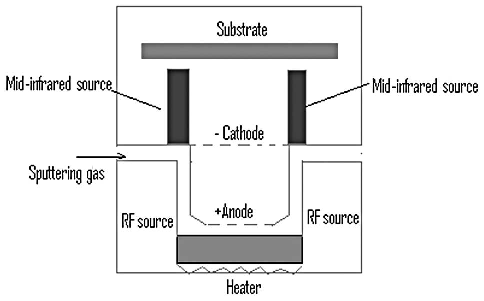
The system shown in Fig. 1 was used in the present study for
nanoparticle preparation. High-moment multifunctional nanoparticles
are synthesized directly from the gas phase by using a physical
vapor nanoparticle-deposition technique.
The target in Fig.
1 is Fe60Co40. As shown in Fig. 1, the heater and radio frequency
(RF) source (13.56 MHz) were simultaneously applied to vaporize the
target. The continual sputtering gas was applied to ensure the
vaporized target atom flowed in the same direction. Electrodes were
also applied to speed up the mobility of the vaporized atoms. Argon
is used as either sputtering gas or cooling media at the same time.
When the supersaturation condition is reached, the vaporized target
atoms form a nucleus and continue to grow into nanoparticles. By
controlling system conditions, including target composition, target
setup, plasma density and gas flow, magnetic nanoparticles are
formed with controlled size, size distribution, composition, phase
and structure. The base pressure of the system was
5×10⁻⁸ Torr. An additional gas tube, for silicon atoms,
does not appear in Fig. 1,
however, the silicon is coated following completion of the
nanoparticle nuclei.
Preparation of sensor system
A sensor is required to combine with the prepared
magnetic nanoparticles. The measuring principle involves
nanoparticles acting as labels, which combine with antibodies. When
the specific biomarker appears, labels with attached antibodies
capture biomarkers. Therefore, another sensor is required to
integrate with nanoparticles and indicate the variations during
measurement.
When the sensor is exposed to the biomarker, it
combines with the biomarker through the antibody. Prior to this,
biomarkers are combined with nanoparticle labels and therefore the
sensor is combined with nanoparticles in this way. The properties
of the sensor vary following combination with the biomarker and
therefore the sensor is used to observe the presence and levels of
the biomarker.
A suitable sensor for the nanoparticles must be
sensitive and measureable. In the present study a SiNW acts as
sensor. The principle is shown in Fig.
2. Fig. 2A demonstrates SiNW
prepared with antibody attached. Fig.
2B demonstrates high moment (FeCo)Si nanoparticles bound with
SiNW through a biomarker.
Sample detection
To avoid the effects of atmosphere and light, all
measurements were performed in a vacuum and in a dark environment.
A passivator layer is likely to be required for future biomedical
applications.
Results
(FeCo)Si nanoparticles
Fig. 3 shows the
TEM image of particles, revealing that the structure is a
core-shell, with cubic shape core. Shell thickness was estimated to
be 3 nm. Fig. 4 demonstrates
selected area electron diffraction patterns of core-shell
structured nanoparticles. Lattice length was measured and indicated
a iron-cobalt bcc structure.
A comparison was made to demonstrate the function of
mid-infrared lighting in particle size distribution. Two groups of
samples were prepared, one assisted with mid-infrared lighting and
the other without lighting. Fig. 5
demonstrates the size distribution of the samples.
Fig. 5A
demonstrates the size distribution of nanoparticles with infrared
lighting. A mean size of 14 nm was observed and the distribution
window was 2%. The line exhibited a normal distribution. Fig. 5B demonstrates the size distribution
of nanoparticles without infrared lighting. The mean size was the
same as nanoparticles with infrared lighting, however, the
distribution window was 10%. Therefore, infrared lighting has an
effect on the size distribution of magnetic samples (Fig. 5). Since size distribution of
particles has an effect on the sensitivity of biosensor, infrared
lighting is utilized to increase sensitivity. To date, this is the
narrowest window for nanoparticles observed.
Sensor system
The preparation of SiNW is not included in this
study due to length limitations. A popular growth method in MBE
(Molecular Beam Epitaxy) was used for preparation (21), assisted by plasma generated by the
RF source inside the MBE system.
The prepared SiNW was 15 nm in diameter and
exhibited an ideal doping profile along its axis. Fig. 6 shows an SEM image, in which drain,
source and gate are annotated.
Comparison of IL-6 detection by SiNW
transistor biosensor and ELISA
To demonstrate the practical application of high
sensitivity SiNW transistor biosensors for biomarker detection,
detection of human IL-6 was performed, following principles used in
ELISA. It was observed that at IL-6 concentrations <1 pg/ml,
ELISA did not generate a sensitive result (Fig. 7).
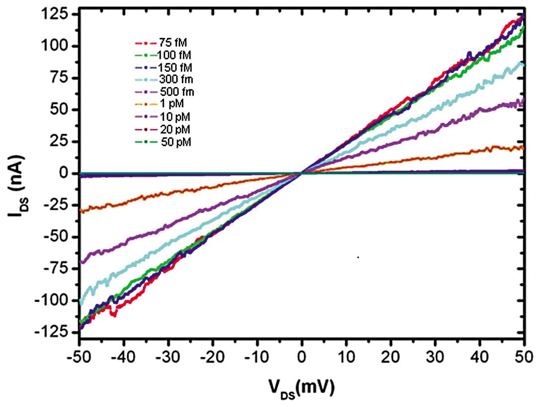
Based on the data in Fig. 8, it is revealed that the novel
method described in the present study detects IL-6 between 75 fM
and 50pM, the most sensitive method reported thus far. When the
dose is fixed, the I-V curve of the transistor appears lineally,
indicative of stable nanoparticle attachment to the nanowire
surface, unaffected by electron flow.
Discussion
Traditional technologies for protein detection are
enzyme-linked immunosorbent assays (ELISA), western blotting,
immunohistochemistry and DNA-based genomics. In the present study,
a novel high sensitivity sensor, composed of a SiNW transistor
combined with high-moment magnetic nanoparticles, was proposed and
demonstrated.
By employing our novel SiNW transistor sensor with
the high-homogenous (FeCo)Si nanoparticles, the sensor was employed
to perform accurate and rapid quantification of IL-6, a
low-abundance protein and potential cancer biomarker, in
unprocessed human serum.
The SiNW transistor sensor system is 20 times more
sensitive than that of the IL-6 ELISA and may be suitable for
quantification of low abundance biomarkers in biological samples.
The SiNW transistor sensor system binds with the same antibody
utilized in the ELISA and therefore may function as an important
supplement to the current commercial ELISA system. Though the SiNW
transistor sensor system has a high sensitivity, the validation of
the specificity of the SiNW transistor sensor system in our
research is insufficient. Additionally, we only detected the
different levels of IL-6 in protein samples, not in patient serum
samples. In our future research we will investigate the method for
using the SiNW transistor sensor system to detect IL-6 in human
serum protein, which has more complex conditions.
In conclusion, the SiNW transistor sensor system is
suitable for quantifying very small amounts of IL-6 in protein
samples. Additionally, it may supplement the current commercial
ELISA system.
Acknowledgements
This study was supported by the National Natural
Science Foundation of China (no. 81141075/H1819) and Medical
Technology Major Projects of Wuxi Hospital Center (YGZX1105).
References
|
1
|
Macy EM, Hayes TE and Tracy RP:
Variability in the measurement of C-reactive protein in healthy
subjects: implications for reference intervals and epidemiological
applications. Clin Chem. 43:52–58. 1997.PubMed/NCBI
|
|
2
|
Engvall E and Perlmann P: Enzyme-linked
immunosorbent assay (ELISA). Quantitative assay of immunoglobulin
G. Immunochemistry. 8:871–874. 1971. View Article : Google Scholar : PubMed/NCBI
|
|
3
|
Alivisatos P: The use of nanocrystals in
biological detection. Nat Biotechnol. 22:47–52. 2004. View Article : Google Scholar : PubMed/NCBI
|
|
4
|
Gao X, Cui Y, Levenson RM, et al: In vivo
cancer targeting and imaging with semiconductor quantum dots. Nat
Biotechnol. 22:969–976. 2004. View
Article : Google Scholar : PubMed/NCBI
|
|
5
|
Soukka T, Paukkunen J, Harma H, Lonnberg
S, Lindroos H and Lovgren T: Supersensitive time-resolved
immunofluorometric assay of free prostate-specific antigen with
nanoparticle label technology. Clin Chem. 47:1269–1278.
2001.PubMed/NCBI
|
|
6
|
Nam JM, Thaxton CS and Mirkin CA:
Nanoparticle-based bio-bar codes for the ultrasensitive detection
of proteins. Science. 301:1884–1886. 2003. View Article : Google Scholar : PubMed/NCBI
|
|
7
|
Wu G, Datar RH, Hansen KM, et al: Bioassay
of prostate-specific antigen (PSA) using microcantilevers. Nat
Biotechnol. 19:856–860. 2001. View Article : Google Scholar : PubMed/NCBI
|
|
8
|
Chen RJ, Bangsaruntip S, Drouvalakis KA,
et al: Noncovalent functionalization of carbon nanotubes for highly
specific electronic biosensors. Proc Natl Acad Sci USA.
100:4984–4989. 2003. View Article : Google Scholar : PubMed/NCBI
|
|
9
|
Chen RJ, Choi HC, Bangsaruntip S, et al:
An investigation of the mechanisms of electronic sensing of protein
adsorption on carbon nanotube devices. J Am Chem Soc.
126:1563–1568. 2004. View Article : Google Scholar : PubMed/NCBI
|
|
10
|
Bosch FX, Ribes J, Diaz M and Cleries R:
Primary liver cancer: worldwide incidence and trends.
Gastroenterology. 127:S5–S16. 2004. View Article : Google Scholar : PubMed/NCBI
|
|
11
|
Scheller J and Rose-John S: Interleukin-6
and its receptor: from bench to bedside. Med Microbiol Immunol.
195:173–183. 2006. View Article : Google Scholar : PubMed/NCBI
|
|
12
|
Pedersen BK and Febbraio MA: Muscle as an
endocrine organ: focus on muscle-derived interleukin-6. Physiol
Rev. 88:1379–1406. 2008. View Article : Google Scholar : PubMed/NCBI
|
|
13
|
Mora LB, Buettner R, Seigne J, et al:
Constitutive activation of Stat3 in human prostate tumors and cell
lines: direct inhibition of Stat3 signaling induces apoptosis of
prostate cancer cells. Cancer Res. 62:6659–6666. 2002.PubMed/NCBI
|
|
14
|
Bromberg JF, Wrzeszczynska MH, Devgan G,
et al: Stat3 as an oncogene. Cell. 98:295–303. 1999. View Article : Google Scholar
|
|
15
|
Swift DC, Gammel JT and Clegg SM:
Treatment of compounds and alloys in radiation hydrodynamics
simulations of ablative laser loading. Phys Rev E Stat Nonlin Soft
Matter Phys. 69:564012004. View Article : Google Scholar : PubMed/NCBI
|
|
16
|
Lee GH, Huh SH, Jeong JW, et al:
Structural and magnetic properties of bimetallic FeCo nanoclusters.
Korean Phys Soc. 42:367–370. 2003.
|
|
17
|
Wang ZH, Choi CJ, Kim BK, et al:
Microstructure and magnetic property of Fe-Co nanoparticles
prepared by chemical vapor condensation process. J Alloys
Compounds. 351:319–323. 2005. View Article : Google Scholar
|
|
18
|
Dong XL, Zhang ZD, Jin SR and Kim BK:
Characterization and magnetic properties of Fe-Co ultrafine
particles. J Magn Magn Mater. 210:143–144. 2000. View Article : Google Scholar
|
|
19
|
Basu S and Chakravorty D: Optical
properties of nanocomposites with iron core-iron oxide shell
structure. J Non Cryst Solids. 352:380–385. 2006. View Article : Google Scholar
|
|
20
|
Ong PL, Mahmood S, Zhang T, et al:
Synthesis of cobalt ferrite nanoparticles by pulsed laser
deposition in a diffusion cloud chamber. Surf Sci. 254:1909–1914.
2008. View Article : Google Scholar
|
|
21
|
Yanfeng J and Yamin Z: Influence of gold
particle size on melting temperature of VLS grown silicon nanowire.
J Semicond. 31:120022010. View Article : Google Scholar
|