Introduction
Cataracts have proven to be a difficult entity for
clinical study, partly due to the difficulty in developing an
objective, accurate and reproducible lens evaluation system.
Evaluation systems based on visual acuity fail to detect early lens
changes and are confounded by the presence of other ocular
abnormalities. Evaluation systems based on clinical impressions
often have large inter- and intra-observer variability. Optical
photography, however, has small variability and is more objective
than other methods (1–4). One of the advantages of Scheimpflug
photography is the large depth of field resulting in an extremely
clear image. Since 1966, Scheimpflug photography has been used for
the documentation and measurement of the anterior eye segment
(5). The first prototypes
constructed by Brown were never produced as commercial versions
(6). An SL-45 camera using film as
data-recording medium was manufactured in 1979 by Topcon Optical
Company (Japan) and for several years it was the only Scheimpflug
camera available (3). In 1984,
Zeiss introduced the SLC system, which was the first video-based
Scheimpflug camera (7). Apart from
these independent camera systems, Sasaki et al(8) designed a photographic unit that may
be attached to the Topcon SL-6E slit lamp microscope. Rapid
progression in digital imaging and computerized image analysis
stimulated the development of Scheimpflug cameras, including the
Oxford Modular Cataract Image Analysis System (case 2000) and the
Nidek Eye Analysis System 1000 (9,10).
In 1996, the photographic camera part of the Topcon SL-45B (which
was also the first rotating Scheimpflug camera) was replaced with a
CCD camera unit, enabling direct image acquisition and
computer-assisted analyses (11).
Pentacam is a newly developed anterior eye segment
analysis system (Oculus Company, Wetzlar, Germany; Fig. 1) and uses the Scheimpflug imaging
principle. It uses a diode with an ultraviolet-free blue light with
a wavelength of 475 nm as the light source and analyzes lens
density through a reflecting image. The system has 25,000 measuring
points and thus, the image is extremely clear. Moreover, it
performs accurate anterior segment analysis, including anterior
chamber depth, anterior chamber angle and anterior chamber volume.
It also performs anterior eye segment three-dimensional model
rebuilding.
In the present study, this system was used to detect
senile nuclear cataract lens density, with the purpose of providing
a quantitative measurement of lens density for the long-term
clinical observation of cataracts.
Patients and methods
Patients
Between January and June 2006, ophthalmic
examinations were performed on outpatients with cataracts at the
Peking University Eye Center (Beijing, China). Subjects with
corneal opacity, glaucoma, eye trauma and other eye diseases,
including diabetic retinopathy, were excluded. Informed consent was
obtained from all the patients. The study was approved by the
Ethics Committee of Peking University Third Hospital.
Imaging
Simple nuclear cataract eyes and transparent eyes
from these patients were imaged by color slit lamp photography
(Zeiss, Yena, Germany) and Scheimpflug photography was performed
with the Anterior Segment Analysis System (Scheimpflug Imaging
System; Pentacam, Oculus Company, Wetzlar, Germany) through pupils
maximally dilated with tripicamide ophthalmic compound (0.5%
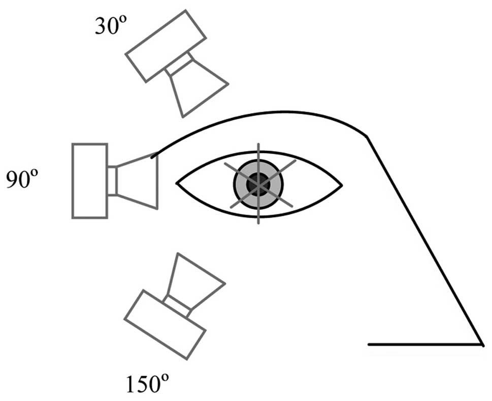
tropicamide and 0.5% phenylephrine) by one technician. Color slit
lamp images of the lenses were captured from the temporal side with
the beam at a 45º angle with fixed width and length of the slit.
Slit images of the lenses were obtained from the temporal side
(12) at three orientations (30,
90 and 150º) and meridional section images of each lens were
obtained (Fig. 2) (13). If blockage of the eyelid was
marked, adhesive tape was used to lift the eyelids in order to
reduce the blockage. Of 238 patients with a mean age of ~70 years
(range, 40–94 years), 422 eyes were evaluated. There were 234 male
and 188 female eyes.
Patients were examined and nuclear hardness was
graded by a doctor using the Emery-Little classification system
(Fig. 3). Another doctor obtained
the lens density values without knowledge of the grading of nuclear
hardness. The values were taken at the optical axis at various
depths measured from the anterior capsule. The optical axis was
determined automatically by the software (Pentacam lens
densitometry program; Oculus, Wetzlar, Germany). In total, eight
depth measurements were taken at 0.5-mm intervals using pointers.
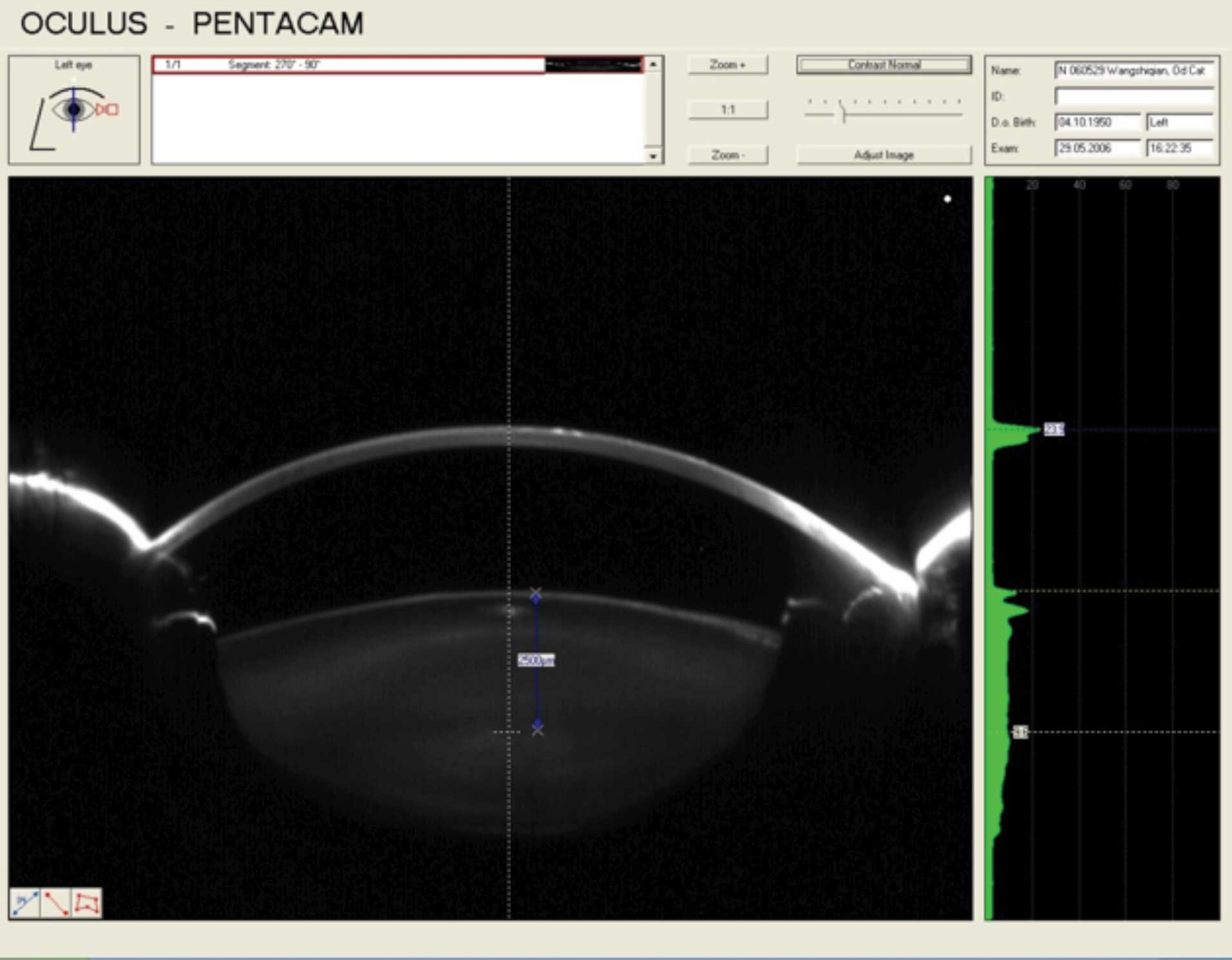
Notably, interference of the reflection of the Scheimpflug
photography system light source was not completely avoided when
obtaining the density values; thus, if the depth point fell on the
reflection of the light source the value was dismissed (Fig. 4).
Statistical analysis
A paired t-test was applied to test the significance
of differences in the three orientation groups and was repeated at
each depth point. One-way analysis of variance was applied to test
the significance of differences among five grading groups at each
depth point of the optical axis. Statistical analyses were
performed with SPSS 11.0 statistical software (SPSS, Inc., Chicago,
IL, USA). P<0.05 was considered to indicate a statistically
significant difference.
Results
Mean density of the three orientation
groups at various depths of the optical axis
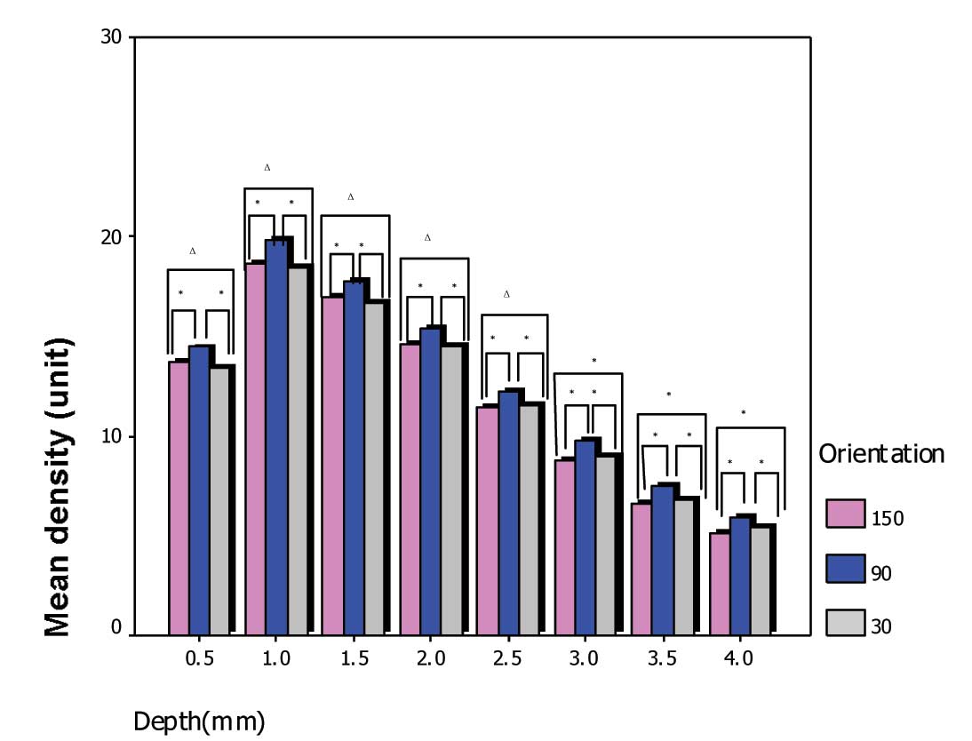
All the groups showed almost the same density value
at each depth point. The number of cases examined and the mean
density of the three orientation groups at each depth point of the
optical axis are shown in Table I.
There was a statistically significant difference between the 90º
group and the two other orientation groups (both P<0.05). At
90º, the mean density value was larger. No significant difference
in density value was noted between the 30 and 150º groups prior to
the 3-mm depth point and the three groups showed a decline in
density beyond the 1.0-mm depth point (Fig. 5).
 | Table IMean density of the three orientation
groups at each depth point of the optical axis. |
Table I
Mean density of the three orientation
groups at each depth point of the optical axis.
| 150º | 90º | 30º |
|---|
|
|
|
|
|---|
| Depth, mm | n | Mean | SD | n | Mean | SD | n | Mean | SD |
|---|
| 0.5 | 412 | 13.712 | 9.2856 | 411 | 14.460 | 10.5877 | 405 | 13.439 | 9.3138 |
| 1.0 | 408 | 18.621 | 8.3560 | 403 | 19.800 | 9.8208 | 408 | 18.465 | 8.5854 |
| 1.5 | 414 | 16.964 | 7.5371 | 412 | 17.770 | 8.2408 | 412 | 16.720 | 6.007 |
| 2.0 | 421 | 14.564 | 6.0807 | 421 | 15.410 | 7.1814 | 417 | 14.483 | 5.9297 |
| 2.5 | 421 | 11.445 | 4.3968 | 422 | 12.277 | 4.5001 | 421 | 11.570 | 4.2786 |
| 3.0 | 421 | 8.815 | 3.1971 | 422 | 9.730 | 3.2123 | 421 | 8.994 | 3.0838 |
| 3.5 | 421 | 6.592 | 2.2155 | 422 | 7.543 | 2.2943 | 421 | 6.856 | 2.2014 |
| 4.0 | 421 | 5.177 | 1.5762 | 422 | 5.909 | 2.0259 | 421 | 5.442 | 2.0488 |
Mean density of the five grades at each
depth point of the optical axis at 90º
Of 422 eyes, there were 35 with grade 0, 96 with
grade 1, 127 with grade 2, 127 with grade 3 and 23 with grade 4; in
14, the value was not determined due to loss of color in the slit
lamp photographs (Fig. 6). The
mean densities of the various grades are shown in Table II.
| Table IIMean density of the five grades at
each depth point of the optical axis at 90º. |
Table II
Mean density of the five grades at
each depth point of the optical axis at 90º.
| 0.5 mm | 1.0 mm | 1.5 mm | 2.0 mm | 2.5 mm | 3.0 mm | 3.5 mm | 4.0 mm |
|---|
|
|
|
|
|
|
|
|
|
|---|
| Grade | Mean | SD | Mean | SD | Mean | SD | Mean | SD | Mean | SD | Mean | SD | Mean | SD | Mean | SD |
|---|
| 0 | 10.960 | 3.1226 | 13.397 | 4.1579 | 11.457 | 3.4342 | 10.146 | 2.8129 | 9.320 | 2.3337 | 8.526 | 2.0535 | 7.809 | 1.7255 | 6.509 | 3.4418 |
| 1 | 13.878 | 9.8328 | 16.451 | 6.4321 | 13.482 | 4.0230 | 12.278 | 3.3263 | 10.827 | 2.7978 | 9.221 | 2.2743 | 7.604 | 1.9366 | 5.886 | 1.7248 |
| 2 | 13.399 | 6.8027 | 18.642 | 5.3914 | 15.696 | 4.0446 | 14.224 | 3.6382 | 12.343 | 3.2979 | 10.376 | 3.0379 | 8.079 | 2.2737 | 6.413 | 1.9247 |
| 3 | 14.148 | 10.3247 | 22.246 | 9.5370 | 22.961 | 8.6140 | 19.763 | 7.4675 | 14.325 | 5.5429 | 10.029 | 3.6553 | 7.056 | 2.2554 | 5.328 | 1.3853 |
| 4 | 31.848 | 22.6109 | 35.513 | 22.0768 | 28.396 | 15.3646 | 19.774 | 17.4216 | 11.535 | 7.4943 | 8.626 | 5.2924 | 6.526 | 3.8077 | 5.391 | 2.9915 |
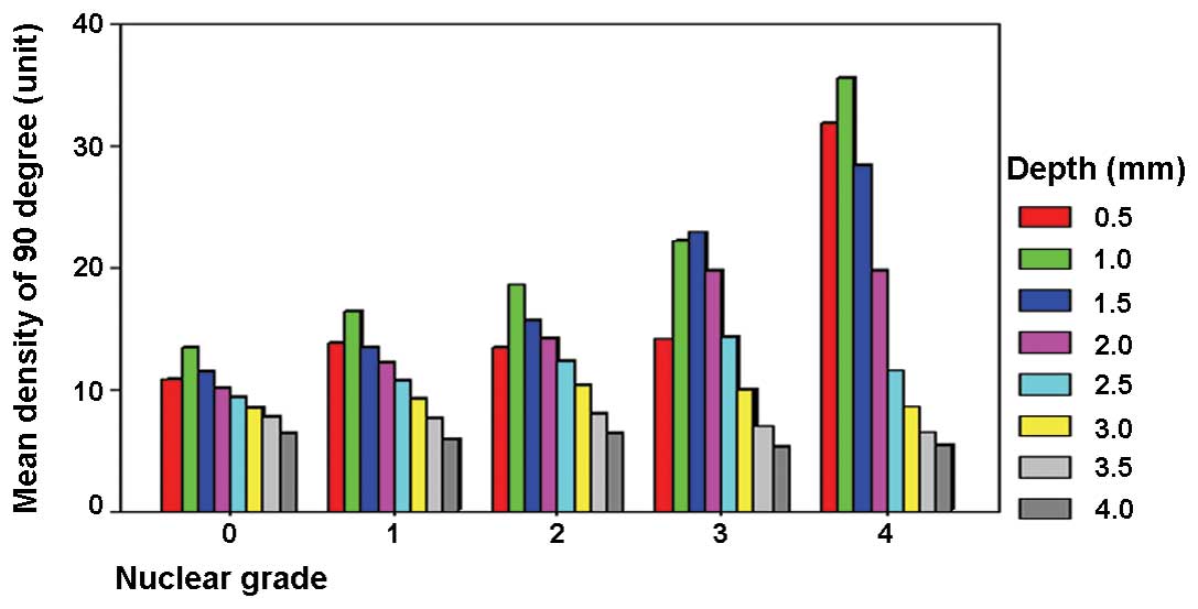
Mean density of five grade groups at
various depths of the optical axis at 90º (depth and nuclear
grade)
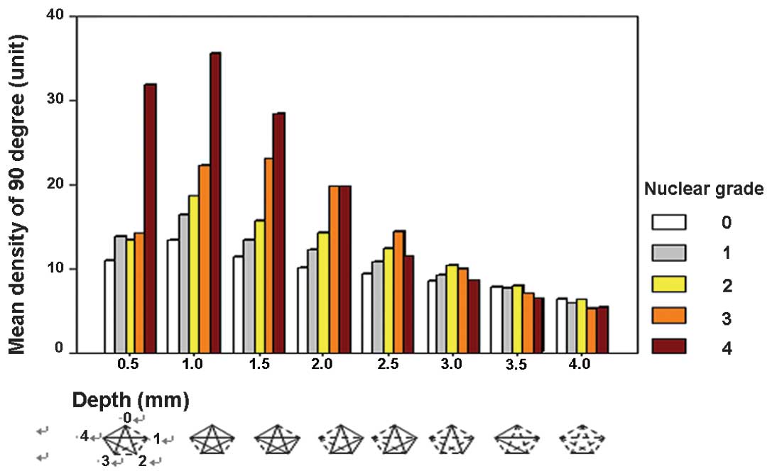
Differences in density of the five grade groups at
each depth point of the optical axis at 90º are shown in Figs. 7 and 8. At the 0.5-mm depth point, there were
no significant differences in density among grades 1, 2 and 3 (all
P>0.05). However, there was a significant difference between
grade 0 and the other groups, as well as between grade 4 and the
other groups (P<0.05). These data suggest that in grade 4
lenses, the density is increased in the superficial part of the
lens, however, the differences among grades 1, 2 and 3 were not
obvious.
There were significant differences among almost all
grades at the 1.0- and 1.5-mm depth points (all P<0.05) and the
mean density showed a tendency to increase with increasing nuclear
grade.
There were significant differences among grades 0,
1, 2 and 3 at the 2.0- and 2.5-mm depth points (P<0.05) and the
mean density showed a tendency to increase with increasing nuclear
grade. However, no significant differences were noted between grade
4 and the other grades (P>0.05).
Beyond the 3.0-mm depth point, values in all the
grade groups were low and no significant differences were noted
(all P>0.05).
Discussion
Previous studies have been performed to show the
correlation between the lens peak and mean density and clinical
nuclear grade (1), as well as lens
density and age (2,12), and follow-up observation of lens
densitometry (14). Results of
this study demonstrated that the standard deviation of density
values of three orientation groups at various depths of the optical
axis was large. One explanation is that each orientation group
included all nuclear grades, thus the range of the values was
large. Of the density values of the five grade groups at various
depths, the standard deviation of grade 4 was large and this was
likely due to the small number of cases. Important factors that
affect the range of values are the positioning of the patient's
head and the fixation of the examined eye. The blockage of eyelids
and eyelashes also affects the range of values. In addition, the
thickness of lens is different; however, values were taken from the
optical axis at various depths measured from the anterior capsule.
With the Scheimpflug Anterior Segment Analysis System, a reflection
of the system light source near the anterior capsule was
consistently found when the density value was abnormally large.
This affected the accuracy of determination of the mean density and
peak density of the whole lens. In order to be as accurate as
possible, if the depth point fell on the light source reflection,
this value was dismissed.
The current data indicated that the 90º orientation
values were the most reliable, which is possibly due to minimal
blockage by the eyelids and eyelashes. Thus, the values of the 90º
group were used for the statistical analysis.
The thickness of the lens is 4–5 mm. In theory, the
density should distribute symmetrically from the center. In this
study, all nuclear grades showed a reduction in density beyond the
1.0-mm depth point and this tendency was more obvious as the grade
increased. An important reason for this observation is that the
Scheimpflug analysis system analyzes the lens density through a
reflected image. A high level of back-scatter light in the anterior
nucleus decreases the amount of light transmitted to the posterior
parts of the nucleus, thus, making them appear less dense. In
addition, heavily pigmented nuclear cataracts may absorb a large
amount of back-scattered light and thus, appear less dense. In
practical terms, the anterior part of the lens is the least
affected by any of these changes and is the easiest to analyze
(15). The results also showed
that beyond the 3.0-mm depth point, values in all the grade groups
were low, without significant differences.
Semiautomatic camera alignment and focusing is one
advantage of the anterior eye segment analysis system used in the
present study. It is a highly automated method of photography and
analysis that is simple to operate without extensive operator
experience. The 180º autorotation of the system captures
high-quality images of the whole anterior eye segment and the
software determines the lens density at any point in any meridional
section. Important factors that affect reproducibility of the data
includes the positioning of the patient's head, fixation of the
examined eye and blockage by the eyelid and eyelashes. As the
measuring depth increased, the effect of the physics of optics was
unavoidable, which affected the reliability of the values.
In conclusion, the Anterior Segment Analysis System
may be used to detect senile nuclear cataract lens density and
provide a quantitative measurement of lens density for long-term
clinical observation of cataracts.
References
|
1
|
Adamsons I, Taylor KI, Enger C and Taylor
HR: A new method for documenting lens opacities. Am J Ophthalmol.
111:65–70. 1991. View Article : Google Scholar : PubMed/NCBI
|
|
2
|
Bosem ME, Sample PA, Martinez GA, Lusky M
and Weinreb RN: Age-related changes in the human lens: a comparison
of Sheimpflug photography and lens density index. J Cataract
Refract Surg. 20:70–73. 1994. View Article : Google Scholar : PubMed/NCBI
|
|
3
|
Wegener A, Hockwin O, Laser H and Strack
C: Comparison of the Nidek EAS 1000 system and the Topcon SL-45 in
clinical application. Ophthalmic Res. 24(Suppl 1): 55–62. 1992.
View Article : Google Scholar : PubMed/NCBI
|
|
4
|
Sakamoto Y, Sasaki K, Nakamura Y and
Watanabe N: Reproducibility of data obtained by a newly developed
anterior eye segment analysis system, EAS-1000. Ophthalmic Res.
24(Suppl 1): 10–20. 1992. View Article : Google Scholar : PubMed/NCBI
|
|
5
|
Drews RC: Depth of field in slit lamp
photography: an optical solution using the Scheimpflug principle.
Ophthalmologica. 148:143–150. 1964. View Article : Google Scholar : PubMed/NCBI
|
|
6
|
Brown N: Slit-image photography. Trans
Ophthalmol Soc UK. 89:397–408. 1970.
|
|
7
|
Hockwin O, Laser H and Wegener A:
Investigations of rat eyes with diabetic cataract and naphthalene
cataract by Zeiss- Scheimpflug measuring system SLC. Graefes Arch
Clin Exp Ophthalmol. 224:502–506. 1986. View Article : Google Scholar : PubMed/NCBI
|
|
8
|
Sasaki K, Sakamoto Y, Shibata T, et al:
New camera for lens photography. J Ophthalmol Opt Soc (Jpn).
6:40–41. 1985.
|
|
9
|
Sparrow JM, Brown NA, Shun-Shin GA and
Bron AJ: The Oxford modular cataract image analysis system. Eye
(Lond). 4:638–648. 1990. View Article : Google Scholar : PubMed/NCBI
|
|
10
|
Kojima M, Wegener A and Hockwin O: Imaging
characteristics of three cameras using the Scheimpflug principle.
Ophthalmic Res. 22(Suppl 1): 29–35. 1990. View Article : Google Scholar
|
|
11
|
Dragomirescu V and Hockwin O: Rotating
slit image camera TOPCON SL 45. New developments for simultaneous
image acquisition by photographic and CCD-assisted on-line
documentation. Ophthalmic Res. 28(Suppl 2): 102–108.
1996.PubMed/NCBI
|
|
12
|
Fujisawa K and Sasaki K: Changes in light
scattering intensity of the transparent lenses of subjects selected
from population-based surveys depending on age: analysis through
Scheimpflug images. Ophthalmic Res. 27:89–101. 1995. View Article : Google Scholar
|
|
13
|
Kashima K, Unser M, Datiles MB, Trus BL
and Edwards PA: Minimum views required to characterize cataracts
when using the Scheimpflug camera. Graefes Arch Clin Exp
Ophthalmol. 231:687–691. 1993. View Article : Google Scholar : PubMed/NCBI
|
|
14
|
Müller-Breitenkamp U, Laser H and Hockwin
O: Objectified measurements of eye lens transparency in a volunteer
group of advanced age, carried out over a period of 3.5 years.
Ophthalmic Res. 24(Suppl 1): 40–46. 1992.PubMed/NCBI
|
|
15
|
Foo KP and Maclean H: Measured changes in
cataract over six months: sensitivity of the Nidek EAS-1000.
Ophthalmic Res. 28(Suppl 2): 32–36. 1996. View Article : Google Scholar : PubMed/NCBI
|