Introduction
Asthma is a chronic inflammatory airway disease
involving several different types of cell. It is considered an
allergic disease with characteristic airway inflammation, airway
hyper-responsiveness (AHR) and airway remodeling (1–3). At
present, anti-inflammatories are the predominant therapeutic option
for patients with asthma (4,5).
Previous studies (6–8) have shown that airway remodeling
results in the progressive loss of lung function. Airway remodeling
includes epithelial denudation, subepithelial fibrosis, mucus gland
hypertrophy, myofibroblast and smooth muscle cell proliferation,
and angiogenesis (9–11). The expression levels of α-smooth
muscle actin (α-SMA) (12,13) and proliferating cell nuclear
antigen (PCNA) (14,15) are indicative of asthmatic cell
proliferation.
Adenosine triphosphate (ATP), a key mediator of
acute and chronic inflammation, can be released in substantial
quantities from various cell types following cellular stress or
tissue injury (16–19). Extracellular ATP exerts
pro-inflammatory and immunomodulatory effects in the tumor
microenvironment by binding to purinergic P2-receptors, which can
be subdivided into two families: The G-protein-coupled P2YR (P2Y
1–14) and the ligand-gated ion channel, P2X receptors (P2XR), which
include P2X1-7 (20–22). Data from patient and animal models
of allergic bronchial asthma indicate that ATP contributes to the
pathophysiology of allergic airway inflammation (16). However, whether ATP-mediated P2X4R
signaling is activated in allergic asthma remains to be
elucidated.
Among the P2XR subtypes, P2X4R is the most widely
expressed (23). P2X4 receptors
are located in immune and non-immune cells, including alveolar
epithelial cells, alveolar macrophages, platelets, lymphocytes and
mast cells (24–26). P2X4R can affect T cell activation
via Ca2+ influx. ATP-P2X4R signaling is involved in
peripheral nerve injury inflammation, which includes the promotion
of the expression levels of interleukin (IL)-1β, IL-6 and tumor
necrosis factor (TNF)-α via mitogen-activated protein kinase
activation; indicating that P2X4R is involved in the immune
response to nerve inflammation (27). Several studies have demonstrated
that P2X4 receptors are involved in hypersensitivity by acting on
prostaglandin E2 (28–30). ATP-P2X4R signaling is also involved
in pulmonary vascular remodeling, and in the proliferation and
differentiation of airway and alveolar epithelial cell lines in
pulmonary hypertension (31,32).
However, whether P2X4R signaling is involved in allergic airway
inflammation and airway remodeling requires further
investigation.
The aim of the present study was to investigate the
effects of P2X4R in a murine experimental asthma model and the
effects of a P2X4R-specific antagonist, 5-BDBD, on airway
inflammation and airway remodeling. Our findings may ultimately
lead to the development of novel therapeutic targets for allergic
asthma.
Materials and methods
Chemicals and reagents
Rabbit polyclonal anti-P2X4R primary antibody was
purchased from Calbiochem (EMD Millipore, Billerica, MA, USA; cat.
no. PC376) and used for western blotting. Mouse monoclonal
anti-PCNA primary antibody (cat. no. BM0104) and anti-α-SMA primary
antibody (cat. no. BM0002) were purchased from Boster Systems, Inc.
(Wuhan, China). Mouse monoclonal anti-β-actin primary antibody
(cat. no. TA-09) was purchased from ZSGB Biotechnology, Inc.
(Beijing, China). Mouse anti-rabbit (cat. no. sc-2357) and rabbit
anti-mouse (cat. no. sc-358914) IgG-horseradish
peroxidase-conjugated secondary antibodies were purchased from
Santa Cruz Biotechnology Inc. (Santa Cruz, CA, USA). The
P2X4R-specific antagonist, 5-BDBD, was obtained from Tocris
Bioscience (Bristol, UK). The Masson's trichromatic staining kit
and Alcian blue-periodic acid-schiff (AB-PAS) mucosa staining kit
were purchased from Fuzhou Maixin Biotechnology, Co., Ltd. (Fuzhou,
China). ATP and the P2XR1/2/3/5/7 antagonist,
phenyl-isopropyl-amine dopes (PPAD), were all purchased from
Sigma-Aldrich (St. Louis, MO, USA). All other reagents were
obtained from common commercial sources.
Experimental animals
Pathogen-free female BALB/c mice, aged 6–8 weeks and
weighing ~18–20 g, were purchased from the Laboratory Animal
Research Center (Beijing, China). Mice were maintained under 12 h
light/dark cycles at room temperature, with free access to food and
water. All animal experiments and manipulation procedures were
approved by the Ethics Committee for Animal Use and Care of Harbin
Medical University-Daqing (Daqing, China).
Animal groups and allergic sensitization
protocol
A total of 48 BALB/c mice were randomized into the
following five groups: Phosphate-buffered saline (PBS) control
group (n=8), OVA group (n=10), ATP group (n=10), PPAD group (n=10)
and 5-BDBD group (n=10). In the OVA group, the mice were sensitized
to OVA (20 µg per injection), absorbed to 2.0 mg per
injection of aluminum hydroxide (Tianjin Fuchen Chemical Reagents
Factory, Tianjin, China), by intraperitoneal injection on day 1. On
days 8, 15 and 22, the mice were sensitized again to OCA (10
µg per injection), absorbed to 1.0 mg per injection of
aluminum hydroxide, by intraperitoneal injection. From day 23, the
mice were exposed, using inhalation aerosols, of 4% OVA in PBS for
25–30 min, until the onset of bronchial obstruction, daily for 7
days consecutively, according to the methods described by Shen
et al (31) and Vanacker
et al (32) with
modifications. The mice in the ATP, PPAD and 5-BDBD groups were
also subjected to OVA sensitization and asthma induction, in the
same manner. Intranasal application of 50 µl ATP (1 mM)
(33), PPAD (0.1 mM) (34) and 5-BDBD (30 µmol) (35) were performed 3 h prior to each
airway allergen challenge in the ATP, PPAD and 5-BDBD groups,
respectively. All treatments were administered daily for 5 days
consecutively. Mice in the control group were administered with PBS
alone (0.5 ml) by injection, and were challenged with PBS (20 ml).
All animals were humanely sacrificed within 24 h of the final OVA
or PBS exposure.
Morphometric analysis
The lungs were fixed in 4% paraformaldehyde,
embedded in paraffin and sectioned at a thickness of 5 µm.
The sections from each animal were stained with hematoxylin and
eosin for inflammation, Masson's trichrome staining for
collagen-deposition and Alcian blue/periodic acid-Schiff (AB-PAS)
staining for the analysis of goblet cell hyperplasia/mucus
production. All staining procedures were performed in accordance
with the manufacturer's protocol. Pathological changes in the lung
and bronchial tissues were observed and images were captured using
a BX-60 microscope (Olympus, Tokyo, Japan). The tissues were scored
based on the presence of inflammatory cells, using the following
scale: Absent (0), rare (1), mild
(2), moderate (3) or severe (4). Scoring was performed by an observer
in a blinded-manner. Positively stained areas of collagen and mucus
in the lung and bronchial tissues were quantified using
high-resolution images of individual vessels by image analysis,
using a color-recognition algorithm, in Image Pro Plus 6.0 (Media
Cybernetics, Inc., Rockville, MD, USA). The results were expressed
as the percentage of collagen area or mucus area, compared to the
total measured area.
Bronchoalveolar lavage fluid (BALF)
Samples of BALF were collected 24 h following the
final challenge, according to previously described methods. The
lungs were lavaged with 1.6 ml (0.8 ml per lavage) of ice-cold PBS
via the tracheal cannula. The retrieved volume (~75–80%) of the
instilled PBS was then centrifuged at 500 × g at 4°C for 5 min. The
resultant pellet was washed twice with PBS and was resuspended in 1
ml PBS. The total numbers of cells were counted using a
hemocytometer (Bio-Rad Laboratories, Inc., Hercules, CA, USA).
Smears of the BALF cells were stained with Wright and Giemsa
staining fluid (Beijing Solarbio Science & Technology Co.,
Ltd., Beijing, China) for differential cell counting. Using a BX-60
Olympus microscope, the numbers of cells were counted by two
independent investigators, in a blinded-manner. A total of ~200
cells were counted in each of four randomly selected locations.
Western blotting
The lung tissues were homogenized with cold lysis
buffer (Beyotime Institute of Biotechnology, Shanghai, China),
containing 50 mM Tris (pH 7.4), 150 mM NaCl, 1% Triton X-100, 1 mM
EDTA and 2 mM PMSF. Following ultrasonication on ice for 5 sec
using an FA-25 (Fluko Equipment Shanghai Co., Ltd., Shanghai,
China) and centrifugation at 12,500 × g at 4°C for 15 min, the
supernatant was collected, and the protein concentration was
determined using a Bicinchoninic Acid Protein Assay (Pierce
Biotechnology, Inc., Rockford, IL, USA) with bovine serum albumin
(Biosharp, Hefei, China) as a standard. Each sample, containing 20
µg protein, was separated on 8–10% SDS-PAGE gels and
transferred onto nitrocellulose membranes (EMD Millipore).
Following blocking with Tris-buffered saline, containing 20 mM
Tris, 150 mM NaCl (pH 7.6) and 0.1% Tween 20, with 5% nonfat milk
at room temperature, the membranes were incubated overnight at 4°C
with specific antibodies against P2X4R (1:400 dilution), α-SMA
(1:500 dilution), PCNA (1:500 dilution) or β-actin (1:1,000
dilution). This was followed by incubation for 2 h at room
temperature with horseradish peroxidase-conjugated secondary
antibodies (1:5,000 dilution). All protein blots were visualized
using enhanced chemiluminescence reagents (GE Healthcare Life
Sciences). Integrated density values were analyzed using a
computerized image analysis system (Fluor Chen 2.0; Olympus
Corporation) and were normalized to those of β-actin.
Statistical analysis
Data were analyzed using SPSS 15.0 software (SPSS,
Inc., Chicago, IL, USA). Data are expressed as the mean ± standard
error of the mean. Differences between groups were analyzed using
Student's t-test and one-way analysis of variance, followed by
Fisher's least significant difference test. P<0.05 was
considered to indicate a statistically significant difference.
Results
OVA-challenge upregulates the protein
expression of P2X4R in mice
The present study performed western blotting to
compare the protein expression levels of P2X4R in the lung tissues
of the treatment groups (Fig. 1A and
B). As shown in Fig. 1,
OVA-challenge significantly increased the expression of P2X4R,
compared with the control mice.
P2X4R contributes to inflammation of the
bronchi and lung tissues in allergic airway challenge in mice
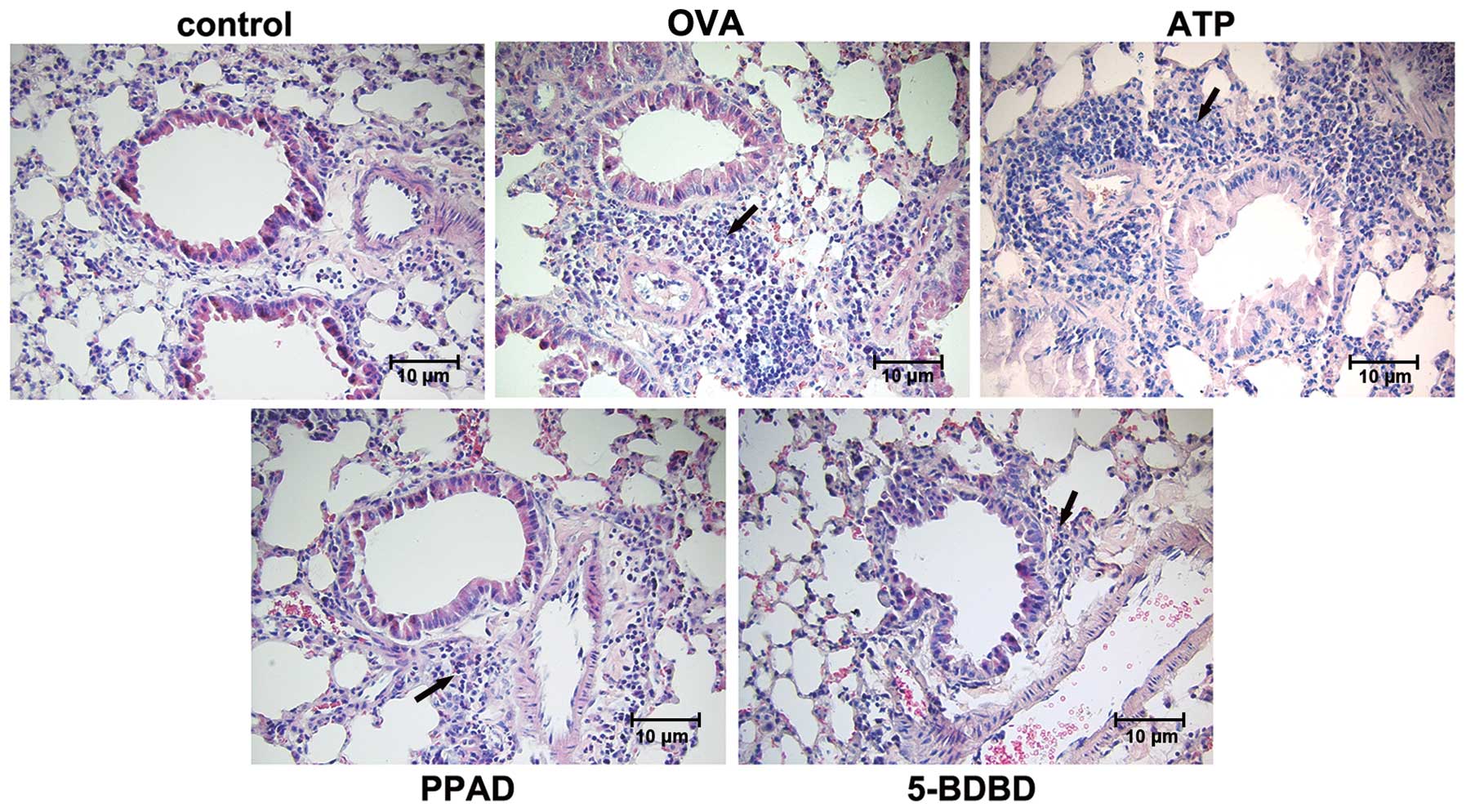
Histological analyses of the tissues of the mice in
the OVA group showed more extensive infiltration of inflammatory
cells around the bronchioles, blood vessels and alveoli. The airway
mucosa showed edema, with bronchiolar wall thickening causing
luminal stenosis, suggesting successful establishment of the
asthmatic model. In the OVA-challenged mice, treatment with ATP
significantly increased OVA-induced inflammation, infiltration, and
pathological changes of the bronchi and lung tissues. By contrast,
treatment of OVA-challenged mice with PPAD (a P2X1, 2, 3, 5 and 7R
antagonist) (36) and 5-BDBD (a
P2X4R antagonist) (37)
significantly reduced the extent of inflammation and cellular
infiltration in the airway. In addition, the pathological changes
of the bronchi and lung tissues were milder, compared with the
changes found in the asthma group without treatment (Fig. 2A and B).
Cell numbers and differential cell
percentages in BALF
The total cell numbers (Fig. 3A) and the percentages of
eosinophils and lymphocytes (Fig.
3B) in the BALF from the asthmatic mice increased
significantly, compared with those in the BALF from the control
mice. The increased numbers of inflammatory cells, including
eosinophils and lymphocytes, were significantly reduced by the
administration of PPAD and 5-BDBD in the asthmatic mice, compared
with the numbers found in the untreated asthmatic mice. The changes
in the percentages of macrophages showed the opposite results.
P2X4R is required for collagen deposition
in allergic airway challenge in mice
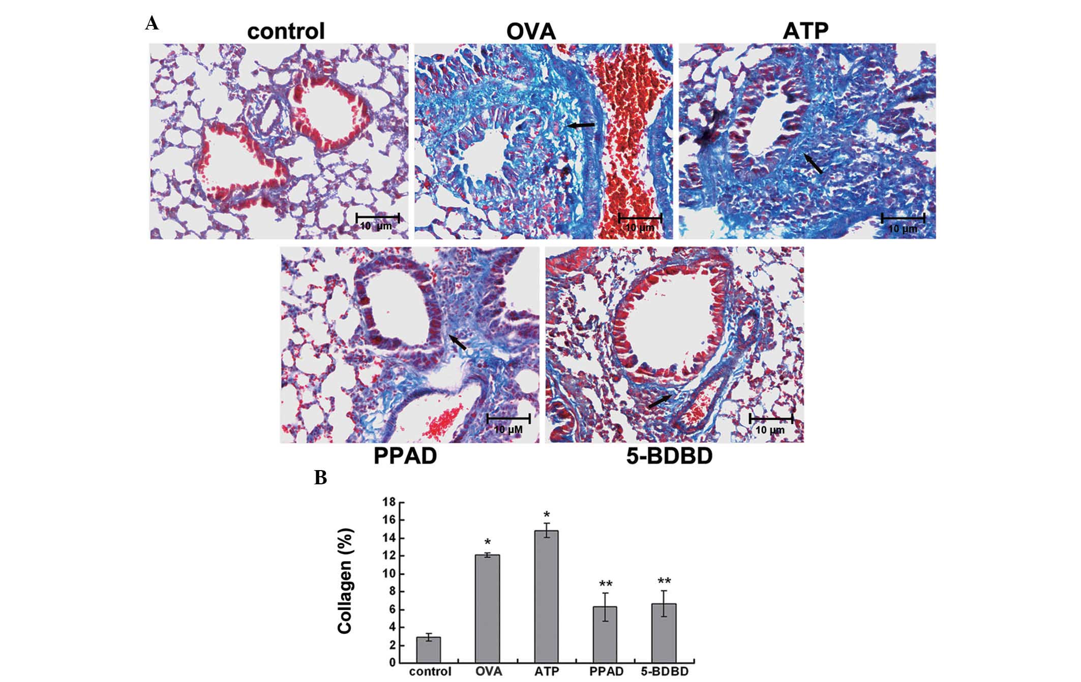
Collagen deposition in airway remodeling was
assessed using Masson's trichrome staining of lung histological
sections. Collagenous matrix was identified by its characteristic
blue color. No obvious collagen fibers were present in the lungs of
mice in the control group. However, deposition of collagen fibers
was increased around the bronchial airways and vessels in the lungs
of the OVA-challenged and ATP-treated mice. By contrast,
intervention with PPAD and 5-BDBD significantly reduced collagen
deposition (Fig. 4A and B).
P2X4R is required for goblet cell
hyperplasia and mucus production in allergic airway challenge in
mice
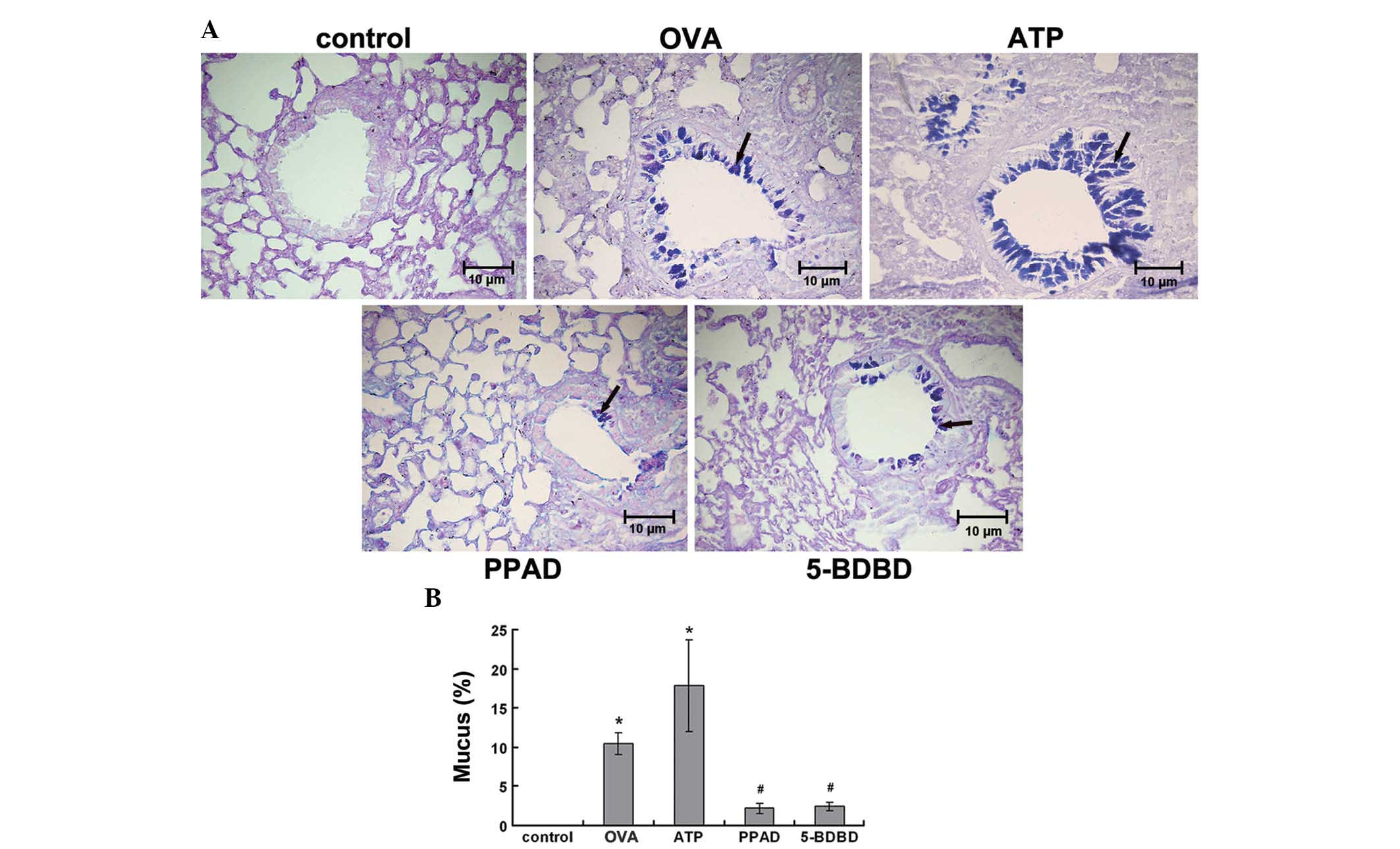
To assess mucus production and the hyperplasia of
goblet cells, lung sections were stained with AB-PAS. In the
OVA-challenged mice, bronchial epithelial mucus secretion and
goblet cell hyperplasia were clearly observed, as a violet color in
the bronchial airways, compared with the control group. The level
of goblet cell hyperplasia in the OVA-challenged mice treated with
ATP was significantly increased, compared with that in the OVA
group. Intervention by PPAD or 5-BDBD significantly reduced goblet
cell hyperplasia (Fig. 5A and
B).
P2X4R regulates the expression of α-SMA
and PCNA in the asthmatic mouse lung
Western blotting was performed to examine the
protein levels of α-SMA (Fig. 6A and
B) and PCNA (Fig. 7A and B) in
each group. The expression levels of α-SMA in the OVA-challenged
mice and ATP-treated mice were significantly lower, compared with
those in the control mice. In addition, the mice treated with
5-BDBD showed significantly increased protein expression levels of
α-SMA, compared with the mice in the OVA and ATP groups. However,
the mice treated with PPAD showed no significant differences in
protein expression, compared with the OVA group. The expression
level of PCNA in the OVA-challenged mice and the ATP-treated mice
were significantly higher than the level in the control mice. In
addition, the mice treated with 5-BDBD showed a significant
reduction in the protein expression levels of PCNA, compared with
the mice in the OVA and ATP groups. Treatment with PPAD had no
significant effects.
Discussion
Allergic asthma is a chronic airway
inflammatory-driven disease, which is characterized by airway
hyper-responsiveness, airway inflammation and varying degrees of
smooth muscle cell proliferation, termed airway remodeling
(36–38). In the present study, airway
inflammation and airway remodeling were examined in
allergen-challenged mice. The pathophysiological mechanisms
underlying airway remodeling remain to be fully elucidated.
Compelling evidence indicates a pathophysiological relevance for
ATP, acting via P2 purinergic receptors, in pain hypersensitivity
and peripheral inflammation (39–41).
Certain studies have suggested that ATP contributes to the
pathophysiology of allergic airway inflammation (14,16,42).
In the present study, treatment of OVA-challenged mice with ATP
significantly increased OVA-induced inflammation, infiltration, and
pathological changes of the bronchi and lung tissues. Increased
collagen deposition, goblet cell hyperplasia/mucus production,
increased expression levels of PCNA expression, and decreased
expression levels of α-SMA are indicative of airway remodeling
(43–46), and were observed in mice belonging
to the OVA-challenged and ATP-treated groups. These results
indicated that ATP may enhance airway inflammation and airway
remodeling in allergic asthma. However, whether P2X4R is activated
in allergic asthma by ATP-mediated signaling has not been
determined.
Among the P2XR subtypes, P2X4R is the most widely
expressed, which is expressed in blood vessels, the lungs, kidneys
and immune cells (47,48). Additionally, P2X4R is involved in
regulating blood pressure and vascular remodeling by ATP-induced
Ca2+ influx and flow-mediated vasodilatation (49,50).
P2X4Rs are also involved in modulating right ventricular
hypertrophy, which occurs due to pulmonary hypertension in
hypobaric hypoxia (51). Asthma
and pulmonary arterial hypertension exhibit three major
pathological features: Inflammation, smooth muscle constriction and
smooth muscle cell proliferation (52). In the present study, the results
demonstrated that treatment with the P2X4R antagonist, 5-BDBD,
prevented the development of the airway and pulmonary inflammation,
and airway remodeling induced by OVA challenge and OVA challenge
coupled with ATP treatment. By contrast, treatment with PPAD showed
no significant effect on the expression levels of α-SMA or PCNA.
These data indicated that ATP-mediated P2X4R activation, but not
P2X1/2/3/5/7R activation, was involved in airway inflammation and
airway remodeling in allergic asthma. This suggests that P2X4R is
involved in the pathogenesis of asthma via an ATP-mediated
signaling pathway. To the best of our knowledge, the present study
provides the first indication that P2X4R signaling is activated in
allergic asthma, and contributes to airway inflammation and
remodeling.
In conclusion, the results of the present study
demonstrated the importance of P2X4R in allergic asthma. ATP-P2X4R
signaling contributes to the pathogenesis of allergic asthma in
mice. OVA-induced allergic asthma in mice was enhanced by the P2X4R
agonist and attenuated by the P2X4R antagonist. The finding
suggested that the ATP-P2X4R pathway may not only contribute to
airway inflammation, but also to airway remodeling in models of
allergic asthma in mice. The exact mechanisms of ATP-P2X4R
signaling responsible for asthma pathogenesis requires further
investigation.
Acknowledgments
This study was supported by the Natural Science
Foundation of China (grant no. 81200011) and the Postdoctoral
Science Foundation of Heilongjiang Province in China (grant no.
LBH-Z12213).
References
|
1
|
Benayoun L and Pretolani M: Airway
remodeling in asthma: Mechanisms and therapeutic perspectives. Med
Sci (Paris). 19:319–326. 2003. View Article : Google Scholar
|
|
2
|
Uhlík J, Simůnková P, Žaloudíková M,
Partlová S, Jarkovský J and Vajner L: Airway wall remodeling in
young and adult rats with experimentally provoked bronchial asthma.
Int Arch Allergy Immunol. 164:289–300. 2014. View Article : Google Scholar : PubMed/NCBI
|
|
3
|
Rhee CK, Kim JW, Park CK, Kim JS, Kang JY,
Kim SJ, Kim SC, Kwon SS, Kim YK, Park SH and Lee SY: Effect of
imatinib on airway smooth muscle thickening in a murine model of
chronic asthma. Int Arch Allergy Immunol. 155:243–251. 2011.
View Article : Google Scholar : PubMed/NCBI
|
|
4
|
Cates C: Inhaled corticosteroids in COPD:
Quantifying risks and benefits. Thorax. 68:499–500. 2013.
View Article : Google Scholar
|
|
5
|
Pascual RM and Peters SP: Airway
remodeling contributes to the progressive loss of lung function in
asthma: An overview. J Allergy Clin Immunol. 116:477–486. 2005.
View Article : Google Scholar : PubMed/NCBI
|
|
6
|
McParland BE, Macklem PT and Pare PD:
Airway wall remodeling: Friend or foe? J Appl Physiol (1985).
95:426–434. 2003. View Article : Google Scholar
|
|
7
|
James AL and Wenzel S: Clinical relevance
of airway remodelling in airway diseases. Eur Respir J. 30:134–155.
2007. View Article : Google Scholar : PubMed/NCBI
|
|
8
|
Royce SG and Tang ML: The effects of
current therapies on airway remodeling in asthma and new
possibilities for treatment and prevention. Curr Mol Pharmacol.
2:169–181. 2009. View Article : Google Scholar : PubMed/NCBI
|
|
9
|
Raissy HH, Kelly HW, Harkins M and Szefler
SJ: Inhaled corticosteroids in lung diseases. Am J Respir Crit Care
Med. 187:798–803. 2013. View Article : Google Scholar : PubMed/NCBI
|
|
10
|
Tagaya E and Tamaoki J: Mechanisms of
airway remodeling in asthma. Allergol Int. 56:331–340. 2007.
View Article : Google Scholar : PubMed/NCBI
|
|
11
|
Warner SM and Knight DA: Airway modeling
and remodeling in the pathogenesis of asthma. Curr Opin Allergy
Clin Immunol. 8:44–48. 2008. View Article : Google Scholar : PubMed/NCBI
|
|
12
|
Roy SG, Nozaki Y and Phan SH: Regulation
of alpha-smooth muscle actin gene expression in myofibroblast
differentiation from rat lung fibroblasts. Int J Biochem Cell Biol.
33:723–734. 2001. View Article : Google Scholar : PubMed/NCBI
|
|
13
|
Stumm CL, Halcsik E, Landgraf RG, Camara
NO, Sogayar MC and Jancar S: Lung remodeling in a mouse model of
asthma involves a balance between tgf-β1 and BMP-7. PloS One.
9:e959592014. View Article : Google Scholar
|
|
14
|
Kesavan R, Potunuru UR, Nastasijević B, T
A, Joksić G and Dixit M: Inhibition of vascular smooth muscle cell
proliferation by gentiana lutea root extracts. PloS One.
8:e613932013. View Article : Google Scholar : PubMed/NCBI
|
|
15
|
Zhao L, Wu J, Zhang X, Kuang H, Guo Y and
Ma L: The effect of shenmai injection on the proliferation of rat
airway smooth muscle cells in asthma and underlying mechanism. BMC
Complement Altern Med. 13:2212013. View Article : Google Scholar : PubMed/NCBI
|
|
16
|
Idzko M, Hammad H, van Nimwegen M, Kool M,
Willart MA, Muskens F, Hoogsteden HC, Luttmann W, Ferrari D, Di
Virgilio F, et al: Extracellular atp triggers and maintains
asthmatic airway inflammation by activating dendritic cells. Nat
Med. 13:913–919. 2007. View
Article : Google Scholar : PubMed/NCBI
|
|
17
|
Khakh BS and North RA: P2x receptors as
cell-surface ATP sensors in health and disease. Nature.
442:527–532. 2006. View Article : Google Scholar : PubMed/NCBI
|
|
18
|
Lommatzsch M, Cicko S, Müller T,
Lucattelli M, Bratke K, Stoll P, Grimm M, Dürk T, Zissel G, Ferrari
D, et al: Extracellular adenosine triphosphate and chronic
obstructive pulmonary disease. Am J Respir Crit Care Med.
181:928–934. 2010. View Article : Google Scholar : PubMed/NCBI
|
|
19
|
Müller T, Vieira RP, Grimm M, Dürk T,
Cicko S, Zeiser R, Jakob T, Martin SF, Blumenthal B, Sorichter S,
et al: A potential role for p2×7r in allergic airway inflammation
in mice and humans. Am J Respir Cell Mol Biol. 44:456–464. 2011.
View Article : Google Scholar
|
|
20
|
Di Virgilio F: Purines, purinergic
receptors and cancer. Cancer Res. 72:5441–5447. 2012. View Article : Google Scholar : PubMed/NCBI
|
|
21
|
Aymeric L, Apetoh L, Ghiringhelli F,
Tesniere A, Martins I, Kroemer G, Smyth MJ and Zitvogel L: Tumor
cell death and ATP release prime dendritic cells and efficient
anticancer immunity. Cancer Res. 70:855–858. 2010. View Article : Google Scholar : PubMed/NCBI
|
|
22
|
la Sala A, Ferrari D, Corinti S, Cavani A,
Di Virgilio F and Girolomoni G: Extracellular ATP induces a
distorted maturation of dendritic cells and inhibits their capacity
to initiate Th1 responses. J Immunol. 166:1611–1617. 2001.
View Article : Google Scholar : PubMed/NCBI
|
|
23
|
Tanaka J, Murate M, Wang CZ, Seino S and
Iwanaga T: Cellular distribution of the P2X4 ATP receptor mRNA in
the brain and non-neuronal organs of rats. Arch Histol Cytol.
59:485–490. 1996. View Article : Google Scholar : PubMed/NCBI
|
|
24
|
Weinhold K, Krause-Buchholz U, Rödel G,
Kasper M and Barth K: Interaction and interrelation of P2X7 and
P2X4 receptor complexes in mouse lung epithelial cells. Cell Mol
Life Sci. 67:2631–2642. 2010. View Article : Google Scholar : PubMed/NCBI
|
|
25
|
Barth K and Kasper M: Membrane
compartments and purinergic signalling: Occurrence and function of
P2X receptors in lung. FEBS J. 276:341–353. 2009. View Article : Google Scholar
|
|
26
|
Wareham K, Vial C, Wykes RC, Bradding P
and Seward EP: Functional evidence for the expression of P2X1, P2X4
and P2X7 receptors in human lung mast cells. British journal of
pharmacology. 157:1215–1224. 2009. View Article : Google Scholar : PubMed/NCBI
|
|
27
|
Inoue K: The function of microglia through
purinergic receptors: Neuropathic pain and cytokine release.
Pharmacol Ther. 109:210–226. 2006. View Article : Google Scholar
|
|
28
|
Ulmann L, Hirbec H and Rassendren F: P2X4
receptors mediate PGE2 release by tissue-resident macrophages and
initiate inflammatory pain. EMBO J. 29:2290–2300. 2010. View Article : Google Scholar : PubMed/NCBI
|
|
29
|
Tsuda M, Kuboyama K, Inoue T, Nagata K,
Tozaki-Saitoh H and Inoue K: Behavioral phenotypes of mice lacking
purinergic P2X4 receptors in acute and chronic pain assays. Mol
Pain. 5:282009. View Article : Google Scholar : PubMed/NCBI
|
|
30
|
Jakobsson PJ: Pain: How macrophages
mediate inflammatory pain via ATP signaling. Nat Rev Rheumatol.
6:679–681. 2010. View Article : Google Scholar : PubMed/NCBI
|
|
31
|
Shen HH, Xu F, Zhang GS, Wang SB and Xu
WH: CCR3 monoclonal antibody inhibits airway eosinophilic
inflammation and mucus overproduction in a mouse model of asthma.
Acta Pharmacol Sin. 27:1594–1599. 2006. View Article : Google Scholar : PubMed/NCBI
|
|
32
|
Vanacker NJ, Palmans E, Kips JC and
Pauwels RA: Fluticasone inhibits but does not reverse
allergen-induced structural airway changes. Am J Respir Crit Care
Med. 163:674–679. 2001. View Article : Google Scholar : PubMed/NCBI
|
|
33
|
Wu T, Dai M, Shi XR, Jiang ZG and Nuttall
AL: Functional expression of P2X4 receptor in capillary endothelial
cells of the cochlear spiral ligament and its role in regulating
the capillary diameter. Am J Physiol Heart Circ Physiol.
301:H69–H78. 2011. View Article : Google Scholar : PubMed/NCBI
|
|
34
|
Krugel U, Schraft T, Regenthal R, Illes P
and Kittner H: Purinergic modulation of extracellular glutamate
levels in the nucleus accumbens in vivo. Int J Dev Neurosci.
22:565–570. 2004. View Article : Google Scholar : PubMed/NCBI
|
|
35
|
Chen K, Zhang J, Zhang W, Zhang J, Yang J,
Li K and He Y: ATP-P2X4 signaling mediates NLRP3 inflammasome
activation: A novel pathway of diabetic nephropathy. Int J Biochem
Cell Biol. 45:932–943. 2013. View Article : Google Scholar : PubMed/NCBI
|
|
36
|
Mauad T, Bel EH and Sterk PJ: Asthma
therapy and airway remodeling. J Allergy Clin Immunol.
120:997–1009. 2007. View Article : Google Scholar : PubMed/NCBI
|
|
37
|
Broide DH: Immunologic and inflammatory
mechanisms that drive asthma progression to remodeling. J Allergy
Clin Immunol. 121:560–570. 2008. View Article : Google Scholar : PubMed/NCBI
|
|
38
|
Bousquet J, Jeffery PK, Busse WW, Johnson
M and Vignola AM: Asthma. From bronchoconstriction to airways
inflammation and remodeling. Am J Respir Crit Care Med.
161:1720–1745. 2000. View Article : Google Scholar : PubMed/NCBI
|
|
39
|
Di Virgilio F: Purinergic signalling
between axons and microglia. Novartis Found Symp. 276:253–258.
2006. View Article : Google Scholar : PubMed/NCBI
|
|
40
|
Donnelly-Roberts D, McGaraughty S, Shieh
CC, Honore P and Jarvis MF: Painful purinergic receptors. J
Pharmacol Exp Ther. 324:409–415. 2008. View Article : Google Scholar
|
|
41
|
Jarvis MF: The neural-glial purinergic
receptor ensemble in chronic pain states. Trends Neurosci.
33:48–57. 2010. View Article : Google Scholar
|
|
42
|
Riteau N, Gasse P, Fauconnier L, Gombault
A, Couegnat M, Fick L, Kanellopoulos J, Quesniaux VF, Marchand-Adam
S, Crestani B, et al: Extracellular ATP is a danger signal
activating P2X7 receptor in lung inflammation and fibrosis. Am J
Respir Crit Care Med. 182:774–783. 2010. View Article : Google Scholar : PubMed/NCBI
|
|
43
|
Chiappara G, Gagliardo R, Siena A,
Bonsignore MR, Bousquet J, Bonsignore G and Vignola AM: Airway
remodelling in the pathogenesis of asthma. Curr Opin Allergy Clin
Immunol. 1:85–93. 2001. View Article : Google Scholar
|
|
44
|
Kudo M, Ishigatsubo Y and Aoki I:
Pathology of asthma. Front Microbiol. 4:2632013. View Article : Google Scholar : PubMed/NCBI
|
|
45
|
Moir LM, Leung SY, Eynott PR, McVicker CG,
Ward JP, Chung KF and Hirst SJ: Repeated allergen inhalation
induces phenotypic modulation of smooth muscle in bronchioles of
sensitized rats. Am J Physiol Lung Cell Mol Physiol. 284:L148–L159.
2003. View Article : Google Scholar
|
|
46
|
Labonté I, Hassan M, Risse PA, Tsuchiya K,
Laviolette M, Lauzon AM and Martin JG: The effects of repeated
allergen challenge on airway smooth muscle structural and molecular
remodeling in a rat model of allergic asthma. Am J Physiol Lung
Cell Mol Physiol. 297:L698–L705. 2009. View Article : Google Scholar : PubMed/NCBI
|
|
47
|
Kim MJ, Turner CM, Hewitt R, Smith J,
Bhangal G, Pusey CD, Unwin RJ and Tam FW: Exaggerated renal
fibrosis in P2X4 receptor-deficient mice following unilateral
ureteric obstruction. Nephrol Dial Transplant. 29:1350–1361. 2014.
View Article : Google Scholar : PubMed/NCBI
|
|
48
|
Soto F, Garcia-Guzman M, Gomez-Hernandez
JM, Hollmann M, Karschin C and Stühmer W: P2X4: An ATP-activated
ionotropic receptor cloned from rat brain. Proc Natl Acad Sci USA.
93:3684–3688. 1996. View Article : Google Scholar : PubMed/NCBI
|
|
49
|
Kellner R, Matzkies F, Sailer D and Berg
G: Potassium substitution in fasting. Z Ernahrungswiss. 16:73–76.
1977.In German. View Article : Google Scholar : PubMed/NCBI
|
|
50
|
Yamamoto K, Sokabe T, Matsumoto T,
Yoshimura K, Shibata M, Ohura N, Fukuda T, Sato T, Sekine K, Kato
S, et al: Impaired flow-dependent control of vascular tone and
remodeling in P2X4-deficient mice. Nat Med. 12:133–137. 2006.
View Article : Google Scholar
|
|
51
|
Ohata Y, Ogata S, Nakanishi K, Kanazawa F,
Uenoyama M, Hiroi S, Tominaga S and Kawai T: Expression of P2X4R
mRNA and protein in rats with hypobaric hypoxia-induced pulmonary
hypertension. Circ J. 75:945–954. 2011. View Article : Google Scholar : PubMed/NCBI
|
|
52
|
Said SI, Hamidi SA and Gonzalez Bosc L:
Asthma and pulmonary arterial hypertension: Do they share a key
mechanism of pathogenesis? Eur Respir J. 35:730–734. 2010.
View Article : Google Scholar : PubMed/NCBI
|


