Introduction
Osteochondroma is one of the most common types of
benign tumor in skeletal bones, including long bones, and jaws at
the condylar or coronoid process. Pedunculated osteochondromas
contain a stalk, and are long and slender, whereas sessile
osteochondromas are flat (1,2). In
addition, several osteochondromas exhibit a cauliflower-like shape.
Histologically, there is a fibrous perichondrium, which covers the
cartilage cap and exhibits continuity with the periosteum of the
underlying bone marrow. Bizarre parosteal osteochondromatous
proliferation (BPOP) is a rare, benign osteocartilaginous lesion,
which can occur in the hands, feet, zygoma, maxilla and mandible.
The histological features of BPOP include osteocartilaginous
interfaces, a scattering of bizarre enlarged chondrocytes and
hypercellular spindle cells (1,3).
Previous studies have shown that BPOP arises from periosteal
tissues through cartilaginous metaplasia, and can be confused with
other benign and malignant lesions of the bone, including
osteochondroma (1,3). Magnetic resonance imaging (MRI) and
computed tomography (CT) examinations show that the central part of
the exophytic bone lesion exhibits continuity with the underlying
bone marrow, which is regarded as a typical feature of
osteochondroma, compared with BPOP. In addition, MRI shows that the
cartilage cap is visible in osteochondroma, and the signal
characteristics of the bony regions of the lesion are typical of
normal bone (4).
Previous studies have shown that inversion of
chromosome 7, and translocation between chromosomes 1 and 2, were
observed in BPOP (5,6). The inversion of chromosome 7 has also
been observed in osteochondroma (7). In addition, exostosin
glycosyltransferase (EXT)1 and EXT2 mutations are contributing
factors in many patients with multiple osteochondromatosis
(8,9). Although the differential diagnosis
between BPOP and osteochondroma can be achieved based on clinical
and radiological examinations, the different biological
characteristics, including the high rate of recurrence in BPOP
(6,10), and the different molecular
mechanism between osteochondroma and BPOP remain to be fully
elucidated. In the present study, differentially expressed genes
between BPOP and osteochondroma were obtained from the Gene
Expression Omnibus (GEO) online database, following which the
associations among these genes were analyzed using the Database for
Annotation, Visualization, and Integrated Discovery (DAVID) online
bioinformatics software to examine the different molecular
mechanisms between BPOP and osteochondroma.
Materials and methods
Obtaining differentially expressed
genes between BPOP and osteochondroma from the GEO database
In the present study, the expression microarray data
of differentially expressed genes between BPOP and osteochondroma
were obtained from the GEO online database (11). Firstly, on accessing the GEO online
database (http://www.ncbi.nlm.nih.gov/gds/), ‘osteochondroma’
was entered into the search box to identify the associated dataset
(no. GSE19357). Detailed information regarding this dataset were
provided on the website. Briefly, total RNA was extracted from four
frozen bone tumor biopsies, comprising two cases of osteochondroma
and two cases of BPOP. cDNA was generated, fragmented and
end-labelled. The fragmented and biotinylated cDNA was hybridized
to the GeneChipR Human Gene 1.0 ST Arrays (Affymetrix, Inc., Santa
Clara, CA, USA). In the present study, the GEO data analysis tool,
GEO2R, was used to identify the differentially expressed genes
between BPOP and osteochondroma. A total of 400 differentially
expressed genes (P<0.001) were obtained and used for further
analysis. The expression values for certain differentially
expressed genes (ACAN, Col2a1, ZFP36, FABP4, PPARG, TGFβ3, FMOD,
DKK3, KREMEN1 and ROR2) were obtained and recorded from the GEO
data profiles.
Systematic evaluation of
differentially expressed genes between BPOP and osteochondroma
using DAVID bioinformatics online software
DAVID bioinformatics online software integrates
several useful and well-regarded heterogeneous annotation
resources, including the Gene Ontology (GO) and Kyoto Encyclopedia
of Genes and Genomes database, and can perform large-scale gene
data analysis to systematically group enriched genes into various
functional subgroups based on their biological and functional
characteristics (12,13). In the present study, the
associations among the differentially expressed genes between BPOP
and osteochondroma were systematically analyzed using DAVID
bioinformatics online software. Briefly, on accessing the DAVID
website (https://david.ncifcrf.gov/), analysis
was initiated, followed by copying the names of the 400
differentially expressed genes into ‘box A’ and selecting the
official gene symbol as the gene identifier type. Homo
sapiens was selected as the annotation, and the list was
submitted using the ‘Submit List’ option to perform systematic
analysis of the differentially expressed genes between BPOP and
osteochondroma based on the analytic modules of DAVID.
Results
Differentially expressed genes between
BPOP and osteochondroma
In the present study, several differentially
expressed genes were obtained between BPOP and osteochondroma from
the GEO online database based on the analysis using GEO2R, and 400
differentially expressed genes (P<0.001) were selected for
further analysis. Among these genes, it was found that two
chondrocyte markers, collagen type II, α I (Col2a1) and aggrecan
(ACAN), showed higher expression levels in BPOP (Fig. 1). By contrast, two inflammatory
signaling molecules, fatty acid binding protein 4 and peroxisome
proliferator-activated receptor γ, showed lower expression levels
in BPOP, whereas ZFP36 ring finger protein, an inflammatory
signaling molecule, exhibited a higher expression level in BPOP
(Fig. 2). The transforming growth
factor (TGF) β3 and fibromodulin genes in the TGFβ signaling
pathway, and the dickkopf WNT signaling pathway inhibitor 3,
kringle containing transmembrane protein and RTK-like orphan
receptor 2 genes in Wnt signaling pathway showed higher expression
levels in BPOP (Figs. 3 and
4).
Differentially expressed genes between
BPOP and osteochondroma are enriched in various biological process
and signaling pathway subgroups
The 400 differentially expressed genes were enriched
in various subgroups based on GO terms or pathways, determined
through analysis using the DAVID bioinformatics online software.
For the GO term ‘biological process’, the 400 differentially
expressed genes were enriched into 256 subgroups. The three
signaling pathway subgroups, ‘transforming growth factor β receptor
signaling pathway’, ‘BMP signaling pathway’ and ‘Wnt receptor
signaling pathway’ were identified (Table I). This suggested that these
signaling pathways may exhibit different roles between BPOP and
osteochondroma. In addition, several subgroups were associated with
environmental stimulation, including ‘response to chemical
stimulus’, ‘response to wounding’, ‘response to hormone stimulus’,
‘regulation of inflammatory response’ and ‘response to stress’
(Table I). This suggested that
environmental stimulation and inflammation may contribute to BPOP
and osteochondroma.
 | Table I.Differentially expressed genes between
human bizarre parosteal osteochondromatous proliferation and
osteochondroma, enriched into different biological process
subgroups. |
Table I.
Differentially expressed genes between
human bizarre parosteal osteochondromatous proliferation and
osteochondroma, enriched into different biological process
subgroups.
| Term | P-value | Genes |
|---|
| Transforming growth
factor β receptor signaling pathway | 1.31E-04 | FMOD, FOS, JUN,
COL3A1, TGFB3, TGFBR3, TGFB2, ACVR1 |
| Collagen fibril
organization | 1.91E-04 | COL3A1, ACAN,
COL12A1, COL2A1, ADAMTS3, TGFB2 |
| Lipid
homeostasis | 3.82E-04 | LPL, INS-IGF2, LCAT,
PPARG, LIPG, FABP4, SCARB1 |
| Positive regulation
of ossification | 9.87E-04 | ACVR2A, ADRB2, TGFB3,
TGFB2, ACVR1 |
| Response to chemical
stimulus | 0.00355 | ADCY4, TF, INS-IGF2,
TACR1, S100A9, SNCA, COL3A1, PPARG, TGFB3, AQP7, TGFB2, FOS,
PRKAR2B, DEFA1B, PLOD2, LCAT, IPCEF1, SEMA3C, SLC30A5, SCARB1,
TIE1, SEMA3A, NDUFS2, EGR1, APOLD1, LIFR, MMP14, ADIPOQ, ITPR1,
S100A12, PCK1, RPS6KA5, BTG2, SQLE, JUN, LIPG, FABP4, TGFBR3,
DEFA1, KLF4 |
| Response to
wounding | 0.005836 | ITGAL, TF, S100A8,
INS-IGF2, TACR1, S100A9, COL3A1, TGFB3, AFAP1L2, MECOM, LMAN1,
S100A12, TGFB2, CD9, VWF, FOS, SCARB1, CFD, ACVR1, AOC3 |
| BMP signaling
pathway | 0.009385 | ACVR2A, CHRDL1,
TGFBR3, UBE2D1, ACVR1 |
| Response to hormone
stimulus | 0.01025 | ADCY4, INS-IGF2,
TACR1, PPARG, TGFB3, MMP14, ADIPOQ, TGFB2, PCK1, PRKAR2B, FOS,
BTG2, LCAT, TGFBR3, FABP4 |
| Response to
stress | 0.010368 | ITGAL, TF, S100A8,
INS-IGF2, TACR1, SNCA, COL3A1, S100A9, PPARG, TGFB3, AFAP1L2,
INTS3, LMAN1, TGFB2, CD9, FOS, LGALS3BP, DEFA1B, PLOD2, IPCEF1,
LTF, SCARB1, CFD, NDUFS2, SHPRH, APOLD1, CIDEA, MMP14, MECOM,
ADIPOQ, EEPD1, ITPR1, S100A12, RPS6KA5, VWF, RECQL, EYA1, BPI,
ADRB2, BTG2, JUN, DEFA3, ROR2, DEFA1, CTSG, ACVR1, AOC3 |
| Wnt receptor
signaling pathway | 0.01325 | DKK3, CCDC88C,
KREMEN1, ROR2, FZD3, FRZB, FZD4, TCF7L1 |
| Regulation of
inflammatory response | 0.014485 | ZFP36, ADRB2,
INS-IGF2, PPARG, FABP4, ADIPOQ |
For the GO term ‘molecular function’, the 400
differentially expressed genes were enriched into 57 subgroups. It
was found that several subgroups were associated with extracellular
matrix, including ‘glycosaminoglycan binding’, ‘polysaccharide
binding’ and ‘extracellular matrix structural constituent’
(Table II). This suggested that
the extracellular matrix may be different between BPOP and
osteochondroma. In addition, growth factors exhibited different
roles between BPOP and osteochondroma.
 | Table II.Differentially expressed genes between
human bizarre parosteal osteochondromatous proliferation and
osteochondroma, enriched into different molecular function
subgroups. |
Table II.
Differentially expressed genes between
human bizarre parosteal osteochondromatous proliferation and
osteochondroma, enriched into different molecular function
subgroups.
| Term | P-value | Genes |
|---|
| Growth factor
binding | 3.50E-05 | ACVR2A, LTBP1, KLB,
INS-IGF2, COL3A1, LIFR, TGFB3, TGFBR3, COL2A1, FGFBP2, ACVR1 |
| Glycosaminoglycan
binding | 8.50E-05 | LPL, HAPLN1, COMP,
LIPG, ACAN, TGFBR3, DCN, ADAMTS3, EPYC, THBS2, PRELP, THBS4 |
| Polysaccharide
binding | 1.99E-04 | LPL, HAPLN1, COMP,
LIPG, ACAN, TGFBR3, DCN, ADAMTS3, EPYC, THBS2, PRELP, THBS4 |
| Calcium ion
binding | 8.86E-04 | ITGAL, REPS2, LTBP1,
S100A8, S100A9, SNCA, ARSJ, MMP7, SPOCK1, EDIL3, CALU, TMEM37,
PLCB4, SLC25A25, NPTX2, COMP, AIF1L, ANO2, THBS2, THBS3, THBS4,
MATN2, SVEP1, PADI4, MMP14, ITPR1, S100A12, ATP2A2, S100B, SULF1,
NOTCH4, RCN3, AOC3 |
| Heparin binding | 0.00378 | LPL, COMP, LIPG,
TGFBR3, ADAMTS3, THBS2, PRELP, THBS4 |
| Transforming growth
factor β receptor activity | 0.004672 | ACVR2A, LTBP1,
TGFBR3, ACVR1 |
| Extracellular matrix
structural constituent | 0.006326 | LAMA1, COMP, COL3A1,
ACAN, COL12A1, COL2A1, PRELP |
| SMAD binding | 0.011707 | FOS, JUN, COL3A1,
TGFBR3, ACVR1 |
| Transforming growth
factor β receptor binding | 0.041652 | TGFB3, TGFBR3,
TGFB2 |
| Lipid binding | 0.054158 | LPL, RBP7, EPB41,
NCF4, SNCA, PPARG, APOLD1, ITPR1, ZCCHC14, BPI, SDPR, FABP4,
SCARB1, ARAP2, APOL4 |
In addition to GO term enrichment, DAVID online
software was also used to enrich large gene lists into signaling
pathway subgroups. In the present study, it was found that the 400
differentially expressed genes were enriched into different
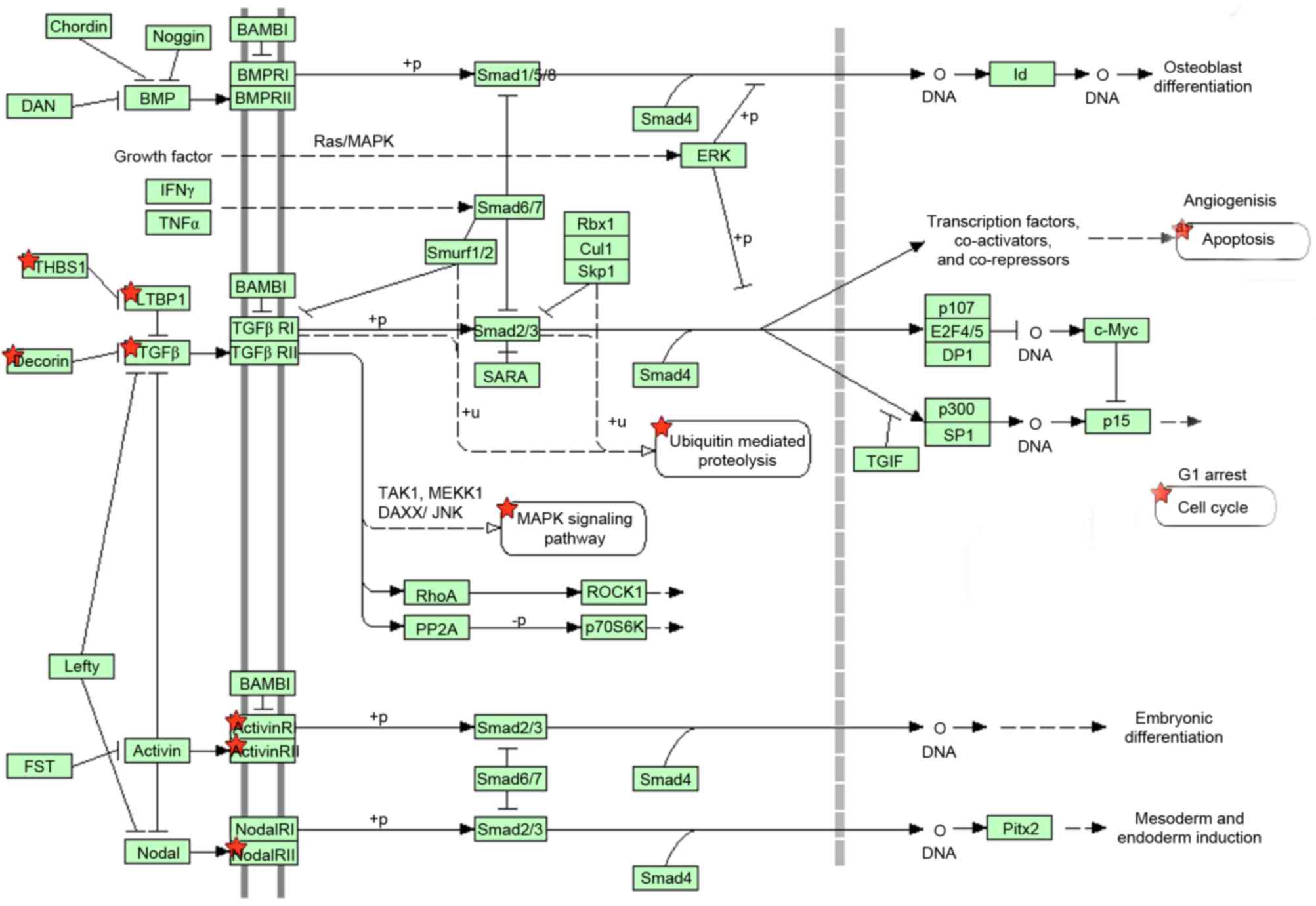
signaling pathway subgroups. There were 10 genes, including activin
A receptor, type I (ACVR1) activin A receptor, type 2A (ACVR2A),
cartilage oligomeric matrix protein (COMP), decorin (DCN), latent
TGFβ binding protein 1 (LTBP1), thrombospondin (THBS)2, THBS3,
THBS4, TGFβ2 and TGFβ3, enriched in the TGFβ signaling pathway. The
position of these 10 genes in the TGFβ signaling pathway are shown
in Fig. 5.
Discussion
BPOP is a rare disease, which usually presents as a
parosteal mass in the short bones of the hands, feet and jaws. BPOP
usually presents as a mushroom-shaped mass, and is often confused
with osteochondroma (1).
Histopathologically, cartilage is present at the margins of the
lesion in BPOP, and the marginal cartilage is fibrocartilage,
compared with the hyaline cartilage usually present in the
cartilage cap of osteochondromas (1,14).
Therefore, it is possible to distinguish a BPOP from an
osteochondroma based on histopathological findings. In addition,
MRI and CT examinations show that the central region of the
exophytic bone lesion in osteochondroma exhibits continuity with
the underlying bone marrow, which is not the case in BPOP (15). This can also assist in
distinguishing a BPOP from an osteochondroma.
BPOP has been reported to have a relatively high
rate of recurrence following surgical resection (10), which is different to that of
osteochondroma, and suggests a different molecular mechanism
between osteochondroma and BPOP. In the present study, it was found
that BPOP exhibited more cartilage characteristics, compared with
BPOP, and the TGFβ receptor signaling pathway, BMP signaling
pathway and Wnt signaling pathway may have different roles between
osteochondroma and BPOP. In addition, the results showed that
several genes were associated with environmental stimulation and
inflammation, for example ‘response to chemical stimulus’,
‘response to wounding’, ‘regulation of inflammatory response’ and
‘response to stress’, which may contribute to BPOP or
osteochondroma. The findings also showed that the extracellular
matrix potentially differs between BPOP and osteochondroma, which
may contribute to the different biological characteristics between
BPOP and osteochondroma.
In conclusion, several differentially expressed
genes between human BPOP and osteochondroma were obtained from the
GEO online database. These differentially expressed genes was
enriched into different subgroups based on analysis using DAVID
online software, which included the ‘transforming growth factor β
receptor signaling pathway’, ‘BMP signaling pathway’, ‘Wnt receptor
signaling pathway’, ‘response to chemical stimulus’, ‘regulation of
inflammatory response’, ‘response to stress’, ‘glycosaminoglycan
binding’, ‘polysaccharide binding’, ‘extracellular matrix
structural constituent’ and ‘growth factors binding’. Taken
together, the results of the present study suggested that there are
different gene regulatory mechanisms between BPOP and
osteochondroma. Environmental stimulation and inflammation may
contribute to BPOP or osteochondroma, and differences in
extracellular matrix may contribute to the different biological
characteristics between BPOP and osteochondroma.
Acknowledgements
The present study work was supported by Nanchong
applied technology research and development program (grant no.
15A0019).
References
|
1
|
Kim SM, Myoung H, Lee SS, Kim YS and Lee
SK: Bizarre parosteal osteochondromatous proliferation in the
lingual area of the mandibular body versus osteochondroma at the
mandibular condyle. World J Surg Oncol. 14:352016. View Article : Google Scholar : PubMed/NCBI
|
|
2
|
Roychoudhury A, Bhatt K, Yadav R, Bhutia O
and Roychoudhury S: Review of osteochondroma of mandibular condyle
and report of a case series. J Oral Maxillofac Surg. 69:2815–2823.
2011. View Article : Google Scholar : PubMed/NCBI
|
|
3
|
Kumar A, Khan SA, Kumar Sampath V and
Sharma MC: Bizarre parosteal osteochondromatous proliferation
(Nora's lesion) of phalanx in a child. BMJ Case Rep. 2014:pii:
bcr2013201714. 2014.
|
|
4
|
Kitsoulis P, Galani V, Stefanaki K,
Paraskevas G, Karatzias G, Agnantis NJ and Bai M: Osteochondromas:
Review of the clinical, radiological and pathological features. In
Vivo. 22:633–646. 2008.PubMed/NCBI
|
|
5
|
Sakamoto A, Imamura S, Matsumoto Y,
Harimaya K, Matsuda S, Takahashi Y, Oda Y and Iwamoto Y: Bizarre
parosteal osteochondromatous proliferation with an inversion of
chromosome 7. Skeletal Radiol. 40:1487–1490. 2011. View Article : Google Scholar : PubMed/NCBI
|
|
6
|
Nilsson M, Domanski HA, Mertens F and
Mandahl N: Molecular cytogenetic characterization of recurrent
translocation breakpoints in bizarre parosteal osteochondromatous
proliferation (Nora's lesion). Hum Pathol. 35:1063–1069. 2004.
View Article : Google Scholar : PubMed/NCBI
|
|
7
|
Panagopoulos I, Bjerkehagen B, Gorunova L,
Taksdal I and Heim S: Rearrangement of chromosome bands 12q14~15
causing HMGA2-SOX5 gene fusion and HMGA2 expression in
extraskeletal osteochondroma. Oncol Rep. 34:577–584.
2015.PubMed/NCBI
|
|
8
|
Tanteles GA, Nicolaou M, Neocleous V,
Shammas C, Loizidou MA, Alexandrou A, Ellina E, Patsia N, Sismani
C, Phylactou LA and Christophidou-Anastasiadou V: Genetic screening
of EXT1 and EXT2 in Cypriot families with hereditary multiple
osteochondromas. J Genet. 94:749–754. 2015. View Article : Google Scholar : PubMed/NCBI
|
|
9
|
Sarrión P, Sangorrin A, Urreizti R,
Delgado A, Artuch R, Martorell L, Armstrong J, Anton J, Torner F,
Vilaseca MA, et al: Mutations in the EXT1 and EXT2 genes in Spanish
patients with multiple osteochondromas. Sci Rep. 3:13462013.
View Article : Google Scholar : PubMed/NCBI
|
|
10
|
Ting BL and Jupiter JB: Recurrent bizarre
parosteal osteochondromatous proliferation of the ulna with erosion
of the adjacent radius: Case report. J Hand Surg Am. 38:2381–2386.
2013. View Article : Google Scholar : PubMed/NCBI
|
|
11
|
Clough E and Barrett T: The gene
expression omnibus database. Methods Mol Biol. 1418:93–110. 2016.
View Article : Google Scholar : PubMed/NCBI
|
|
12
|
da Huang W, Sherman BT and Lempicki RA:
Systematic and integrative analysis of large gene lists using DAVID
bioinformatics resources. Nat Protoc. 4:44–57. 2009. View Article : Google Scholar : PubMed/NCBI
|
|
13
|
Huang DW, Sherman BT, Tan Q, Kir J, Liu D,
Bryant D, Guo Y, Stephens R, Baseler MW, Lane HC and Lempicki RA:
DAVID Bioinformatics Resources: Expanded annotation database and
novel algorithms to better extract biology from large gene lists.
Nucleic Acids Res. 35:(Web Server Issue). W169–W175. 2007.
View Article : Google Scholar : PubMed/NCBI
|
|
14
|
Abramovici L and Steiner GC: Bizarre
parosteal osteochondromatous proliferation (Nora's lesion): A
retrospective study of 12 cases, 2 arising in long bones. Hum
Pathol. 33:1205–1210. 2002. View Article : Google Scholar : PubMed/NCBI
|
|
15
|
Torreggiani WC, Munk PL, Al-Ismail K,
O'Connell JX, Nicolaou S, Lee MJ and Masri BA: MR imaging features
of bizarre parosteal osteochondromatous proliferation of bone
(Nora's lesion). Eur J Radiol. 40:224–231. 2001. View Article : Google Scholar : PubMed/NCBI
|


