Introduction
In the process of orthodontics treatment, mechanical
force is loaded on the tooth, resulting in the production of
inflammatory mediators in periodontal tissues, and the surrounding
alveolar bone is remodeled. According to the pressure-tension
theory (1), bone resorption occurs
on the side of root under pressure, while bone formation occurs on
the other side of the root under tension. The bone remodeling in
the dental and periodontal tissues enables the tooth to move in the
alveolar bone (2,3). A previous study demonstrated
alternations of the alveolar bone density due to active bone
remodeling during orthodontic treatment, the reduction of alveolar
bone density results from less bone mineral content as a result of
resorption of pre-existing bone tissue and formation of new bone
tissue during the process of bone remodeling (4). Therefore, it is likely that bone
mineral density (BMD) reflects progress of bone remodeling during
orthodontic treatment. Additionally, some cell factors and proteins
including tumor necrosis factor ligand superfamily member-11
(RANKL), osteoprotegerin (OPG), lubricin and tumor necrosis
factor-related apoptosis-inducing ligand were also proved to
influence the bone remodeling in orthodontic tooth movement (OTM)
(5–7).
Hydrogen sulfide (H2S) can be produced by
at least three enzymes: Cystathionine β-synthetase (CBS),
cystathionine γ-lyase (CSE) and 3-mercaptopyruvate
sulfurtransferase in mammals (8,9). As
a gasotransmitter, H2S serves a vital role in various
physiological and pathophysiological processes, including
vasodilation, neurotransmission, inflammation, anti-inflammation
and hypoxia sensing (10–14). In rat periodontal tissues,
H2S increases the osteoclast activity and upregulates
RANKL expression levels (15,16).
It was reported that human periodontal ligament stem cells
(PDLSCs), expressed both CBS and CSE and produced H2S,
which maintained the osteogenic capacity of PDLSCs (17). Our previous study indicated that
human PDLSCs produced H2S via CSE, and the mechanical
stimuli increased the mRNA expression levels of CSE. Another study
suggested that mechanical stimuli promoted the production of
H2S in human PDLSCs (18). In has also been demonstrated that
H2S could promote osteogenic differentiation of human
PDLSCs by activating the p38-mitogen-activated protein kinase and
the extracellular signal-regulated kinase signaling pathways
(19).
However, the effect of H2S on alveolar
bone remodeling has not been extensively investigated, as most
studies are based on in vitro investigation without in
vivo evidence. The aim of this study was to explore the effects
of H2S on alveolar bone formation and resorption in an
OTM mouse model. Results of this study may provide an understanding
on H2S mechanisms of action on orthodontic tooth
movement and a potential approach to accelerate orthodontic
treatment.
Materials and methods
Animals
A total of 60 8-week old male C57BL6/J mice were
used in this study in accordance with a protocol approved by the
Animal Use and Care Committee of Tongji University (Shanghai,
China). This study was granted ethical approval by Medical Ethics
Committee of Tongji University, Shanghai, China. Mice were housed
under specific pathogen-free conditions with a controlled
temperature (22±1°C), humidity (40–60%) and a 12-h light/dark
cycle. The animals were given soft diet and water, ad
libitum. The general condition and weight of each animal in
this study were monitored during all experiments. In the present
study, mice were divided into four groups, including a control
(n=15), a sodium hydrosulfide (NaHS; n=15), a DL-propargylglycine
(PAG; n=15) and a combination group (PAG+NaHS; n=15). Both NaHS (a
H2S donor, Sigma-Aldrich; Merck KGaA, Darmstadt,
Germany) and PAG (a CSE inhibitor, Sigma-Aldrich; Merck KGaA) were
administered intraperitoneally at a dose of 10 mg/kg/day for 14
days, and a micro-computed tomography (micro-CT) scan was performed
on day 14. Following the sacrifice of mice with an overdose of
anesthetic (10% chloralic hydras, 400 mg/kg, Sigma-Aldrich; Merck
KGaA), tissues were collected for further histological,
immunohistochemical and molecular biology analyses. Briefly,
tissues were fixed in 4% paraformaldehyde at 4°C for 24 h, followed
by incubation with 10% ethylenediaminetetraacetic Acid (pH 7.4) for
decalcification (4°C for 4 weeks). Sections (5 µm thick) were cut
by a microtome (Leica RM2235, Leica Microsystems Ltd., Wetzlar,
Germany) in a mesiodistal direction parallel to the long axis of
mesial root of the first molar. Sections that included mesial roots
and the alveolar bone of the first molar were selected for
titrate-resistant acid phosphatase (TRAP) and immunohistochemical
staining.
Establishment of the OTM model
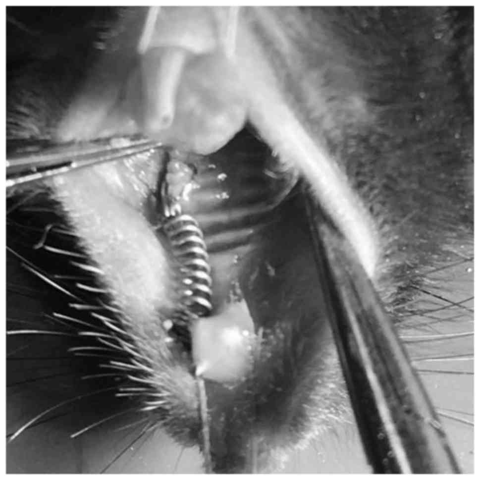
An OTM mouse model was set up as described in a
previous study (20). Orthodontic
force was generated by a nickel-titanium coiled spring (0.2 mm in
thickness, 1 mm in diameter and 1 mm in length, Smart Technology
Co, Ltd., Beijing, China) bonded between the maxillary right first
molar and the incisors with flowable restorative resin (3M ESPE,
St. Paul, MN, USA), producing ~35 g force (21) (Fig.
1). A dynamometer (YDM Corporation, Tokyo, Japan) was used to
measure the force accurately. The distance between the first and
the second molar in mice was identified as OTM.
Micro-CT scanning
Microarchitecture of the maxillary was scanned and
the distance between the maxillary first and the second molar was
measured using micro-CT (SCANCO Medical AG, Bruttisellen,
Switzerland). The BMD of alveolar bone in the root furcation area
was analyzed quantitatively by a self-contained 3D analysis
software of micro-CT (SCANCO Medical AG, Bruttisellen,
Switzerland), by assessing the bone volume over total volume
(BV/TV).
TRAP immunohistochemistry
TRAP staining (37°C, 1 h) was performed to identify
and quantify osteoclasts using a TRAP ELISA kit (cat. no. 387A,
Sigma-Aldrich; Merck KGaA) according to the manufacturer's
protocol. The osteoclasts of the alveolar bone located between the
first and second molar were counted under a confocal microscope
(NIKON ECLIPSE 80i, Nikon Corporation, Tokyo, Japan).
RNA extraction and reverse
transcription polymerase chain reaction (RT-qPCR)
Total RNA was extracted from the alveolar bone
surrounding the upper region of the first molar using TRIzol
reagent (Sigma-Aldrich; Merck KGaA). A RT-qPCR Kit (Takara Bio,
Inc., Otsu, Japan) was used to reverse transcribe the RNA to
complementary (c)DNA, according to the manufacturer's protocol, the
conditions of reactions were: 30°C for 10 min, 42°C for 30 min,
99°C for 5 min and 4°C for 5 min. Relative mRNA levels of alkaline
phosphatase (ALP), osteocalcin (OCN), OPG and
RANKL were determined using a SYBR Green PCR Master mix and
an ABI 7500 Real-Time PCR Detection System (Roche Diagnostics,
Basel, Switzerland). Primer sequences are listed in Table I. The conditions of reactions were:
Initial denaturation at 94°C for 3 min and followed by 35 cycles of
denaturation at 95°C for 10 sec, and annealing at 57°C for 30 sec.
Reactions were run in triplicate and mean-averaged the results.
Relative fold changes were calculated using the method of
2−ΔΔCq (22). GAPDH was
used as the housekeeping gene.
 | Table I.Primer sequences of ALP, OCS, OPG,
RNKL and GAPDH. |
Table I.
Primer sequences of ALP, OCS, OPG,
RNKL and GAPDH.
| Gene | Primer Sequence
(5′-3′) | Length (bp) |
|---|
| ALP | F:
ACACTCGGCCGATCGGGACT | 20 |
|
| R:
CCGCCACCCATGATCACGTCG | 21 |
| OCN | F:
TAGTGAACAGACTCCGGCGCTA | 22 |
|
| R:
TGTAGGCGGTCTTCAAGCCAT | 21 |
| OPG | F:
GTGGAATAGATGTCACCCTGTGT | 23 |
|
| R:
TTTGGTCCCAGGCAAACTGT | 20 |
| RANKL | F:
CAGAAGATGGCACTCACTGCA | 21 |
|
| R:
CACCATCGCTTTCTCTGCTCT | 21 |
| GAPDH |
F:AGCAGTCCCGTACACTGGCAAAC | 23 |
|
|
R:TCTGTGGTGATGTAAATGTCCTCT | 24 |
Immunohistochemistry
Following routine deparaffinage with 100% xylene for
20 min at room temperature, and hydration with alcohol gradient, 3%
H2O2 was used to inactivate endogenous
enzymes for 30 min at room temperature. Antigen repair was
performed with Trypsin (Sigma-Aldrich; Merck KGaA) for 20 min at
room temperature. Sections were incubated with primary antibodies
(ALP, cat. no. SAB3700030; OCN, cat. no. SAB1306277; OPG, cat. no.
SAB4502041, and RANKL, cat. no. SAB4503430; all 1:200,
Sigma-Aldrich; Merck KGaA) at 37°C for 1 h. Sections were also
incubated with second antibody (cat. no. B3640, 1:200,
Sigma-Aldrich; Merck KGaA) at 37°C for 1 h. Positive staining was
visualized with the DAB (D8001, Sigma-Aldrich; Merck KGaA) and
counterstained with hematoxylin.
Statistical analysis
Data are expressed as the mean ± standard deviation.
One-way analysis of variance was performed followed by Tukey's test
using the SPSS software (version 20.0, IBM Corp., Armonk, NY, USA).
P<0.05 was considered to indicate a statistically significant
difference.
Results
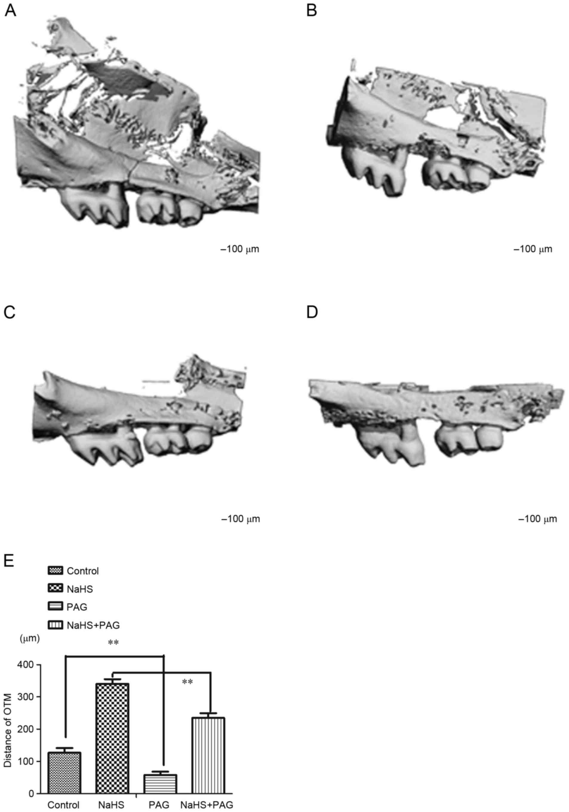
Effect of H2S on OTM
When compared with the control animals (Fig. 2A; 127±15 µm) NaHS resulted in a
significant increase in the OTM (Fig.
2B; 341±14 µm, P<0.01). There was a significant decrease in
the OTM in the PAG-treated group (Fig.
2C; 58±10 µm, P<0.01) compared with the control. Notably,
NaHS rescued the decreased OTM induced by PAG (Fig. 2D; 235±6 µm, P<0.01 and Fig. 2E; 58±10 µm, P<0.01).
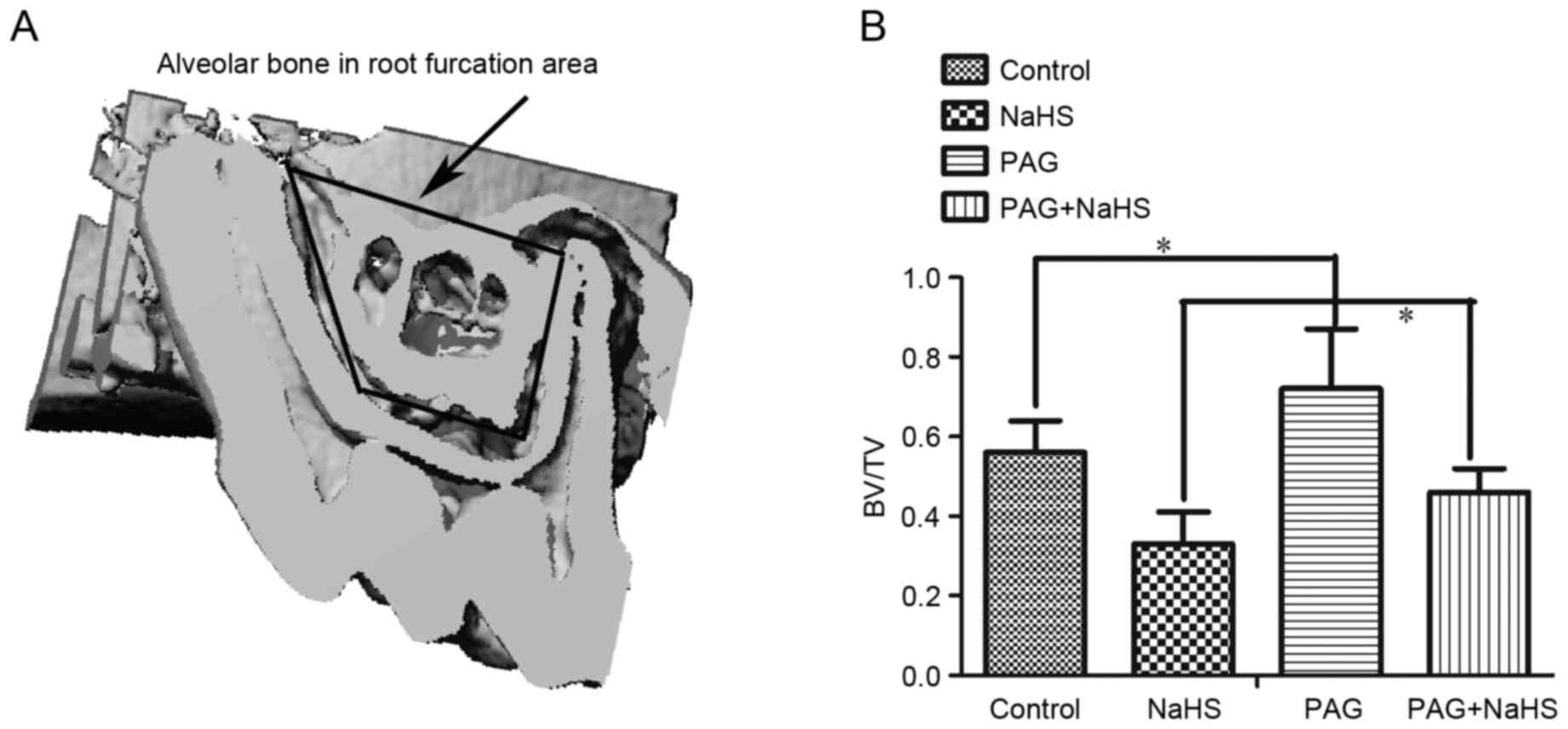
Effect of H2S on BMD
Compared with the control group (56±8%), there was a
significant decrease in the BMD measured in the root furcation area
in the NaSH group (33±7%, P<0.05). The PAG group (72±15%,
P<0.05) demonstrated significantly higher BMD than the control
group (56±8%). Finally, NaHS (46±6%, P<0.05) down-regulated the
increased BMD-induced by PAG (72±15%; Fig. 3).
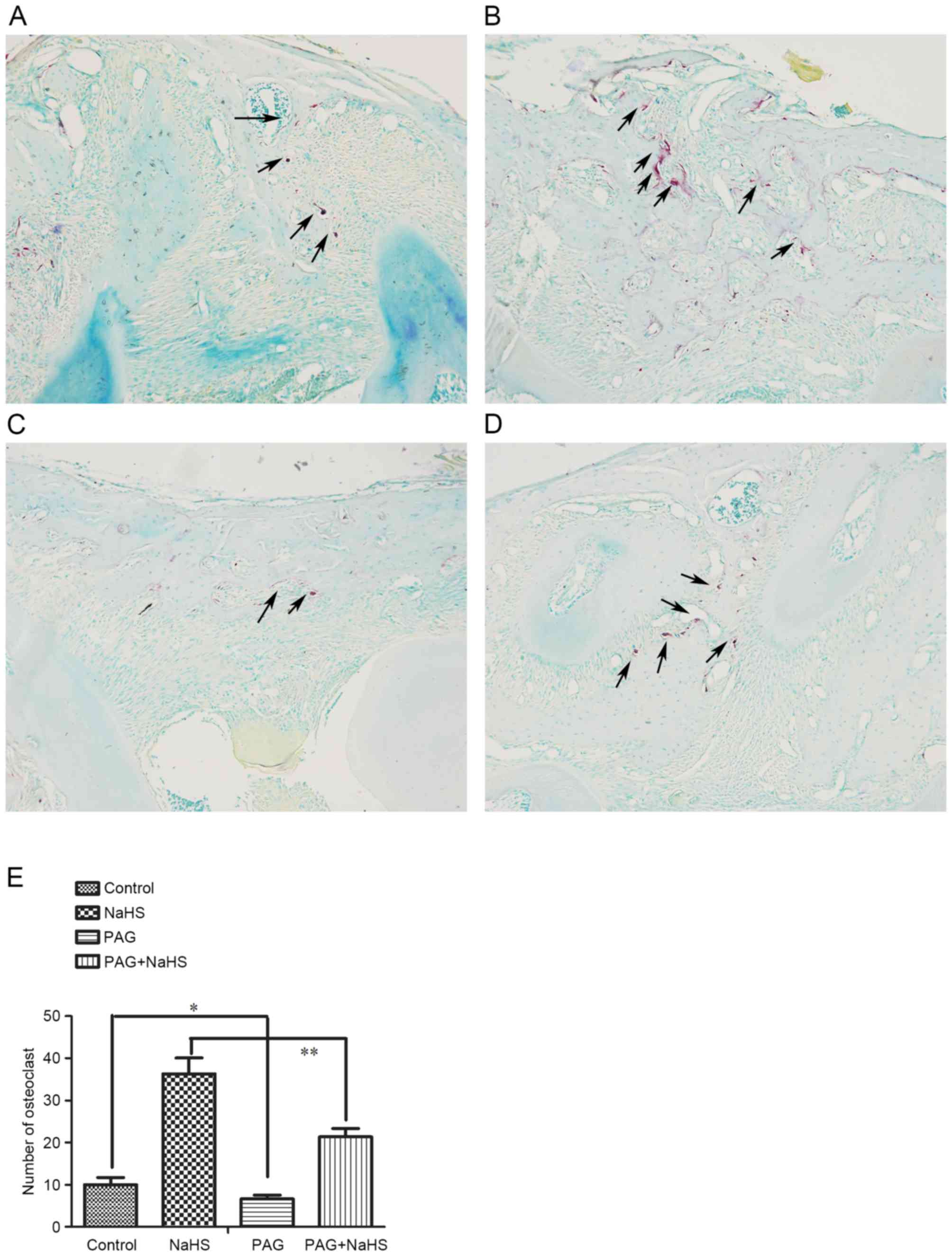
Effect of H2S on osteoclast
numbers
To explore the effect of H2S on bone
resorption, the osteoclasts in the alveolar bone between the first
and the second molar were identified and quantified with TRAP
staining (Fig. 4A-D). Treatment of
NaHS (Fig. 3A and B; 36±4,
P<0.05) increased the number of osteoclasts whereas PAG (7±1,
P<0.05) decreased their number, compared with the control group
(Fig. 4C and E; 10±2). In
addition, the PAG+NaHS group (21±2, P<0.01) indicated a
significant increase in the number of osteoclasts compared with the
PAG group (7±1; Fig. 4D and
E).
Effect of H2S on mRNA and
protein expression levels of OCN, ALP, RANKL, OPG and
RANKL/OPG
To further understand the effect of H2S
on bone resorption and bone formation, mRNA and protein expression
levels of OCN, ALP, RANKL and OPG in the alveolar bone were
investigated. Results demonstrated that both the treatment of NaHS
alone and NaHS combined with PAG significantly up-regulated the
mRNA expression levels of OCN, ALP and RANKL, whereas it
down-regulated the mRNA expression levels of OPG. However, PAG
administration decreased significantly the mRNA expression levels
of OCN, ALP, and RANKL and increased the mRNA expression levels of
OPG (Fig. 5A-D). The mRNA
expression ratio of RANKL/OPG was up-regulated by NaHS and
down-regulated by PAG (Fig. 5E).
Similarly, both the treatment of NaHS alone and NaHS combined with
PAG significantly up-regulated the protein expression levels of
OCN, ALP and RANKL whereas down-regulated OPG protein expression
levels. Additionally, PAG administration decreased significantly
the protein expression levels of OCN, ALP and RANKL, and increased
OPG protein expression levels (Fig.
6).
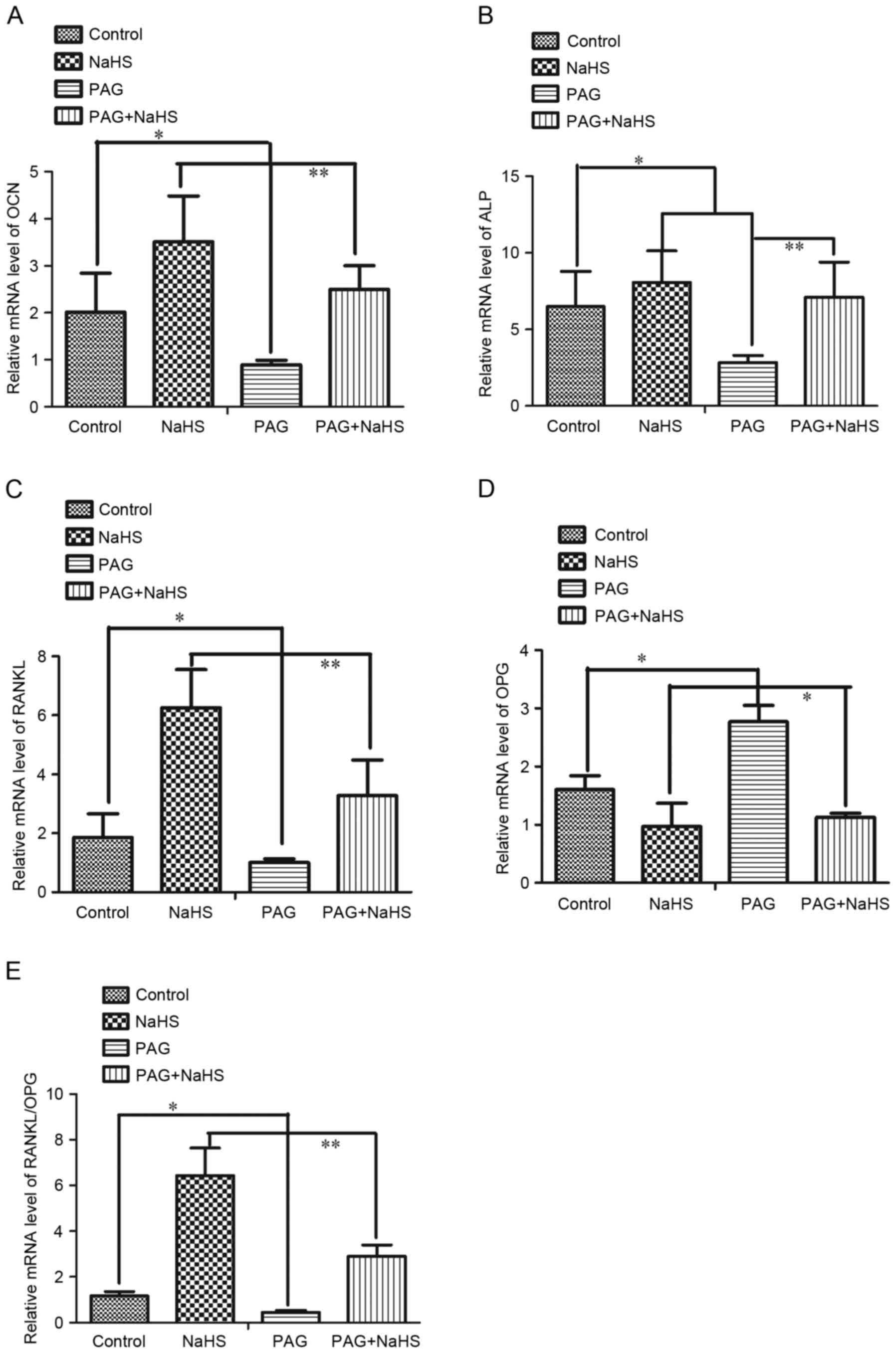 | Figure 5.mRNA expression levels of (A) OCN,
(B) ALP, (C) RANKL, (D) OPG and (E) ratio of RANKL/OPG in the
alveolar bone surrounding the upper first molar region. Data are
presented as the mean ± standard deviation. *P<0.05;
**P<0.01. OTM, orthodontic tooth movement; NaHS, sodium
hydrosulfide; PAG, propargylglycine; ALP, alkaline phosphatase;
OCN, osteocalcin; OPG, osteoprotegerin; RANKL, tumor necrosis
factor ligand superfamily member-11. |
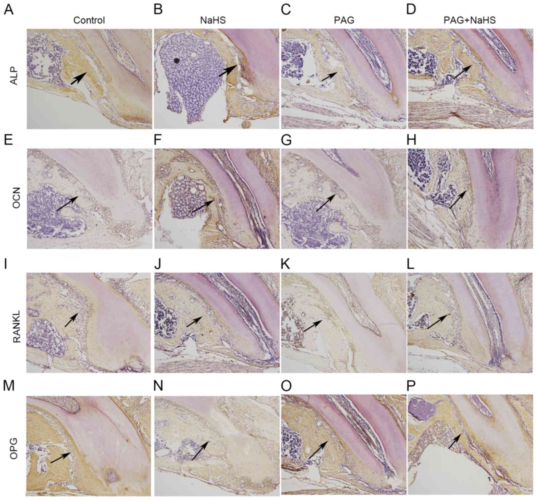 | Figure 6.Protein expression levels of ALP in
the (A) control, (B) NaHS, (C) PAG and (D) PAG+NaHS groups; of OCN
in the (E) control, (F) NaHS, (G) PAG and (H) PAG+NaHS groups; of
RANKL in the (I) control, (J) NaHS, (K) PAG and (L) PAG+NaHS
groups; of OPG in the (M) control, (N) NaHS, (O) PAG and (P)
PAG+NaHS groups. Original magnification, ×100. Arrows indicate
positive staining. OTM, orthodontic tooth movement; NaHS, sodium
hydrosulfide; PAG, propargylglycine; ALP, alkaline phosphatase;
OCN, osteocalcin; OPG, osteoprotegerin; RANKL, tumor necrosis
factor ligand superfamily member-11. |
Discussion
Orthodontic treatment usually lasts 24 to 30 months
(23). The long treatment duration
could increase the risk of caries development, root resorption and
periodontal problems (24–26). Accelerating OTM and shortening the
time of treatment has become a crucial research area. OTM occurs in
the presence of a mechanical stimulus sequenced by remodeling of
the alveolar bone and periodontal ligament (1). It was reported that cyclical forces
of 60Hz could increase the rate of OTM (27). Periodontally accelerated osteogenic
orthodontics has also been proved to be a safe technique to
accelerate tooth movement by reducing the treatment time (28). It is well known that the OTM can be
controlled by the size of the applied force and the biological
responses from the periodontal ligament, such as the release of
inflammatory mediators including cytokines, growth factors,
neurotransmitters and arachidonic acid metabolites (2). One of the cytokines, RANKL is
considered to be involved in the acceleration of tooth movement by
binding to the RANK on osteoclasts stimulating osteoclastogenesis
(29). OPG, a circulating
glycoprotein, binds to RANKL and inhibits osteoclast activities by
competitively inhibiting the RANKL-RANK interaction (30). The balance between RANKL and OPG
levels determines osteoclast activation, skeletal calcium release
and bone remodeling (31).
H2S, a gasotransmitter, serves many
physiological and pathophysiological roles in maintaining the
normal activity of various organs in the neuronal, cardiovascular,
gastrointestinal and respiratory systems (10,11,32,33).
A previous study indicated that bone marrow mesenchymal stem cells
could produce H2S, regulating osteogenic differentiation
and cell self-renewal, and that H2S deficiency could
lead to defects in their differentiation (34). Our previous in vitro study
also demonstrated that H2S could promote osteogenic
differentiation by up-regulating the expression ratio of OPG/RANKL
expression in human PDLSCs (18).
It was also demonstrated that H2S significantly
increased the expression of ALP, OCN, runt-related transcription
factor 2 and collagen 1 in the human PDLSCs with tension force
stimulation, and promoted osteogenic differentiation of human
PDLSCs by activating the p38-MAPK and ERK signaling pathways
(19,35). In the present study, exogenous
H2S could up-regulate the expression of ALP and OCN, the
ratio of RANKL/OPG and the number of osteoclasts in the alveolar
bone. ALP has been commonly studied as an early marker of
osteogenic differentiation in tension force experiments and OCN is
a key transcription factor in the modulation of osteogenic
differentiation and bone formation (19). The results of the present study
revealed that H2S could up-regulate the process of
osteogenesis, osteoclastic activities and bone remodeling. These
findings are consistent with our previous study, where mechanical
stimulation and H2S could enhance the osteogenic
differentiation (18,19,35).
These results were also in accordance with some studies
illustrating that H2S could enhance RANKL-induced
osteoclast differentiation in vitro and induce osteoclast
differentiation in vivo (17,36).
It is generally known that remodeling and modeling
of alveolar bone is considered as the most important aspect in
orthodontic tooth movement (37,38).
In the present study, non-invasive micro-CT was used to monitor the
migration of tooth and the internal conditions of the alveolar
bone, such as bone density, during orthodontic and H2S
treatment. Data of the present study indicated that systemic
administration of NaHS accelerated the OTM and reduced the alveolar
bone density, providing evidence supporting that H2S
could promote osteogenesis and osteoclastogenesis differentiation,
and might facilitate the tooth movement during orthodontic
treatment. In addition, it was observed that PAG decreased the OTM
in the mouse model. This change might be associated with a
reduction of endogenous H2S.
It is worth noting that the present study was based
on an OTM mouse model, and is different from clinical work. The
mechanism of how H2S regulates OTM is still unclear.
However, the present study may provide a novel understanding on how
to accelerate the tooth movement and shorten the treatment
time.
In conclusion, the present study provided an in
vivo evidence that H2S increased the rate of tooth
movement by promoting osteogenesis and osteoclastogenesis
differentiation in alveolar bone. These findings could encourage
further studies looking for a potential therapeutic value of
H2S for accelerating the orthodontic treatment.
Acknowledgements
This study was supported by the National Science
Foundation of China (grant no. 81371177) and the Shanghai Science
and Technology Committee (grant no. 17140903700).
References
|
1
|
Melsen B: Biological reaction of alveolar
bone to orthodontic tooth movement. Angle Orthod. 69:151–158.
1999.PubMed/NCBI
|
|
2
|
Krishnan V and Davidovitch Z: Cellular,
molecular, and tissue-level reactions to orthodontic force. Am J
Orthod Dentofacial Orthop. 129:469.e1–32. 2006. View Article : Google Scholar
|
|
3
|
Masella RS and Meister M: Current concepts
in the biology of orthodontic tooth movement. Am J Orthod
Dentofacial Orthop. 129:458–468. 2006. View Article : Google Scholar : PubMed/NCBI
|
|
4
|
Zahrowski JJ: Optimizing orthodontic
treatment in patients taking bisphosphonates for osteoporosis. Am
Orthod Dentofacial Orthop. 135:361–374. 2009. View Article : Google Scholar
|
|
5
|
Pichler K, Loreto C, Leonardi R, Reuber T,
Weinberg AM and Musumeci G: RANKL is downregulated in bone cells by
physical activity (treadmill and vibration stimulation training) in
rat with glucocorticoid-induced osteoporosis. Histol Histopathol.
28:1185–1196. 2013.PubMed/NCBI
|
|
6
|
Leonardi R, Loreto C, Talic N, Caltabiano
R and Musumeci G: Immunolocalization of lubricin in the rat
periodontal ligament during experimental tooth movement. Acta
Histochem. 114:700–704. 2012. View Article : Google Scholar : PubMed/NCBI
|
|
7
|
Cardile V, Musumeci G, Sicurezza E, Caggia
S, Rusu MC, Leonardi R and Loreto C: Expression of TRAIL and its
receptors DR5 and DcR2 in orthodontic tooth movement. Histol
Histopathol. 28:933–940. 2013.PubMed/NCBI
|
|
8
|
Kabil O, Motl N and Banerjee R: H2S and
its role in redox signaling. Biochim Biophys Acta. 1844:1355–1366.
2014. View Article : Google Scholar : PubMed/NCBI
|
|
9
|
Shibuya N, Tanaka M, Yoshida M, Ogasawara
Y, Togawa T, Ishii K and Kimura H: 3-Mercaptopyruvate
sulfurtransferase produces hydrogen sulfide and bound sulfane
sulfur in the brain. Antioxid Redox Signal. 11:703–714. 2009.
View Article : Google Scholar : PubMed/NCBI
|
|
10
|
Yang G, Wu L, Jiang B, Yang W, Qi J, Cao
K, Meng Q, Mustafa AK, Mu W, Zhang S, et al: H2S as a physiologic
vasorelaxant: Hypertension in mice with deletion of cystathionine
gamma-lyase. Science. 322:587–590. 2008. View Article : Google Scholar : PubMed/NCBI
|
|
11
|
Abe K and Kimura H: The possible role of
hydrogen sulfide as an endogenous neuromodulator. J Neurosci.
16:1066–1071. 1996.PubMed/NCBI
|
|
12
|
Papapetropoulos A, Pyriochou A, Altaany Z,
Yang G, Marazioti A, Zhou Z, Jeschke MG, Branski LK, Herndon DN,
Wang R and Szabó C: Hydrogen sulfide is an endogenous stimulator of
angiogenesis. Proc Natl Acad Sci USA. 106:pp. 21972–21977. 2009;
View Article : Google Scholar : PubMed/NCBI
|
|
13
|
Li L, Bhatia M, Zhu YZ, Zhu YC, Ramnath
RD, Wang ZJ, Anuar FB, Whiteman M, Salto-Tellez M and Moore PK:
Hydrogen sulfide is a novel mediator of lipopolysaccharide-induced
inflammation in the mouse. FASEB J. 19:1196–1198. 2005.PubMed/NCBI
|
|
14
|
Olson KR, Healy MJ, Qin Z, Skovgaard N,
Vulesevic B, Duff DW, Whitfield NL, Yang G, Wang R and Perry SF:
Hydrogen sulfide as an oxygen sensor in trout gill chemoreceptors.
Am J Physiol Regul Integr Comp Physiol. 295:R669–R680. 2008.
View Article : Google Scholar : PubMed/NCBI
|
|
15
|
Irie K, Ekuni D, Tomofuji T, Endo Y,
Kasuyama K, Yaegaki K and Morita M: Combined effects of hydrogen
sulfide and lipopolysaccharide on osteoclast differentiation in
rats. J Periodontol. 83:522–527. 2012. View Article : Google Scholar : PubMed/NCBI
|
|
16
|
Irie K, Ekuni D, Yamamoto T, Morita M,
Yaegaki K, Ii H and Imai T: A single application of hydrogen
sulphide induces a transient osteoclast differentiation with RANKL
expression in the rat model. Arch Oral Biol. 54:723–729. 2009.
View Article : Google Scholar : PubMed/NCBI
|
|
17
|
Su Y, Liu D, Liu Y, Zhang C, Wang J and
Wang S: Physiologic levels of endogenous hydrogen sulfide maintain
the proliferation and differentiation capacity of periodontal
ligament stem cells. J Periodontol. 86:1276–1286. 2015. View Article : Google Scholar : PubMed/NCBI
|
|
18
|
Liao C and Hua Y: Effect of hydrogen
sulphide on the expression of osteoprotegerin and receptor
activator of NF-κB ligand in human periodontal ligament cells
induced by tension-force stimulation. Arch Oral Biol. 58:1784–1790.
2013. View Article : Google Scholar : PubMed/NCBI
|
|
19
|
Jiang Z and Hua Y: Hydrogen sulfide
promotes osteogenic differentiation of human periodontal ligament
cells via p38-MAPK signaling pathway under proper tension
stimulation. Arch Oral Biol. 72:8–13. 2016. View Article : Google Scholar : PubMed/NCBI
|
|
20
|
Cao H, Kou X, Yang R, Liu D, Wang X, Song
Y, Feng L, He D, Gan Y and Zhou Y: Force-induced Adrb2 in
periodontal ligament cells promotes tooth movement. J Dent Res.
93:1163–1169. 2014. View Article : Google Scholar : PubMed/NCBI
|
|
21
|
Yan Y, Liu F, Kou X, Liu D, Yang R, Wang
X, Song Y, He D, Gan Y and Zhou Y: T cells are required for
orthodontic tooth movement. J Dent Res. 94:1463–1470. 2015.
View Article : Google Scholar : PubMed/NCBI
|
|
22
|
Schmittgen TD and Livak KJ: Analyzing
real-time PCR data by the comparative C(T) method. Nat Protoc.
3:1101–1108. 2008. View Article : Google Scholar : PubMed/NCBI
|
|
23
|
Long H, Pyakurel U, Wang Y, Liao L, Zhou Y
and Lai W: Interventions for accelerating orthodontic tooth
movement: A systematic review. Angle Orthod. 83:164–171. 2013.
View Article : Google Scholar : PubMed/NCBI
|
|
24
|
Nimeri G, Kau CH, Abou-Kheir NS and Corona
R: Acceleration of tooth movement during orthodontic treatment-a
frontier in orthodontics. Prog Orthod. 14:422013. View Article : Google Scholar : PubMed/NCBI
|
|
25
|
Kau CH, Kantarci A, Shaughnessy T,
Vachiramon A, Santiwong P, de la Fuente A, Skrenes D, Ma D and
Brawn P: Photobiomodulation accelerates orthodontic alignment in
the early phase of treatment. Prog Orthod. 14:302013. View Article : Google Scholar : PubMed/NCBI
|
|
26
|
Kau CH: A radiographic analysis of tooth
morphology following the use of a novel cyclical force device in
orthodontics. Head Face Med. 7:142011. View Article : Google Scholar : PubMed/NCBI
|
|
27
|
Nishimura M, Chiba M, Ohashi T, Sato M,
Shimizu Y, Igarashi K and Mitani H: Periodontal tissue activation
by vibration: Intermittent stimulation by resonance vibration
accelerates experimental tooth movement in rats. Am J Orthod
Dentofacial Orthop. 133:572–583. 2008. View Article : Google Scholar : PubMed/NCBI
|
|
28
|
Wilcko WM, Wilcko T, Bouquot JE and
Ferguson DJ: Rapid orthodontics with alveolar reshaping: Two case
reports of decrowding. Int J Periodontics Restorative Dent.
21:9–19. 2001.PubMed/NCBI
|
|
29
|
Ciacli C and Puşchiţă M: RANKL/RANK/OPG
molecular complex-control factors in bone remodeling in psoriatic
arthritis. Rev Med Chir Soc Med Nat Iasi. 115:354–360.
2011.PubMed/NCBI
|
|
30
|
Simonet WS, Lacey DL, Dunstan CR, Kelley
M, Chang MS, Lüthy R, Nguyen HQ, Wooden S, Bennett L, Boone T, et
al: Osteoprotegerin: A novel secreted protein involved in the
regulation of bone density. Cell. 89:309–319. 1997. View Article : Google Scholar : PubMed/NCBI
|
|
31
|
Karsenty G: The genetic transformation of
bone biology. Genes Dev. 13:3037–3051. 1999. View Article : Google Scholar : PubMed/NCBI
|
|
32
|
Souza LK, Araújo TS, Sousa NA, Sousa FB,
Nogueira KM, Nicolau LA and Medeiros JV: Evidence that d-cysteine
protects mice from gastric damage via hydrogen sulfide produced by
d-amino acid oxidase. Nitric Oxide. 64:1–6. 2017. View Article : Google Scholar : PubMed/NCBI
|
|
33
|
Ivanciuc T, Sbrana E, Ansar M, Bazhanov N,
Szabo C, Casola A and Garofalo RP: Hydrogen sulfide is an antiviral
and antiinflammatory endogenous gasotransmitter in the airways.
Role in respiratory syncytial virus infection. Am J Respir Cell Mol
Biol. 55:684–696. 2016. View Article : Google Scholar : PubMed/NCBI
|
|
34
|
Liu Y, Yang R, Liu X, Zhou Y, Qu C,
Kikuiri T, Wang S, Zandi E, Du J, Ambudkar IS and Shi S: Hydrogen
sulfide maintains mesenchymal stem cell function and bone
homeostasis via regulation of Ca(2+) channel sulfhydration. Cell
Stem Cell. 15:66–78. 2014. View Article : Google Scholar : PubMed/NCBI
|
|
35
|
Qin J and Hua Y: Effects of hydrogen
sulfide on the expression of alkaline phosphatase, osteocalcin and
collagen type I in human periodontal ligament cells induced by
tension force stimulation. Mol Med Rep. 14:3871–3877. 2016.
View Article : Google Scholar : PubMed/NCBI
|
|
36
|
Itou T, Maldonado N, Yamada I, Goettsch C,
Matsumoto J, Aikawa M, Singh S and Aikawa E: Cystathionine γ-lyase
accelerates osteoclast differentiation: Identification of a novel
regulator of osteoclastogenesis by proteomic analysis. Arterioscler
Thromb Vasc Biol. 34:626–634. 2014. View Article : Google Scholar : PubMed/NCBI
|
|
37
|
Viecilli RF, Katona TR, Chen J, Hartsfield
JK Jr and Roberts WE: Orthodontic mechanotransduction and the role
of the P2X7 receptor. Am J Orthod Dentofacial Orthop.
135:694.e1–16; discussion 694–695. 2009. View Article : Google Scholar
|
|
38
|
Krishnan V and Davidovitch Z: On a path to
unfolding the biological mechanisms of orthodontic tooth movement.
J Dent Res. 88:597–608. 2009. View Article : Google Scholar : PubMed/NCBI
|