Introduction
Cutaneous melanoma is the most dangerous type of
skin cancer. In the United States, the five-year survival rates is
98% among those with localized disease and 17% among those in whom
spread has occurred. Apparently, metastatis is the most important
factor affecting prognosis of melanoma patients. Study on melanoma
metastasis mechanism would beneficial to the development of novel
therapeutic strategies to extend the overall survival of
patients.
Certain signaling pathways have been involved in
metastasis of cutaneous melanoma, such as integrin alpha V beta 3
signaling (1) and signal
transducer and activator of transcription 3 (STAT3) signaling
(2). Several indicators and
biomarkers associated with metastasis have also been identified in
melanoma. LINE1 hypermethylation in peripheral blood of cutaneous
melanoma patients is associated with metastasis (3). Increased level of circulating U2
small nuclear RNA fragments indicates metastasis in melanoma
patients (4). Chen et al
found that Tip60 may regulate melanoma metastasis and could be a
potential therapeutic target (5).
Thang et al reported that deltex-3-like (DTX3L) stimulates
metastasis of melanoma (6). The
study by De Semir et al (7)
indicated that pleckstrin homology domain-interacting protein
(PHIP) is a marker and mediator of melanoma metastasis. Serpin
family E member 1 (SERPINE1) expression discriminates site-specific
metastasis in human melanoma (8).
Galectin-3 expression favors metastasis in murine melanoma
(9).
Gene expression profiling is a powerful tool to
unveil genes implicated in metastasis of melanoma (10,11).
In present study, those gene expression data were combined and more
differentially expressed genes (DEGs) were identified via
meta-analysis. Feature genes were revealed via support vector
machine (SVM) classification. Meanwhile, a SVM classifier was
acquired and validated. These findings could advance the
understanding about melanoma metastasis and also provide potential
therapeutic targets.
Materials and methods
Gene expression data and
pre-treatment
Two gene expression datasets (GSE46517 and GSE7553)
were retrieved from Gene Expression Omnibus (GEO) with key words
‘cutaneous melanoma’, ‘homo sapiens’ and ‘metastasis’ by the end of
May 13th, 2016. Dataset GSE46517 contained 73 metastatic melanoma
samples and 31 primary melanoma samples. Dataset GSE7553 contained
40 metastatic melanoma samples and 14 primary melanoma samples.
Moreover, gene expression profiles of 481 skin cutaneous melanoma
specimens were downloaded from The Cancer Genome Atlas (TCGA,
https://tcga-data.nci.nih.gov/tcga)
with keyword ‘cutaneous melanoma’, out of which 183 were metastatic
melanoma samples, 71 primary melanoma samples and others not
specified.
Gene expression dataset GSE7553 was acquired with
Affymetrix platform. Background correction and normalization were
performed with package oligo (12) of R. Missing values were
filled with median method. Backgroud correction was carried out
with MSA method. Normalization was achieved with quantiles method.
As for dataset GSE46517 from Agilent platform, probes were mapped
into genes. For probes corresponding to a same gene, the averaged
value was considered as the expression value of the gene. Log2
transformation and normalization with the median were then applied.
Level 3 gene expression data from TCGA were normalized with package
limma (13) of
R.
Screening of differentially expressed
genes (DEGs)
Meta-analysis of the three gene expression dataset
was performed with package MetaDE (14) of R to identify differentially
expressed genes (DEGs). This method firstly tested heterogeneity of
gene expression value in various platforms with three statistic
parameters: tau2, Q value and Qpval. Then it tested
differential expression of genes between different groups with
statistic parameters P-value and false positive rate (FDR). To
ensure the homogeneity of feature genes, tau2=0, Qpval
>0.05 and FDR <0.05 were set as the cut-offs.
Construction of protein-protein
interaction (PPI) network
The PPI information was downloaded from Database of
Protein, Chemical, and Genetic Interactions (BioGRID, http://thebiogrid.org/) (15), Human Protein Reference Database
(HPRD, http://www.hprd.org/) (16) and Database of Interacting Proteins
(DIP, http://dip.doe-mbi.ucla.edu/dip/Main.cgi) (17). The protein products of the DEGs
were mapped into the whole network and the PPI network for the DEGs
was then acquired. The network was then visualized with Cytoscape
(18).
Calculation of betweenness centrality
(BC)
Feature genes were screened out from the DEGs using
betweenness centrality (BC), which reflected hubness of the node in
the PPI network. The BC was calculated as follow:
CB(v)=∑t≠v≠u∈Vσst(v)σst
Where σst is the shortest path from s to
t; σst(v) is number of the shortest path from
s to t passing through node v; BC score
is between 0 and 1, and greater BC score indicates higher
degree of hubness.
Training and validation of SVM
classifier
Gene expresson data from TCGA were chosen as the
training set. Genes were ranked based upon BC value and top 10
genes were selected out to train SVM classifier. An increment of 10
genes were added into the classifier until metastatic melanomas
could be totally separated from primary melanomas. These DEGs were
regarded as feature genes. Gene expression datasets GSE46517 and
GSE7553 were used as the validation set. Sensitivity (Se),
specificity (Sp), positive predictive value (PPV), negative
predictive value (NPV) and area under curve (AUC) were calculated
to evaluate the SVM classifier.
Pathway enrichment analysis
Gene ontology (GO) biological pathways and Kyoto
Encyclopedia of Genes and Genomes (KEGG) pathways related to
feature genes were identified using runHyperGO and runHyperKEGG
from package EMA of R. The significance was calculated using
Fisher's exact test as follow. P-value <0.05 was set as the
threshold.
p=1–∑i=0x–1(Mi)(N–MK–i)(NK)
Where N is the total number of gene; M
is number of gene in the pathway; K is number of feature
gene.
Results
Differentially expressed genes
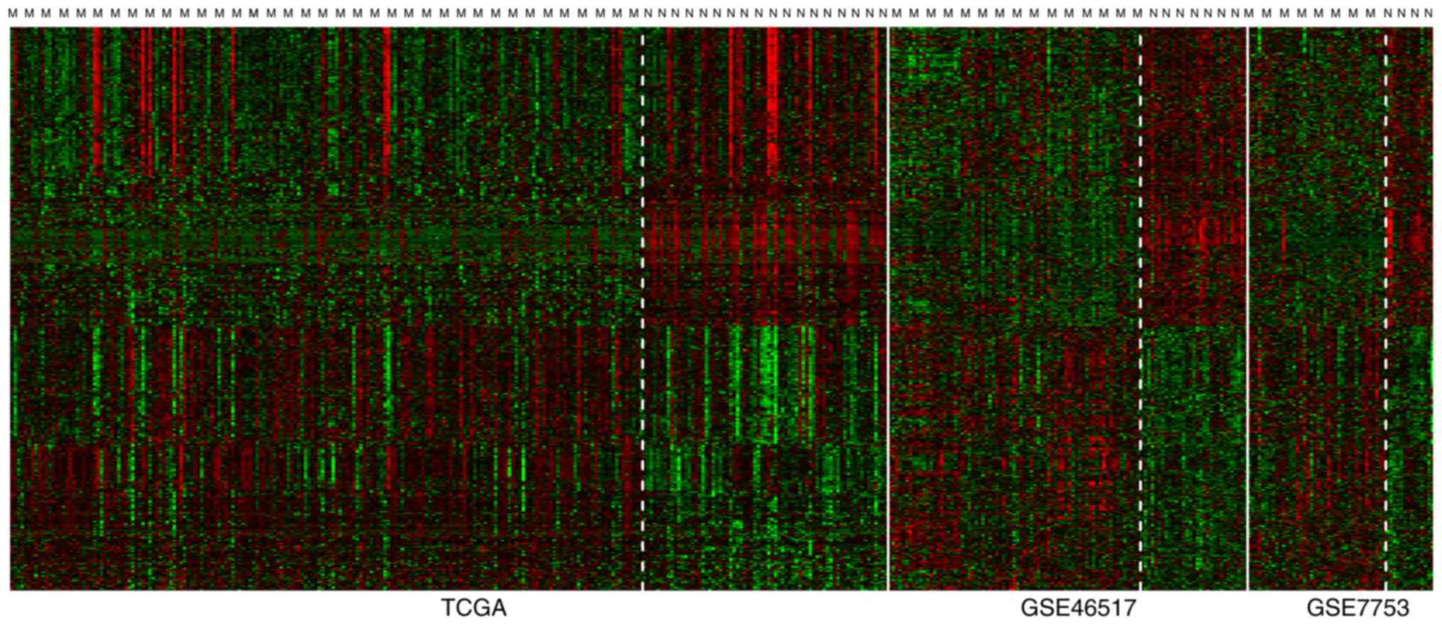
According to the criteria, a total of 798 DEGs were
revealed from the three gene expression datasets. Heat map of the
expression levels of the 798 DEGs is shown in Fig. 1.
PPI network
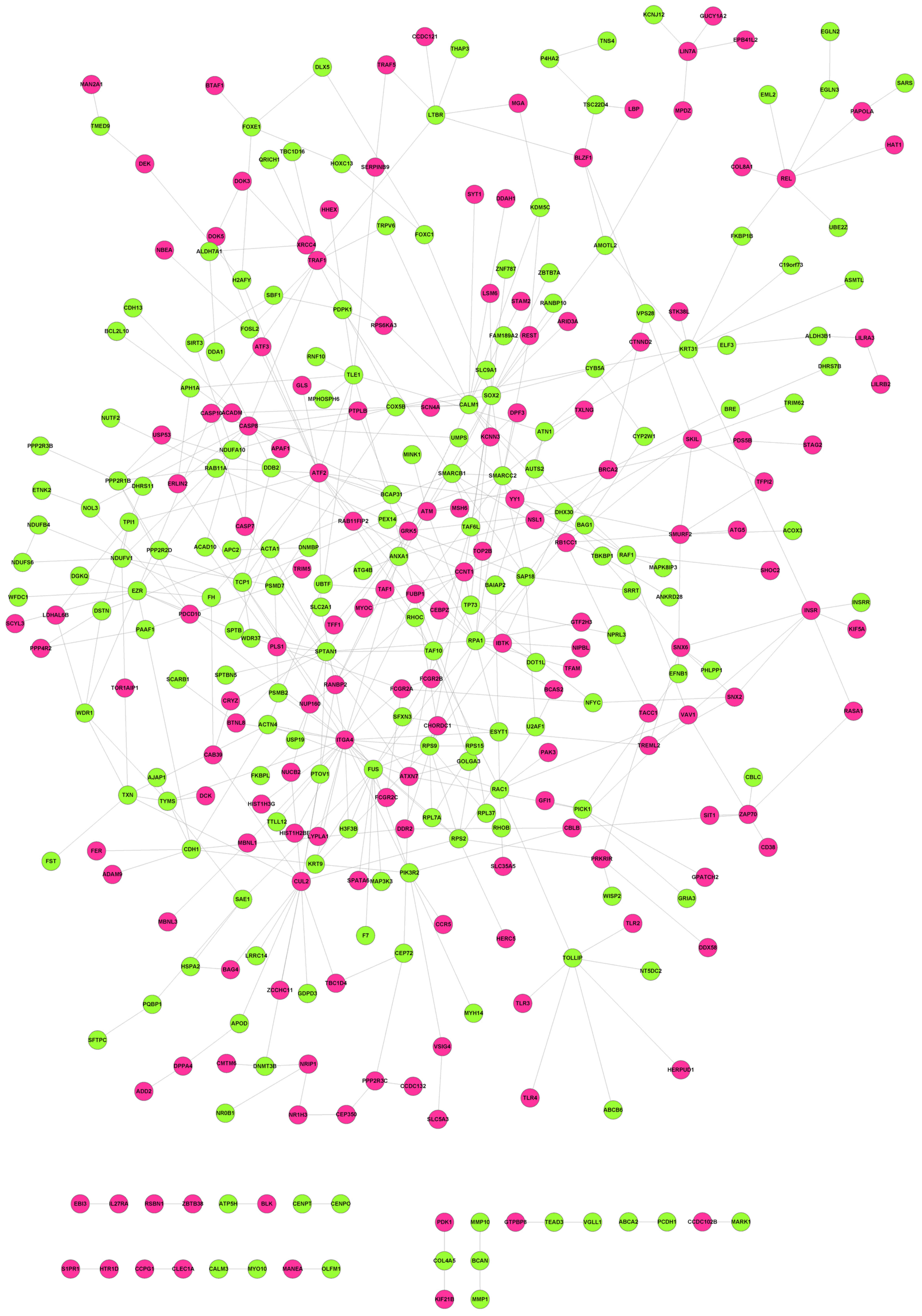
Based upon information from BioGRID, HPRD and DIP, a
PPI network containing 337 nodes and 466 edges was obtained
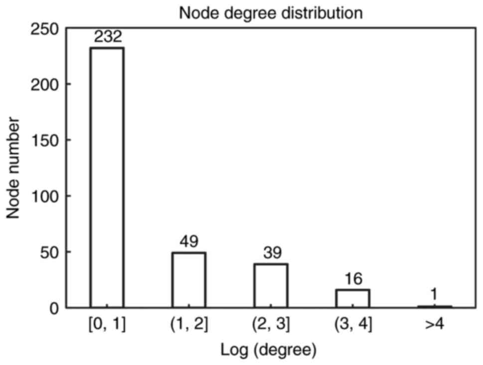
(Fig. 2). Distribution of degree
is shown in Fig. 3. Most genes
(232 genes) showed small degree (Log transformed degree <1)
while only 1 gene had Log transformed degree >4. Therefore, like
most biological networks, this PPI network exhibited scale-free
property. These genes with high degree were hub genes and might
play important roles in the development of diseases.
SVM classifier
Top 10 DEGs ranked by BC are listed in Table I: Integrin subunit alpha 4 (ITGA4),
activating transcription factor 2 (ATF2), SRY-box 2 (SOX2), keratin
31 (KRT31), cullin 2 (CUL2), replication protein A1 (RPA1),
calmodulin 1 (CALM1), FUS RNA binding protein (FUS), angiomotin
like 2 (AMOTL2) and ras-related C3 botulinum toxin substrate 1
(RAC1).
 | Table I.Top 10 genes ranked by betweenness
centrality. |
Table I.
Top 10 genes ranked by betweenness
centrality.
| Gene | BC | Exp | Degree | P-value | FDR | Q | Qp |
tau2 |
|---|
| ITGA4 |
2.06×10⁻¹ | 1 | 25 |
1.00×10⁻²⁰ |
9.22×10⁻¹⁹ | 1.39E+00 |
5.00×10⁻¹ | 0 |
| ATF2 |
1.47×10⁻¹ | 1 | 16 |
2.46×10⁻⁶ |
1.78×10⁻⁴ | 1.54E+00 |
4.64×10⁻¹ | 0 |
| SOX2 |
1.43×10⁻¹ | 0 | 15 |
3.73×10⁻⁴ |
6.85×10⁻³ |
5.21×10⁻² |
9.74×10⁻¹ | 0 |
| KRT31 |
1.36×10⁻¹ | 0 | 10 |
1.00×10⁻²⁰ |
9.22×10⁻¹⁹ |
4.96×10⁻¹ |
7.80×10⁻¹ | 0 |
| CUL2 |
1.22×10⁻¹ | 1 | 16 |
2.69×10⁻⁴ |
5.49×10⁻³ |
1.19×10⁻¹ |
9.42×10⁻¹ | 0 |
| RPA1 |
1.10×10⁻¹ | 0 | 16 |
4.07×10⁻³ |
3.69×10⁻² |
6.28×10⁻¹ |
7.31×10⁻¹ | 0 |
| CALM1 |
9.78×10⁻² | 0 | 13 |
7.63×10⁻⁴ |
1.12×10⁻² |
8.76×10⁻¹ |
6.45×10⁻¹ | 0 |
| FUS |
9.26×10⁻² | 0 | 12 |
6.57×10⁻⁶ |
3.27×10⁻⁴ | 1.95E+00 |
3.77×10⁻¹ | 0 |
| AMOTL2 |
9.25×10⁻² | 0 | 4 |
9.86×10⁻⁶ |
4.24×10⁻⁴ |
8.40×10⁻¹ |
6.57×10⁻¹ | 0 |
| RAC1 |
8.70×10⁻² | 0 | 7 |
2.38×10⁻³ |
2.54×10⁻² | 1.13E+00 |
5.69×10⁻¹ | 0 |
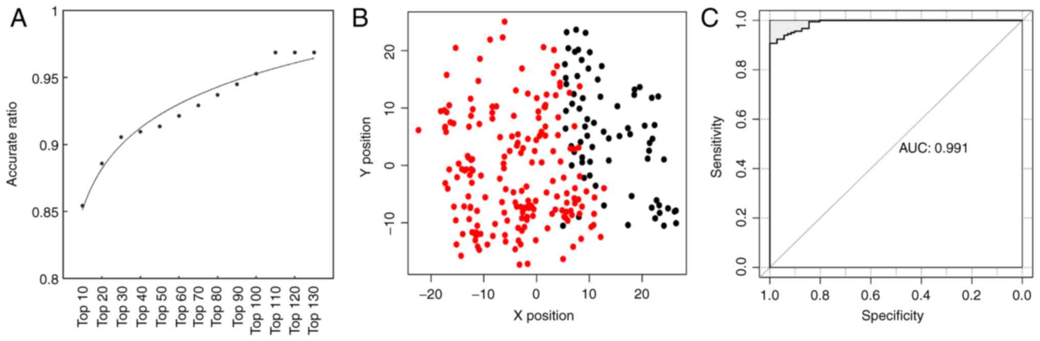
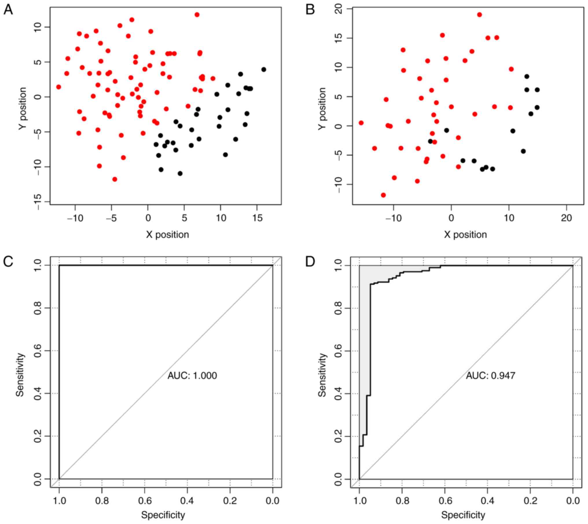
As shown in Fig.
4A, the accuracy increased from 85.4 to 96.8% while more genes
were included in the classifier. Details are shown in Table II. The accuracy rate reached 96.8%
while top 110 DEGs were included in the classifier, and it didn't
increase though more genes were added in. Therefore, the top 110
genes were used to construct the SVM classifier. The scatter plot
of prediction result for the training dataset is shown in Fig. 4B and the receiver operating
characteristic (ROC) curve is shown in Fig. 4C.
| Table II.Prediction results of the SVM
classifier in the training dataset with varying numbers of
differentially expressed genes. |
Table II.
Prediction results of the SVM
classifier in the training dataset with varying numbers of
differentially expressed genes.
| SVM classifier | Correct
prediction | Wrong
prediction | Accuracy rate
(%) | Error rate (%) |
|---|
| Top10 | 217 | 37 | 85.4 | 14.6 |
| Top20 | 225 | 29 | 88.6 | 11.4 |
| Top30 | 230 | 24 | 90.6 |
9.4 |
| Top40 | 231 | 23 | 90.9 |
9.1 |
| Top50 | 232 | 22 | 91.3 |
8.7 |
| Top60 | 234 | 20 | 92.1 |
7.9 |
| Top70 | 236 | 18 | 92.9 |
7.1 |
| Top80 | 238 | 16 | 93.7 |
6.3 |
| Top90 | 240 | 14 | 94.5 |
5.5 |
| Top100 | 242 | 12 | 95.3 |
4.7 |
| Top110 | 246 | 8 | 96.9 |
3.1 |
| Top120 | 246 | 8 | 96.9 |
3.1 |
| Top130 | 246 | 8 | 96.9 |
3.1 |
The SVM classifier was validated using dataset
GSE46517 and GSE7553. The accuracy rates in dataset GSE46517
(Fig. 5A) and GSE7553 (Fig. 5B) were 100 and 94.4%, respectively.
Three non-metastatic melanoma samples from dataset GSE7753 were
predicted wrong. The accuracy, sensitivity, specificity, PPV, NPV
and AUC are listed in Table III.
The receiver operating characteristic (ROC) curves are shown in
Fig. 5C and D.
| Table III.Prediction results of the SVM
classifier in the 3 independent datasets. |
Table III.
Prediction results of the SVM
classifier in the 3 independent datasets.
| Dataset | No. of sample | Accuracy | Sensitivity | Specificity | PPV | NPV | AUROC |
|---|
| TCGA | 254 | 0.968 | 1.000 | 0.987 | 0.958 | 1.000 | 0.991 |
| GSE46517 | 128 | 1.000 | 1.000 | 1.000 | 1.000 | 1.000 | 1.000 |
| GSE7753 | 54 | 0.944 | 1.000 | 0.886 | 0.942 | 1.000 | 0.947 |
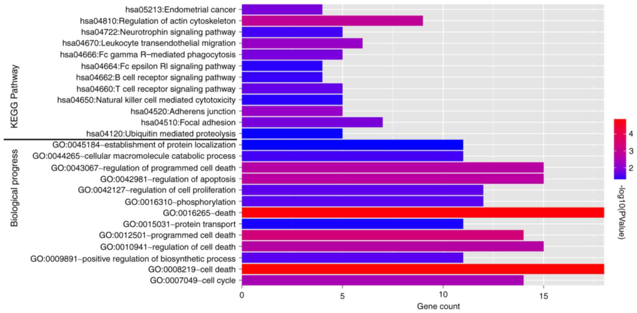
Biological pathways
A total of 11 KEGG pathways (Fig. 6, top) and 13 GO biological pathways
(Fig. 6, bottom) were identified
in the 110 feature genes. Endometrial cancer, regulation of actin
cytoskeleton, focal adhesion, ubiquitin mediated proteolysis,
regulation of apoptosis, and regulation of cell proliferation were
significantly over-represented in the feature genes.
Discussion
Three gene expression datasets were downloaded from
GEO or TCGA. A total of 798 DEGs were identified in metastatic
melanoma compared with non-metastatic melanoma via meta-analysis. A
PPI network containing 337 nodes and 466 edges was constructed. BC
was calculated for each gene in the network. Top 110 feature genes
ranked by BC were included in the SVM classifier. The prediction
accuracies for the three datasets were 96.8, 100, and 94.4%,
respectively. A total of 11 KEGG pathways and 13 GO biological
pathways were significantly over-represented in the 110 feature
genes, such as endometrial cancer, regulation of actin
cytoskeleton, focal adhesion, ubiquitin mediated proteolysis,
regulation of apoptosis, and regulation of cell proliferation.
Among the 110 feature genes, several genes have been involved in
metastasis of melanoma.
ATF2 is a transcription factor and a member of the
leucine zipper family of DNA binding proteins. It confers radiation
resistance to human melanoma cells (19). Lau et al further pointed out
that transcriptional repression of IFNβ1 by ATF2 confers melanoma
resistance to therapy (20). It
also promotes melanoma metastasis by suppressing protein
fucosylation (21). However,
ATF2-derived peptides can inhibit melanoma growth and metastasis
(22). Therefore, ATF2 is a
critical gene in melanoma metastasis and could be a therapeutic
target.
SOX2 is a member of the SRY-related HMG-box (SOX)
family of transcription factors involved in the regulation of
embryonic development and in the determination of cell fate. It
regulates self-renewal and tumorigenicity of human
melanoma-initiating cells (23).
It also contributes to melanoma cell invasion (24). Downregulation of SOX2 in cancer
stem cells suppresses growth and metastasis of lung cancer
(25). We speculated that it could
be a potential therapeutic target for metastatic melanoma.
RAC1 is a GTPase which belongs to the RAS
superfamily of small GTP-binding proteins, which regulate a diverse
array of cellular events, including the control of cell growth,
cytoskeletal reorganization, and the activation of protein kinases.
RAC1 is a critical genes associated with melanoma metastasis. A
previous study has reported that a RAC1 mutant (Pro29 to serine,
P29S) promotes melanocyte proliferation and migration (26). A recent study indicates that RAC1
P29S regulates PD-L1 expression in melanoma (27). Besides, matrix metallopeptidase 14
modulates melanoma cell dissemination and metastasis through
activation of MMP2 and RAC1 (28).
Immunity-related GTPase family M member 1 (IRGM1) enhances B16
melanoma cell metastasis through PI3K-RAC1 mediated epithelial
mesenchymal transition (29).
Certain genes take parts in metastasis of various
cancers and their roles in melanoma metastasis remain to be
uncovered. AMOTL2 is related to angiomotin and is a member of the
motin protein family. It disrupts apical-basal cell polarity and
promotes tumour invasion (30).
Raf-1 proto-oncogene (RAF1) activation in gastrointestinal
carcinoid cells decreases tumor cell adhesion and thus promotes
metastasis (31). Vav guanine
nucleotide exchange factor 1 (VAV1) is a member of the VAV gene
family. Inhibition of VAV1 can attenuate metastasis of pancreatic
cancer (32). Besides, RPA1 is
regarded as a useful prognostic indicator in colon cancer (33) and esophageal carcinoma (34). We thought that it might exert
prognostic effect in melanoma. Further studies on these genes could
expand the knowledge about melanoma metastasis.
In the present study, the biological pathways that
were significantly enriched in the metastasis related feature genes
in melanoma were screened. To find out the pathways involved in the
feature genes in metastatic melanoma, Valsesia et al also
conducted a network-guided investigation (35). They connected the genes of somatic
copy number alterations in metastatic melanoma with significant
biological pathways. The results showed the frequently altered
pathways in melanoma, including angiogenesis and melanogenesis,
while we found that regulation of apoptosis and regulation of cell
proliferation participate in melanoma.
Overall, a multigene SVM predictor of high accuracy
was obtained for metastatic melanoma. Several critical genes linked
with melanoma metastasis were also revealed. Further researches on
the feature genes could improve the understanding about the
molecular mechanisms underlying metastasis and provide potential
therapeutic targets.
References
|
1
|
Bai J, Zhang J, Wu J, Shen L, Zeng J, Ding
J, Wu Y, Gong Z, Li A, Xu S, et al: JWA regulates melanoma
metastasis by integrin alphaVbeta3 signaling. Oncogene.
29:1227–1237. 2010. View Article : Google Scholar : PubMed/NCBI
|
|
2
|
Cao HH, Chu JH, Kwan HY, Su T, Yu H, Cheng
CY, Fu XQ, Guo H, Li T, Tse AK, et al: Inhibition of the STAT3
signaling pathway contributes to apigenin-mediated anti-metastatic
effect in melanoma. Sci Rep. 6:217312016. View Article : Google Scholar : PubMed/NCBI
|
|
3
|
De Araújo ÉS, Kashiwabara AY, Achatz MI,
Moredo LF, De Sá BC, Duprat JP, Rosenberg C, Carraro DM and
Krepischi AC: LINE-1 hypermethylation in peripheral blood of
cutaneous melanoma patients is associated with metastasis. Melanoma
Res. 25:173–177. 2015. View Article : Google Scholar : PubMed/NCBI
|
|
4
|
Kuhlmann JD, Wimberger P, Wilsch K, Fluck
M, Suter L and Brunner G: Increased level of circulating U2 small
nuclear RNA fragments indicates metastasis in melanoma patients.
Clin Chem Lab Med. 53:605–611. 2015. View Article : Google Scholar : PubMed/NCBI
|
|
5
|
Chen G, Cheng Y, Tang Y, Martinka M and Li
G: Role of Tip60 in human melanoma cell migration, metastasis, and
patient survival. J Invest Dermatol. 132:2632–2641. 2012.
View Article : Google Scholar : PubMed/NCBI
|
|
6
|
Thang ND, Yajima I, Kumasaka MY, Iida M,
Suzuki T and Kato M: Deltex-3-like (DTX3L) stimulates metastasis of
melanoma through FAK/PI3K/AKT but not MEK/ERK pathway. Oncotarget.
6:14290–14299. 2015. View Article : Google Scholar : PubMed/NCBI
|
|
7
|
De Semir D, Nosrati M, Bezrookove V, Dar
AA, Federman S, Bienvenu G, Venna S, Rangel J, Climent J, Meyer
Tamgüney TM, et al: Pleckstrin homology domain-interacting protein
(PHIP) as a marker and mediator of melanoma metastasis. Proc Natl
Acad Sci USA. 109:pp. 7067–7072. 2012; View Article : Google Scholar : PubMed/NCBI
|
|
8
|
Klein RM, Bernstein D, Higgins SP, Higgins
CE and Higgins PJ: SERPINE1 expression discriminates site-specific
metastasis in human melanoma. Exp Dermatol. 21:551–554. 2012.
View Article : Google Scholar : PubMed/NCBI
|
|
9
|
Comodo AN, Lacerda Bachi AL, Soares MF,
Franco M and de Paulo Castro Teixeira V: Galectin-3 expression
favors metastasis in murine melanoma. Adv Biosci Biotechnol.
04:55–62. 2013. View Article : Google Scholar
|
|
10
|
Kabbarah O, Nogueira C, Feng B, Nazarian
RM, Bosenberg M, Wu M, Scott KL, Kwong LN, Xiao Y, Cordon-Cardo C,
et al: Integrative genome comparison of primary and metastatic
melanomas. PLoS One. 5:e107702010. View Article : Google Scholar : PubMed/NCBI
|
|
11
|
Riker AI, Enkemann SA, Fodstad O, Liu S,
Ren S, Morris C, Xi Y, Howell P, Metge B, Samant RS, et al: The
gene expression profiles of primary and metastatic melanoma yields
a transition point of tumor progression and metastasis. BMC Med
Genomics. 1:132008. View Article : Google Scholar : PubMed/NCBI
|
|
12
|
Carvalho B, Bengtsson H, Speed TP and
Irizarry RA: Exploration, normalization, and genotype calls of
high-density oligonucleotide SNP array data. Biostatistics.
8:485–499. 2007. View Article : Google Scholar : PubMed/NCBI
|
|
13
|
Smyth GK: Limma: Linear models for
microarray data. Stat Biol Health. 1–420. 2005.
|
|
14
|
Li J and Tseng GC: An adaptively weighted
statistic for detecting differential gene expression when combining
multiple transcriptomic studies. Ann Appl Stat. 5:994–1019. 2011.
View Article : Google Scholar
|
|
15
|
Chatr-Aryamontri A, Breitkreutz BJ,
Oughtred R, Boucher L, Heinicke S, Chen D, Stark C, Breitkreutz A,
Kolas N, O'Donnell L, et al: The BioGRID interaction database: 2015
update. Nucleic Acids Res. 43(Database issue): D470–DS478. 2015.
View Article : Google Scholar : PubMed/NCBI
|
|
16
|
Keshava Prasad TS, Goel R, Kandasamy K,
Keerthikumar S, Kumar S, Mathivanan S, Telikicherla D, Raju R,
Shafreen B, Venugopal A, et al: Human protein reference
database-2009 update. Nucleic Acids Res. 37(Database issue):
D767–D772. 2009. View Article : Google Scholar : PubMed/NCBI
|
|
17
|
Xenarios I, Rice DW, Salwinski L, Baron
MK, Marcotte EM and Eisenberg D: DIP: The database of interacting
proteins. Nucleic Acids Res. 28:289–291. 2000. View Article : Google Scholar : PubMed/NCBI
|
|
18
|
Smoot ME, Ono K, Ruscheinski J, Wang PL
and Ideker T: Cytoscape 2.8: New features for data integration and
network visualization. Bioinformatics. 27:431–432. 2011. View Article : Google Scholar : PubMed/NCBI
|
|
19
|
Ronai Z, Yang YM, Fuchs SY, Adler V,
Sardana M and Herlyn M: ATF2 confers radiation resistance to human
melanoma cells. Oncogene. 16:523–531. 1998. View Article : Google Scholar : PubMed/NCBI
|
|
20
|
Lau E, Sedy J, Sander C, Shaw MA, Feng Y,
Scortegagna M, Claps G, Robinson S, Cheng P, Srivas R, et al:
Transcriptional repression of IFNβ1 by ATF2 confers melanoma
resistance to therapy. Oncogene. 34:5739–5748. 2015. View Article : Google Scholar : PubMed/NCBI
|
|
21
|
Lau E, Feng Y, Claps G, Fukuda MN, Perlina
A, Donn D, Jilaveanu L, Kluger H, Freeze HH and Ronai ZA: The
transcription factor ATF2 promotes melanoma metastasis by
suppressing protein fucosylation. Sci Signal. 8:ra1242015.
View Article : Google Scholar : PubMed/NCBI
|
|
22
|
Bhoumik A, Gangi L and Ronai Z: Inhibition
of melanoma growth and metastasis by ATF2-derived peptides. Cancer
Res. 64:8222–8230. 2004. View Article : Google Scholar : PubMed/NCBI
|
|
23
|
Santini R, Pietrobono S, Pandolfi S,
Montagnani V, D'Amico M, Penachioni JY, Vinci MC, Borgognoni L and
Stecca B: SOX2 regulates self-renewal and tumorigenicity of human
melanoma-initiating cells. Oncogene. 33:4697–4708. 2014. View Article : Google Scholar : PubMed/NCBI
|
|
24
|
Girouard SD, Laga AC, Mihm MC, Scolyer RA,
Thompson JF, Zhan Q, Widlund HR, Lee CW and Murphy GF: SOX2
contributes to melanoma cell invasion. Lab Invest. 92:362–370.
2012. View Article : Google Scholar : PubMed/NCBI
|
|
25
|
Xiang R, Liao D, Cheng T, Zhou H, Shi Q,
Chuang TS, Markowitz D, Reisfeld RA and Luo Y: Downregulation of
transcription factor SOX2 in cancer stem cells suppresses growth
and metastasis of lung cancer. Br J Cancer. 104:1410–1417. 2011.
View Article : Google Scholar : PubMed/NCBI
|
|
26
|
Krauthammer M, Kong Y, Ha BH, Evans P,
Bacchiocchi A, McCusker JP, Cheng E, Davis MJ, Goh G, Choi M, et
al: Exome sequencing identifies recurrent somatic RAC1 mutations in
melanoma. Nat Genet. 44:1006–1014. 2012. View Article : Google Scholar : PubMed/NCBI
|
|
27
|
Vu HL, Rosenbaum S, Purwin TJ, Davies MA
and Aplin AE: RAC1 P29S regulates PD-L1 expression in melanoma.
Pigment Cell Melanoma Res. 28:590–598. 2015. View Article : Google Scholar : PubMed/NCBI
|
|
28
|
Shaverdashvili K, Wong P, Ma J, Zhang K,
Osman I and Bedogni B: MT1-MMP modulates melanoma cell
dissemination and metastasis through activation of MMP2 and RAC1.
Pigment Cell Melanoma Res. 27:287–296. 2014. View Article : Google Scholar : PubMed/NCBI
|
|
29
|
Tian L, Li L, Xing W, Li R, Pei C, Dong X,
Fu Y, Gu C, Guo X, Jia Y, et al: IRGM1 enhances B16 melanoma cell
metastasis through PI3K-Rac1 mediated epithelial mesenchymal
transition. Sci Rep. 5:123572015. View Article : Google Scholar : PubMed/NCBI
|
|
30
|
Mojallal M, Zheng Y, Hultin S, Audebert S,
van Harn T, Johnsson P, Lenander C, Fritz N, Mieth C, Corcoran M,
et al: AmotL2 disrupts apical-basal cell polarity and promotes
tumour invasion. Nat Commun. 5:45572014. View Article : Google Scholar : PubMed/NCBI
|
|
31
|
Greenblatt DY, Kunnimalaiyaan M and Chen
H: Raf-1 activation in gastrointestinal carcinoid cells decreases
tumor cell adhesion. Am J Surg. 193:331–335. 2007. View Article : Google Scholar : PubMed/NCBI
|
|
32
|
Razidlo GL, Magnine C, Sletten AC, Hurley
RM, Almada LL, Fernandez-Zapico ME, Ji B and McNiven MA: Targeting
pancreatic cancer metastasis by inhibition of Vav1, a driver of
tumor cell invasion. Cancer Res. 75:2907–2915. 2015. View Article : Google Scholar : PubMed/NCBI
|
|
33
|
Givalos N, Gakiopoulou H, Skliri M,
Bousboukea K, Konstantinidou AE, Korkolopoulou P, Lelouda M,
Kouraklis G, Patsouris E and Karatzas G: Replication protein A is
an independent prognostic indicator with potential therapeutic
implications in colon cancer. Mod Pathol. 20:159–166. 2007.
View Article : Google Scholar : PubMed/NCBI
|
|
34
|
Dahai Y, Sanyuan S, Hong L, Di Z and Chong
Z: A relationship between replication protein A and occurrence and
prognosis of esophageal carcinoma. Cell Biochem Biophys.
67:175–180. 2013. View Article : Google Scholar : PubMed/NCBI
|
|
35
|
Valsesia A, Rimoldi D, Martinet D,
Ibberson M, Benaglio P, Quadroni M, Waridel P, Gaillard M, Pidoux
M, Rapin B, et al: Network-guided analysis of genes with altered
somatic copy number and gene expression reveals pathways commonly
perturbed in metastatic melanoma. PLoS One. 6:e183692011.
View Article : Google Scholar : PubMed/NCBI
|