Introduction
Osteoarthritis (OA) is a chronic disorder of
synovial joints, in which there is progressive softening and
disintegration of articular cartilage accompanied by other
alterations, including osteophyte growth, cyst formation and
subchondral sclerosis (1). It is
the most common form of arthritis, and is the primary cause of
disability and impaired quality of life in the elderly (2). By the age of 65, ~80% of the
population is likely to suffer from this disease (3), and the knee joint is the most
frequently affected. No treatment, despite considerable medical
necessity, has yet been proven to act as a disease-modifying agent
that may halt or reverse the structural progression of OA. Apart
from lifestyle modifications, the only available non-surgical
treatments are directed at symptoms, primarily to alleviate pain
and improve quality of life (4).
In cases of advanced disease, a recommended option
is total joint arthroplasty (TJA), replacing the articulation with
a prosthesis. However, such surgical treatment is costly (5), and frequently there is a long waiting
list. Although the risk of serious adverse events following TJA is
low, perioperative risks may be high in elderly patients or in
those with comorbidities. Long-term progression may be considered
in patients with a prolonged life expectancy (6).
In recent years, the aforementioned issues have
provoked a search for novel drug therapy regimens. In addition to
pain management through antidepressants, nonsteroidal
anti-inflammatory drugs or opiates, the oral administration of
other substances, including glucosamine, chondroitin sulfate,
methylsulfonylmethane or collagen hydrolysates, has been studied,
demonstrating limited or no benefit. Intra-articular injections of
different substances, including hyaluronic acid derivatives or
platelet-rich plasma, are being evaluated (7). Stem cell and gene therapy are
additional strategies that are currently in development (8,9).
An innovative concept is the creation of novel
cartilage in a damaged joint through the administration of
bioactive substances that promote chondrogenesis. This idea is
based on the fact that the fluid inside the joint contains
mesenchymal cells (MSCs) that are able to differentiate into
chondrocytes (9). Corticosteroids
are one such bioactive substance. When acting alone,
corticosteroids may facilitate tissue atrophy and joint
destruction; however, when acting in synergy with other factors,
they have been demonstrated to increase a number of chondrogenic
markers (10), in addition to
aiding in the accumulation of extracellular matrix, which is
necessary for the formation of cartilage (11). In addition, substances that contain
insulin or certain organic acids favor chondrogenesis (12–14).
A formulation for the regeneration of cartilage has
recently been developed (15). It
is composed of a corticosteroid, insulin and organic acids. The
intra-articular application of this bioactive formulation, termed
BIOF2 in the present study, is a promising strategy to alleviate
and reverse the adverse effects of OA. The present study evaluated
the effects of BIOF2 on gene expression in human cell cultures,
followed by efficacy trials in in vivo models.
Materials and methods
BIOF2
The formulation BIOF2 was provided by Esteripharma
México S.A de C.V., Ciudad de México, México.
Effect of BIOF2 on gene expression in
human synovial fluid cells
Synovial fluid cells were isolated from a sample
from the knee of a 58 years old Mexican women with OA who underwent
articular lavage as part of her treatment in the Clínica-Hospital
Unión in Villa de Álvarez, Colima, México on November, 2015. The
patient voluntarily donated the sample and signed a statement of
informed consent. A total of 200 µl synovial fluid was incubated in
Dulbecco's modified Eagle's medium (DMEM; Gibco; Thermo Fisher
Scientific, Inc., Waltham, MA, USA) supplemented with 10% fetal
bovine serum (FBS; Gibco; Thermo Fisher Scientific, Inc.) and 1%
penicillin/streptomycin (PS) in a 25 cm2 (T-25) cell
culture flask, in a 5% CO2 atmosphere at 37°C for 24 h.
Washes with sterile 1X PBS (pH 7.4; Gibco; Thermo Fisher
Scientific, Inc.) were performed to remove unattached cells, and
DMEM supplemented with 10% FBS and 1% PS was added. Cells were
cultured until cell outgrowths were well-established. At passage 3,
the cells were morphologically homogeneous and exhibited the
appearance of fibroblast-like synoviocytes, with the typical
bipolar configuration visible under inverse microscopy. Following
the same methodology, a previous report stated that >98% of the
cells obtained expressed surface markers for fibroblasts (16). Low passage (3–6) cell
stocks were maintained at −70°C. To perform the differential
expression experiment, cells were seeded in two T-25 flasks in DMEM
supplemented with 10% FBS. When the cells covered 80% of the flask
surface, the medium was substituted with supplemented DMEM (1%
FBS). BIOF2 was added to one flask (5%) and the other flask was
used as a reference. The cells were removed 48 h subsequently for
RNA extraction and to verify the alterations in expression caused
by BIOF2 in certain genes of interest.
Total RNA was extracted from the cells using
TRIzol® reagent (Thermo Fisher Scientific, Inc.),
according to the manufacturer's protocol. Subsequently, 50 ng total
RNA was subjected to a one-step reverse transcription-quantitative
polymerase chain reaction (RT-qPCR) using the
SuperScript® III Platinum® SYBR®
Green One-Step RT-qPCR kit (Invitrogen; Thermo Fisher Scientific,
Inc.). qPCR was performed using the Roche Light Cycler version 1.5
(Roche Applied Science, Penzberg, Germany). The reaction was
performed as follows: 3 min at 50°C, 5 min at 95°C, and 40 cycles
(15 sec at 95°C, 30 sec at 60°C and 60 sec at 95°C), followed by 1
min at 40°C. Previously described primers were used to amplify
GAPDH, macrophage-stimulating protein receptor (MST1R),
transcription factor SOX-9 (SOX9), neurogenic locus notch homolog
protein 2 (NOTCH2), and mimecan (OGN) (17–21).
The human housekeeping gene, GAPDH, was used for internal
normalization. To determine the relative expression of the mRNAs,
qPCR data were analyzed using the comparative ΔΔCq
method, as previously described (22). All data were expressed as a
relative fold difference compared with the expression in cells
unexposed to BIOF2. RT-qPCR analysis was performed in
quadruplicate.
OA animal model evaluation
The new formulation, BIOF2, was evaluated for the
treatment of OA of the knee in three animal models. A total of two
models were created via intra-articular injection of papain in
young animals (BALB/c mice and Californian rabbits). The third
model consisted of an advanced-age BALB/c mice. This type of mouse
has been reported to spontaneously produce OA. Only male animals
were used, since it is known that sex hormones serve an important
role in the development of OA, with female hormones acting as a
protective factor and male hormones exacerbating the disease
(23).
Papain-induced OA mouse model
Male BALB/c mice (Envigo, Huntingdon, UK), between 8
and 10 weeks of age and initially weighing 25–28 g, were used in
the present study. The mice were divided into two groups of 23
animals/group. The two groups had intra-articular papain
administration to the right knee through the patellar ligament,
with a 10 µl solution of papain, as has previously been described
(24). Papain (type IV, double
crystallized; 15 U/mg; Sigma-Aldrich; Merck KGaA, Darmstadt,
Germany) was used at a concentration of 2.0% (w/v) and supplemented
with 0.03 M L-cysteine HCI (Sigma-Aldrich; Merck KGaA). A total of
3 weeks subsequently, 10 µl BIOF2 was administered to the
experimental group and 10 µl sterile saline solution was
administered to the control group. The two groups received
intra-articular administration to the right knee. A total of 8 mice
from each group were sacrificed on day 7 following BIOF2 injection,
followed by 7 mice from each group on day 14, and 8 mice from each
group on day 28. The right knees were removed and processed for
histopathological analysis.
Papain-induced OA rabbit model
A total of eight 4-month-old Californian rabbits
were included in the present study (initially weighing 2.8–3.3 kg).
The knee joints were subjected to an intra-articular injection of a
500 µl solution of papain through the patellar ligament, as
described above. A total of 21 days following the application of
papain, the right knee was subjected to an intra-articular
injection of 500 µl BIOF2 through the patellar ligament and the
left knee joint was injected with 500 µl saline solution. All the
rabbits that received the new formulation were sacrificed on day 28
following treatment, and the knees were histologically
analyzed.
Spontaneous OA mouse model
A total of 16 18-month-old BALB/c mice were included
(initially weighing 30–34 g). This strain of mouse has been
reported to present spontaneous osteoarthritis starting at 12
months of age. However, the histological alterations have been
reported to present variations from one joint to another and from
one individual to another (25).
Therefore, as it was not known which joint of which mouse may be
more damaged, intra-articular injection of the new formulation into
the two knees of the 8 mice in the experimental group was decided
upon. Saline solution was injected into the two knees of each mouse
in the control group, containing 8 mice. Intra-articular
application was performed as in the mouse model described above. A
total of 28 days following treatment, the mice were euthanized and
the knees were histologically analyzed.
The mice were housed in cages, with a maximum of 5
mice/cage. The mice were fed a standard diet (2018S Tekland Global
18% Protein Rodent Diet; Envigo). The rabbits were housed in
individual cages and fed a standard diet (Tekland 8630 Rabbit Diet;
Envigo). Light (12-h light/dark cycle), temperature (23°C) and
humidity (50%) were controlled for all the animals and they had
access to food and water ad libitum. The animals were
sacrificed via sodium pentobarbital overdose.
Histopathological analysis
The knee joints were dissected simultaneously and
were fixed in solutions of 10% formaldehyde for 5 days at 24°C, and
subsequently decalcified in 5% formic acid for 4 days. The
specimens were dehydrated in ethanol, embedded in paraffin wax,
sectioned (5-µm thick), and stained at 24°C for 5, 2 and 1 min,
respectively with hematoxylin/eosin and toluidine blue. As the
degree of toluidine blue staining is influenced by variables,
including staining time, all samples were sectioned and stained
using stringent protocols to minimize variability. Images of the
mouse femoral weight-bearing cartilage were captured. The slices
were evaluated via images captured with an Axiocam MRC-5 model
digital camera (Zeiss GmbH, Jena, Germany) attached to an AxioPlan
2 M model bright field optical microscope (Zeiss GmbH) with a
motorized stage and A-plan ×5, ×10 and ×40 objectives (total
magnification, ×50 for the rabbit cartilage and ×100/x400 for the
mouse cartilage). Images of the entire sample surfaces were scanned
using the Mosai X and Autofocus modules. All images were captured
under the same conditions of light and exposure. One blinded
pathologist performed the analyses using AxioVision software
version 4.0 (Zeiss GmbH, Jena, Germany).
Cartilage thicknesses from the middle part of the
femur was determined (5 measurements for each articular cartilage
sample). The measurements were made exclusively in the mid-zone,
below the surface, in the femoral weight-bearing region. This was
performed as previous reports have stated that in papain-induced
models, this part is the most severely affected area that exhibits
little or no regenerative alterations (26). Cartilage thickness was determined
from zones 1 to 3 (the surface layer to the radial zone) (27).
The relative area of the cartilage and the
chondrocyte counts were determined using a photomicroscope at a
magnification of ×400 from each scanned section (28). The relative area of the cartilage
was calculated using AxioVision software, by drawing a circle with
a diameter extending from zone 1 to zone 3. The chondrocytes within
the circle were counted. This made it possible to obtain relative
articular values, the total number of chondrocytes in that area,
and the chondrocytes/mm2. A total of three circular
areas were quantified at the middle part of the femur for each
joint.
The histological architecture of the cartilage was
evaluated by two blinded observers using a modification of the
published mouse scoring system by Chambers et al (29). A score of 0 represented normal
cartilage; 1 was a roughened articular surface and small
fibrillations; 2 was fibrillation down to the layer immediately
below the superficial layer (zone 2) and a degree of loss of
surface lamina; 3 was the loss of surface lamina and fibrillations
extending down to the calcified cartilage; 4 was major
fibrillations and cartilage erosion down to the subchondral bone; 5
was major fibrillations and erosion of ≤80% of the cartilage; and 6
was a loss of cartilage of >80%.
Statistical analysis
The quantitative data were represented with their
mean and standard deviation, while the ordinal data were presented
through medians. Mean and median values were used for the
descriptive statistics. For the inferential statistics, normal data
distribution was first determined using the Kolmogorov-Smirnov
test. All the data groups were distributed normally and exhibited
homogeneity of variance. Comparisons between two groups was
performed using the Student's t-test. The comparison between the
groups of mice sacrificed at different times (days 7, 14, and 28)
was performed using one-way analysis of variance with post-hoc
Tukey honest significant difference test. Due to the small sample
size, the Mann-Whitney U test was employed to compare the RT-qPCR
values. Mann-Whitney U was additionally used to compare the data of
the histological pattern of the cartilage, as it was determined
through an ordinal scale [the Chambers et al (29) scoring system]. Statistical tests
were performed using IBM SPSS version 20 software (IBM Corp.,
Armonk, NY, USA). A 95% confidence interval was used in all the
tests, and P<0.05 was considered to indicate a statistically
significant difference.
Ethics
The trials complied with the national and
international legal and ethical requirements applicable to
pre-clinical research. The experimental protocols were approved by
the research ethics committee of the Instituto Estatal de
Cancerología de los Servicios de Salud del Estado de Colima
(Colima, México). The animals were manipulated according to
institutional guidelines and the Mexican official norm regulating
laboratory animal use (no. NOM-062-ZOO-1999), in addition to the
Guide for the Care and Use of Laboratory Animals prepared by the
National Academy of Sciences of the USA (2011). All animals were
sacrificed according to the American Veterinary Medical Association
2013 guidelines for the sacrifice of animals.
Results
Alterations in expression caused by
BIOF2 in human synovial fluid cells
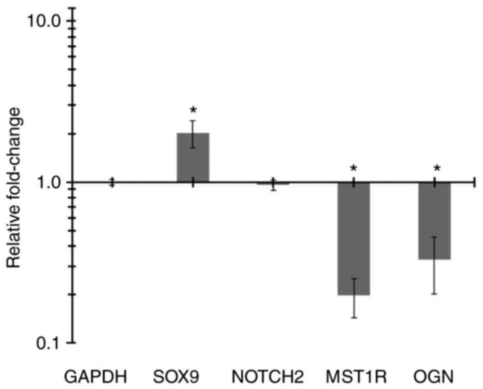
Synovial fluid cells exposed to BIOF2 for 48 h
exhibited increased SOX9 expression by >2-fold (P=0.02) and
decreased MST1R and OGN expression by 5- and 3-fold, respectively
(P=0.02). There were no alterations in NOTCH2 expression compared
with unexposed cells (Fig. 1).
In vivo effects on osteoarthritic
cartilage thickness
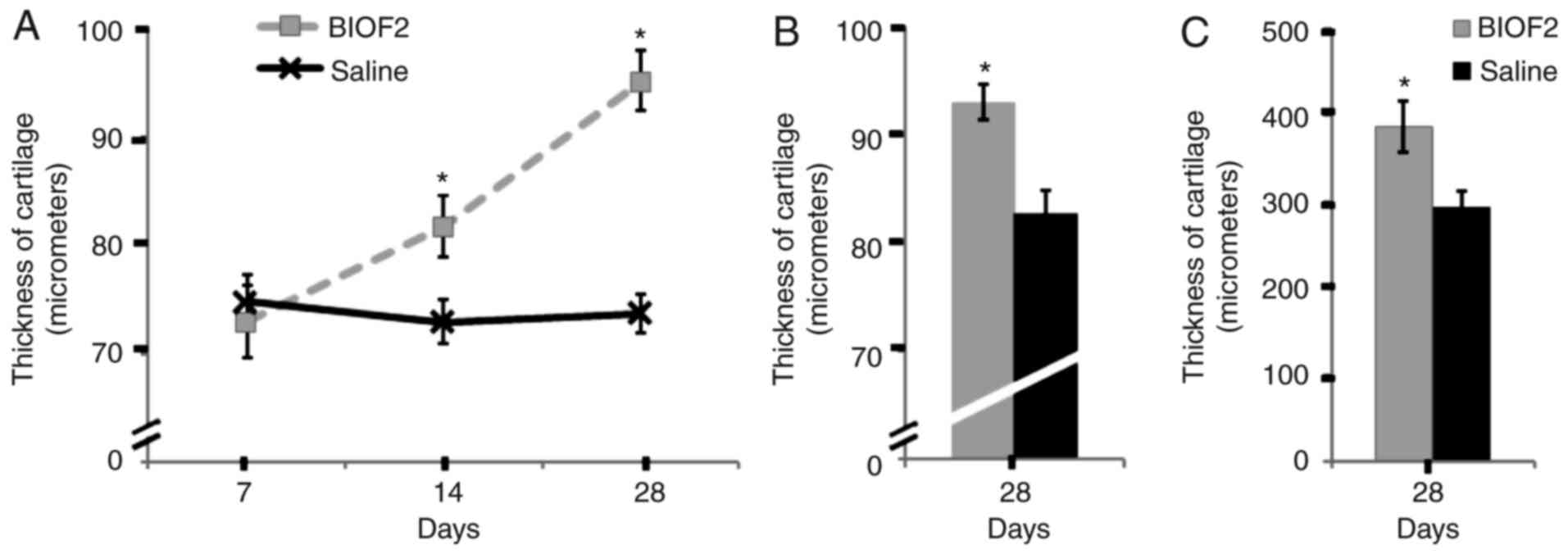
The thickness of cartilage in the medial femur
sections of osteoarthritic model animals was studied in the damaged
articulation 28 days following the application of BIOF2. In the
mouse model of induced articular damage, the intra-articular
administration of saline solution caused no significant alterations
in cartilage thickness at 28 days. By contrast, BIOF2 application
caused increased articular thickness from day 14 of treatment, with
a highly significant difference at day 28, compared with the saline
solution group (P<0.01; Fig.
2A). In the mouse model of spontaneous articular damage
(advanced-age mice) and the OA rabbit model, the cartilage of the
joints treated with BIOF2 was significantly thicker compared with
the controls (P=0.001 and P=0.009 for the mouse and rabbit models,
respectively) (Fig. 2B and C). On
day 28 following the intra-articular application of BIOF2,
cartilage thickness increased by 29, 12, and 31%, respectively,
compared with the controls in the models of induced articular
damage in mice, spontaneous damage in mice and induced damage in
rabbits.
Structure and composition of articular
cartilage in the mouse models
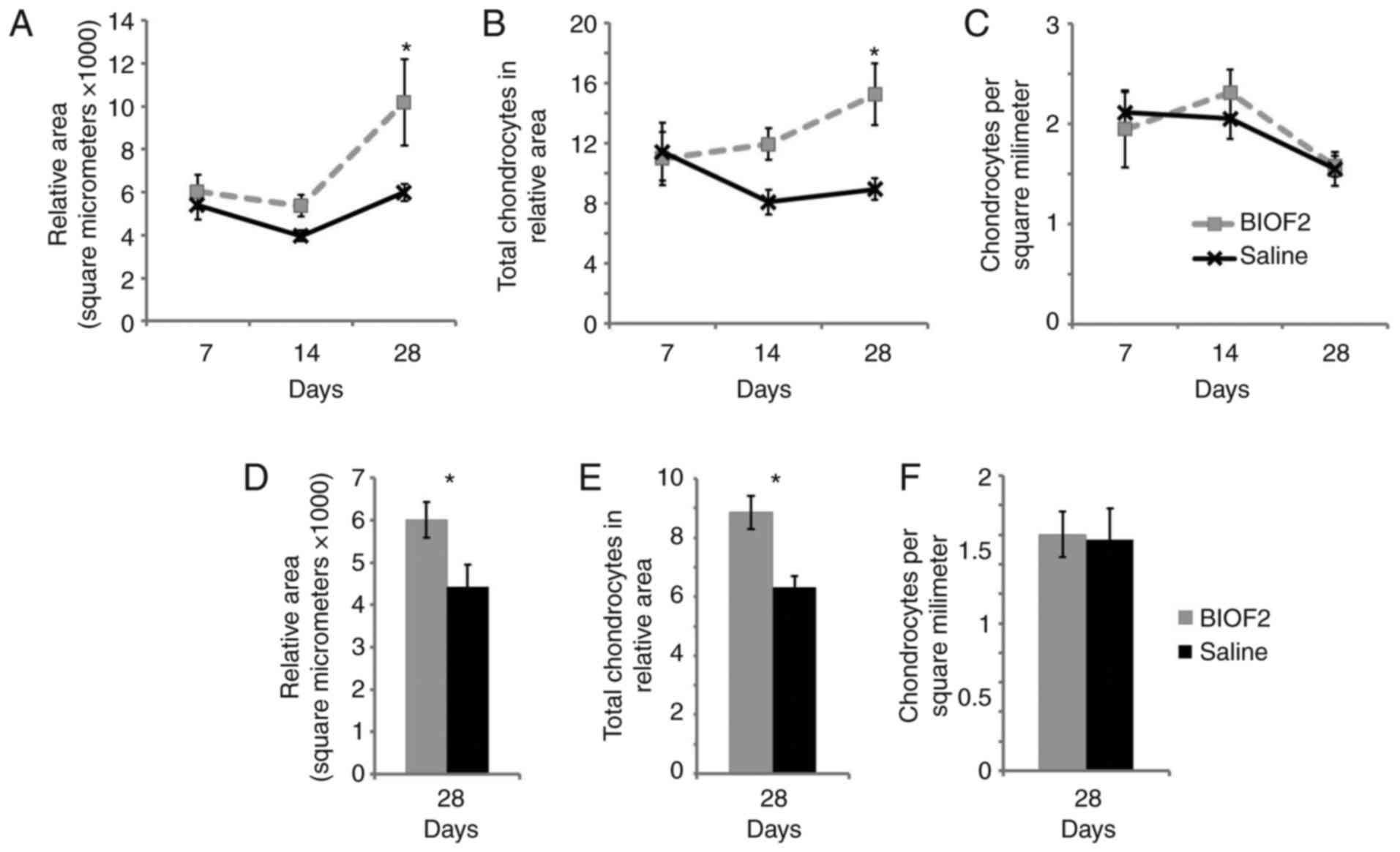
The articular area, total number of chondrocytes and
histological architecture of the cartilages were determined
(Fig. 3). In the papain-induced
articular damage mouse model, BIOF2 application significantly
increased the articular area (P=0.01) and the total number of
chondrocytes (P=0.04) at day 28, compared with the group treated
with saline solution (Fig. 3A and
D). The joints treated with saline solution remained unaltered
throughout the period following treatment. The area and cellularity
increased by an average of 70% compared with the cartilage treated
with saline solution. In the spontaneous OA mouse model, the
results were similar, with a 36% increase in area (P=0.04) and a
40% increase in cellularity (P=0.009), compared with the control
group (Fig. 3B and E). It is
important to note that the number of chondrocytes/mm2
remained unaltered in all the groups (Fig. 3C and F).
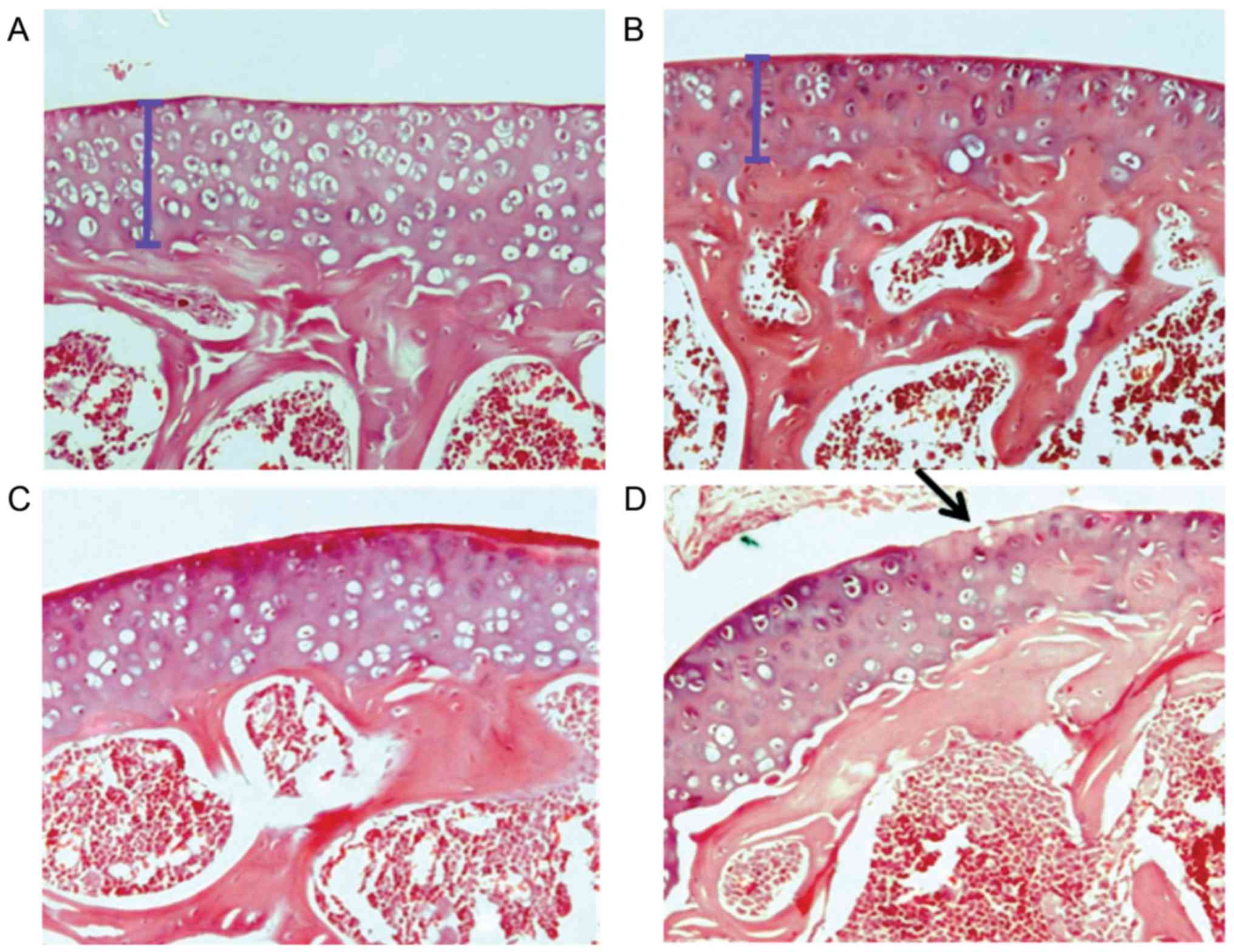
With respect to the qualitative evaluation of the
histological architecture of the cartilage, on day 28 following
BIOF2 application there was a significantly decreased grade of
articular damage, compared with cartilage treated with saline
solution (Fig. 4). According to
the scoring system developed by Chambers et al (29), which has a scale from grades 0 to 6
(0, no damage; 6, >80% loss of cartilage), the medians of the
groups treated with BIOF2 compared with saline solution were 2 vs.
3.5 (P=0.02) and 3 vs. 4 (P=0.04) for the spontaneous OA and
induced OA mouse models, respectively.
Discussion
The present study demonstrated that hyaline
cartilage regeneration may be induced in vivo via the
intra-articular application of the novel bioactive formulation
BIOF2. This was observed in three different animal models. In a
28-day follow-up of one of the animal models it was observed that
the thickness of the cartilage and the number of chondrocytes began
to increase slightly on day 14 post-treatment. However, the most
important histological alterations occurred between days 14 and 28,
and the latter day was when all the morphometric parameters were
highly significant and beneficial in regard to treatment with
BIOF2, compared with saline solution application. It was notable
that the increase in thickness and articular area was accompanied
by a proportional increase in cellularity
(chondrocytes/mm2 was unaltered). This demonstrated that
cartilage growth was not solely due to extracellular matrix
growth.
In addition, upon analyzing the histological
architecture, the joints treated with BIOF2 exhibited a lower grade
of damage compared with the controls, suggesting repair towards a
normal joint morphology. An additional relevant aspect in relation
to the BALB/c mouse animal models, compared with their controls,
was the fact that there was a greater increase in cartilage
thickness (29%) in the induced articular damage model in young mice
compared with the advanced-age mouse model (12%) at post-treatment
day 28. This concurs with clinical findings that have demonstrated
that current techniques for damaged cartilage repair appear to
produce better results in young patients (30). This may be relevant to the
suggested dosage for young or elderly patients in future clinical
trials.
The mechanism through which BIOF2 may regenerate
articular cartilage was examined at the molecular level via
experiments performed on human synovial fluid cells. BIOF2 caused
an increase in the expression of SOX9, a transcription factor that
is essential for chondrocyte differentiation and cartilage
formation (31). SOX9 serves a
notable role in the development and maintenance of the chondrogenic
phenotype. One previous study demonstrated that SOX9 expression was
relatively high in normal cartilage, although its transcript levels
were substantially decreased in OA, accompanied by degradation of
the extracellular matrix; the study additionally suggested that a
reduction of SOX9 transcript levels in osteoarthritic chondrocytes
may be responsible for such loss of the extracellular matrix
(32). Effective chondrogenesis
and inhibition of bone morphogenetic protein 2-induced osteogenesis
and endochondral ossification have additionally been demonstrated
to be achieved by directing MSCs towards the chondrocyte lineage
with SOX9 (33). A further
hypothesis for correcting OA is the overexpression of SOX9 through
gene therapy, possibly combined with cell therapy (34,35).
This indicates that the increase in SOX9 by BIOF2 is a consistent
mechanism with the therapeutic effect observed in the present
study.
The results of the present study demonstrated that
BIOF2 caused a decrease in OGN and MST1R expression. OGN has been
reported to be elevated in OA synovial fluid samples and may induce
the mineralization and calcification of cartilage (37). Additionally, MST1R has been
previously identified to be associated with osteoclastogenesis,
osteolysis and inflammation (38,39).
The decreased expression of these genes is consistent with a
therapeutic effect of BIOF2 on OA. Although previous studies have
demonstrated that the Notch pathway is active during chondrogenesis
(40,41), the present study identified no
increase in NOTCH2. However, NOTCH2 expression was measured only
once, and the possibility of its modification at another evaluation
time may not be excluded, which is a limitation of the present
analysis. Gene expression alterations (including SOX9 and
cytokines) in the joint tissue following in vivo application
of BIOF2 are an important aspect to study in future experiments in
animal models or in clinical trials.
The results of the present study have led to the
proposal of an innovative strategy to clinically repair
osteoarthritic lesions through BIOF2-induced cartilage
regeneration. This therapeutic strategy is novel and different from
other OA treatment alternatives, including viscosupplementation,
gene therapy or cell therapy. The novel strategy is based on
modifying the intra-articular microenvironment to stimulate
articular regeneration, by generating molecular and morphological
alterations in synovial fluid cells and chondrocytes. The synovium
is considered to be a candidate source of cells for cartilage.
Compared with MSCs from other sources, synovium-derived stem cells
have a higher capacity for chondrogenic differentiation (42). Fibrous synovium cells (with the
typical bipolar configuration), including the cells cultivated in
the present study, have previously been observed to release large
numbers of MSCs (43). It is
likely that BIOF2 produces molecular alterations, including SOX9
upregulation, in synovial MSCs that favor SOX9 migration to the
cartilage and differentiation into chondrocytes. In addition, BIOF2
appears to generate SOX9 elevation in chondrocytes, thus reversing
the alterations associated with OA.
There are numerous problems associated with current
OA treatment strategies. In advanced disease cases, the
recommendation is to substitute the joint with a prosthesis.
However, the surgical treatment is costly (5), with long waiting lists, and it may be
risky for elderly patients or those with comorbidities (6). Conservative treatment with oral
medications, including glucosamine, chondroitin sulfate,
methylsulfonylmethane or collagen hydrolysates, has exhibited
limited or no benefit. Intra-articular injections of different
substances are additionally being evaluated. The intra-articular
administration of corticosteroids produces a short-term
improvement. However, the repeated use of corticosteroids may
facilitate tissue atrophy, joint destruction or cartilage
degeneration (44). Hyaluronic
acid derivatives produce apparent effectiveness 5–13 weeks
following treatment. They are inferior to steroids in the
short-term, although they provide greater improvement over an
increased length of time (45).
Platelet-rich plasma is under consideration as an innovative and
promising tool with an effectiveness pattern comparable to the
intra-articular administration of hyaluronic acid (7). Stem cell therapy is an additional
strategy whose aim is to support the process of self-healing of the
knee joint cartilage damage resulting from OA symptoms (1). The intra-articular application of
stem cells, facilitating their differentiation into chondrocytes,
is a process that includes the separation of the cells by
centrifugation and other purification steps, with the aim of
increasing cartilage buildup. Clinical data on the effectiveness of
stem cell therapy remains insufficient and certain authors have
expressed concerns regarding the issues of dosage, intervention
timing, mode, route of delivery and the type of stem cells in
clinical studies (8,9). It is hypothesized that the
BIOF2-induced regeneration strategy has the potential to ameliorate
the problems or limitations of current OA treatments.
In conclusion, the application of BIOF2 was
effective for the treatment of OA in animal models, possibly as a
result of increased SOX9 expression in articular cells. This
regeneration strategy merits further study as a realistic research
focus in the near future, particularly in clinical trials.
Acknowledgements
The present study was completed using equipment
resources obtained through grant nos. 270485 and 272792 from the
INFRAESTRUCTURA-CONACYT-2016 and FOSISS-CONACYT-2016, respectively.
Dr Juan Paz and Dr Brenda Paz-Michel declare that they are the
inventors of the experimental formulation used in the present study
(patent no. US9089580 B1).
References
|
1
|
Vaishya R, Pariyo GB, Agarwal AK and Vijay
V: Non-operative management of osteoarthritis of the knee joint. J
Clin Orthop Trauma. 7:170–176. 2016. View Article : Google Scholar : PubMed/NCBI
|
|
2
|
Karsdal MA, Michaelis M, Ladel C, Siebuhr
AS, Bihlet AR, Andersen JR, Guehring H, Christiansen C, Bay-Jensen
AC and Kraus VB: Disease-modifying treatments for osteoarthritis
(DMOADs) of the knee and hip: Lessons learned from failures and
opportunities for the future. Osteoarthritis Cartilage.
24:2013–2021. 2016. View Article : Google Scholar : PubMed/NCBI
|
|
3
|
Raeissadat SA, Rayegai SM, Hassanbadi H,
Fathi M, Ghorbani E, Babaee M and Azma K: Knee osteoarthritis
injection choices: Platelet-rich plasma (PRP) verses hyaluronic
acid (A one year randomized clinical trial). Clin Med Insights
Arthritis Musculoskelet Disord. 8:1–8. 2015. View Article : Google Scholar : PubMed/NCBI
|
|
4
|
Bannuru RR, Osani M, Vaysbrot EE and
McAlindon TE: Comparative safety profile of hyaluronic acid
products for knee osteoarthritis: A systematic reviewand network
meta-analysis. Osteoarthritis Cartilage. 24:2022–2024. 2016.
View Article : Google Scholar : PubMed/NCBI
|
|
5
|
Herrera-Espiñeira C, Escobar A,
Navarro-Espigares JL, Castillo Jde D, García-Pérez L and
Godoy-Montijano A: Total knee and hip prosthesis: Variables
associated with costs. Cir Cir. 81:207–213. 2013.PubMed/NCBI
|
|
6
|
Hawker GA, Badley EM, Borkhoff CM,
Croxford R, Davis AM, Dunn S, Gignac MA, Jaglal SB, Kreder HJ and
Sale JE: Which patients are most likely to benefit from total joint
arthroplasty? Arthritis Rheum. 65:1243–1252. 2013. View Article : Google Scholar : PubMed/NCBI
|
|
7
|
Montañez-Heredia E, Irízar S, Huertas PJ,
Otero E, Del Valle M, Prat I, Díaz-Gallardo MS, Perán M, Marchal JA
and Hernandez-Lamas Mdel C: Intra-Articular injections of
Platelet-Rich plasma versus hyaluronic acid in the treatment of
osteoarthritic knee pain: A randomized clinical trial in the
context of the Spanish national health care system. Int J Mol Sci.
17(pii): E10642016. View Article : Google Scholar : PubMed/NCBI
|
|
8
|
Richter W: Cell-based cartilage repair:
Illusion or solution for osteoarthritis. Curr Opin Rheumatol.
19:451–456. 2007.PubMed/NCBI
|
|
9
|
Uth K and Trifonov D: Stem cell
application for osteoarthritis in the knee joint: A minireview.
World J Stem Cells. 6:629–636. 2014. View Article : Google Scholar : PubMed/NCBI
|
|
10
|
Jakobsen RB, Østrup E, Zhang X, Mikkelsen
TS and Brinchmann JE: Analysis of the effects of five factors
relevant to in vitro chondrogenesis of human mesenchymal stem cells
using factorial design and high throughput mRNA-profiling. PLoS
One. 9:e966152014. View Article : Google Scholar : PubMed/NCBI
|
|
11
|
Tangtrongsup S and Kisiday JD: Effects of
dexamethasone concentration and timing of exposure on
chondrogenesis of equine bone Marrow-derived mesenchymal stem
cells. Cartilage. 7:92–103. 2016. View Article : Google Scholar : PubMed/NCBI
|
|
12
|
Scioli MG, Bielli A, Gentile P, Cervelli V
and Orlandi A: Combined treatment with platelet-rich plasma and
insulin favours chondrogenic and osteogenic differentiation of
human adipose-derived stem cells in three-dimensional collagen
scaffolds. J Tissue Eng Regen Med. 11:2398–2410. 2017. View Article : Google Scholar : PubMed/NCBI
|
|
13
|
Yao Y, Zhai Z and Wang Y: Evaluation of
insulin medium or chondrogenic medium on proliferation and
chondrogenesis of ATDC5 cells. Biomed Res Int. 2014:5692412014.
View Article : Google Scholar : PubMed/NCBI
|
|
14
|
Gerstenfeld LC, Toma CD, Schaffer JL and
Landis WJ: Chondrogenic potential of skeletal cell populations:
Selective growth of chondrocytes and their morphogenesis and
development in vitro. Microsc Res Tech. 43:156–173. 1998.
View Article : Google Scholar : PubMed/NCBI
|
|
15
|
Paz-García J and Paz-Michel BA:
Formulation for regeneration of bone, cartilage, teeth, and
periodontium and treatment of tumors and cysts US Patent US9089580
B1. July 28–2015, issued February 4, 2015.
|
|
16
|
Casnici C, Lattuada D, Tonna N, Crotta K,
Storini C, Bianco F, Truzzi MC, Corradini C and Marelli O:
Optimized ‘in vitro’ culture conditions for human rheumatoid
arthritis synovial fibroblasts. Mediators Inflamm. 2014:7020572014.
View Article : Google Scholar : PubMed/NCBI
|
|
17
|
Li XW, Tuergan M and Abulizi G: Expression
of MAPK1 in cervical cancer and effect of MAPK1 gene silencing on
epithelial-mesenchymal transition, invasion and metastasis. Asian
Pac J Trop Med. 8:937–943. 2015. View Article : Google Scholar : PubMed/NCBI
|
|
18
|
Mayer S, Hirschfeld M, Jaeger M, Pies S,
Iborra S, Erbes T and Stickeler E: RON alternative splicing
regulation in primary ovarian cancer. Oncol Rep. 34:423–430. 2015.
View Article : Google Scholar : PubMed/NCBI
|
|
19
|
Biason-Lauber A, Konrad D, Meyer M,
DeBeaufort C and Schoenle EJ: Ovaries and female phenotype in a
girl with 46,XY karyotype and mutations in the CBX2 gene. Am J Hum
Genet. 84:658–663. 2009. View Article : Google Scholar : PubMed/NCBI
|
|
20
|
O'Neill CF, Urs S, Cinelli C, Lincoln A,
Nadeau RJ, León R, Toher J, Mouta-Bellum C, Friesel RE and Liaw L:
Notch2 signaling induces apoptosis and inhibits human MDA-MB-231
xenograft growth. Am J Pathol. 171:1023–1036. 2007. View Article : Google Scholar : PubMed/NCBI
|
|
21
|
Shin JE, Oh JH, Kim YK, Jung JY and Chung
JH: Transcriptional regulation of proteoglycans and
glycosaminoglycan chain-synthesizing glycosyltransferases by UV
irradiation in cultured human dermal fibroblasts. J Korean Med Sci.
26:417–424. 2011. View Article : Google Scholar : PubMed/NCBI
|
|
22
|
Pottosin I, Delgado-Enciso I,
Bonales-Alatorre E, Nieto-Pescador MG, Moreno-Galindo EG and
Dobrovinskaya O: Mechanosensitive Ca2+-permeable
channels in human leukemic cells: Pharmacological and molecular
evidence for TRPV2. Biochim Biophys Acta. 1848:51–59. 2015.
View Article : Google Scholar : PubMed/NCBI
|
|
23
|
Ma HL, Blanchet TJ, Peluso D, Hopkins B,
Morris EA and Glasson SS: Osteoarthritis severity is sex dependent
in a surgical mouse model. Osteoarthritis Cartilage. 15:695–700.
2007. View Article : Google Scholar : PubMed/NCBI
|
|
24
|
van der Kraan PM, Vitters EL, van de Putte
LB and van den Berg WB: Development of osteoarthritic lesions in
mice by ‘metabolic’ and ‘mechanical’ alterations in the knee
joints. Am J Pathol. 135:1001–1014. 1989.PubMed/NCBI
|
|
25
|
Stoop R, van der Kraan PM, Buma P,
Hollander AP, Billinghurst RC, Poole AR and van den Berg WB: Type
II collagen degradation in spontaneous osteoarthritis in C57Bl/6
and BALB/c mice. Arthritis Rheum. 42:2381–2389. 1999. View Article : Google Scholar : PubMed/NCBI
|
|
26
|
Moriizumi T, Yamashita N and Okada Y:
Papain-induced changes in the guinea pig knee joint with special
reference to cartilage healing. Virchows Arch B Cell Pathol Incl
Mol Pathol. 51:461–474. 1986. View Article : Google Scholar : PubMed/NCBI
|
|
27
|
Hardin JA, Cobelli N and Santambrogio L:
Consequences of metabolic and oxidative modifications of cartilage
tissue. Nat Rev Rheumatol. 11:521–529. 2015. View Article : Google Scholar : PubMed/NCBI
|
|
28
|
Fazelipour S, Tootian Z, Matini E and
Hadipour-Jahromy M: Histomorphometric alteration of knee articular
cartilage and serum alkaline phosphatase in young female mice by
chronic supplementation with soybean. Phytother Res. 25:886–891.
2011. View
Article : Google Scholar : PubMed/NCBI
|
|
29
|
Chambers MG, Cox L, Chong L, Suri N, Cover
P, Bayliss MT and Mason RM: Matrix metalloproteinases and
aggrecanases cleave aggrecan in different zones of normal cartilage
but colocalize in the development of osteoarthritic lesions in
STR/ort mice. Arthritis Rheum. 44:1455–1465. 2001. View Article : Google Scholar : PubMed/NCBI
|
|
30
|
Musumeci G, Loreto C, Castorina S, Imbesi
R, Leonardi R and Castrogiovanni P: Current concepts in the
treatment of cartilage damage. A review. Ital J Anat Embryol.
118:189–203. 2013.PubMed/NCBI
|
|
31
|
Bi W, Deng JM, Zhang Z, Behringer RR and
de Crombrugghe B: Sox9 is required for cartilage formation. Nat
Genet. 22:85–89. 1999. View
Article : Google Scholar : PubMed/NCBI
|
|
32
|
Goldberg VM, Buckwalter J, Halpin M,
Jiranek W, Mihalko W, Pinzur M, Rohan B, Vail T, Walker P, Windsor
R and Wright T: Recommendations of the OARSI FDA Osteoarthritis
Devices Working Group. Osteoarthritis Cartilage. 19:509–514. 2011.
View Article : Google Scholar : PubMed/NCBI
|
|
33
|
Liao J, Hu N, Zhou N, Lin L, Zhao C, Yi S,
Fan T, Bao W, Liang X, Chen H, et al: Sox9 potentiates BMP2-induced
chondrogenic differentiation and inhibits BMP2-induced osteogenic
differentiation. PLoS One. 9:e890252014. View Article : Google Scholar : PubMed/NCBI
|
|
34
|
Tao K, Frisch J, Rey-Rico A, Venkatesan
JK, Schmitt G, Madry H, Lin J and Cucchiarini M: Co-overexpression
of TGF-β and SOX9 via rAAV gene transfer modulates the metabolic
and chondrogenic activities of human bone marrow-derived
mesenchymal stem cells. Stem Cell Res Ther. 7:202016. View Article : Google Scholar : PubMed/NCBI
|
|
35
|
Wan R, Hu J, Zhou Q, Wang J, Liu P and Wei
Y: Application of co-expressed genes to articular cartilage: New
hope for the treatment of osteoarthritis (Review). Mol Med Rep.
6:16–18. 2012.PubMed/NCBI
|
|
36
|
Balakrishnan L, Nirujogi RS, Ahmad S,
Bhattacharjee M, Manda SS, Renuse S, Kelkar DS, Subbannayya Y, Raju
R, Goel R, et al: Proteomic analysis of human osteoarthritis
synovial fluid. Clin Proteomics. 11:62014. View Article : Google Scholar : PubMed/NCBI
|
|
37
|
De Ceuninck F, Marcheteau E, Berger S,
Caliez A, Dumont V, Raes M, Anract P, Leclerc G, Boutin JA and
Ferry G: Assessment of some tools for the characterization of the
human osteoarthritic cartilage proteome. J Biomol Tech. 16:256–265.
2005.PubMed/NCBI
|
|
38
|
Akiyama M, Nakahama K and Morita I: Impact
of docosahexaenoic acid on gene expression during
osteoclastogenesis in vitro-a comprehensive analysis. Nutrients.
5:3151–3162. 2013. View Article : Google Scholar : PubMed/NCBI
|
|
39
|
Cully M: Bone diseases: MST1R inhibitor
prevents bone osteolysis. Nat Rev Drug Discov. 16:164–165. 2017.
View Article : Google Scholar : PubMed/NCBI
|
|
40
|
Watanabe M, Tezuka Y, Matsuno K, Miyatani
S, Morimura N, Yasuda M, Fujimaki R, Kuroda K, Hiraki Y, Hozumi N
and Tezuka K: Suppresion of differentiation and proliferation of
each chondrogenic cells by Notch. J Bone Miner Metab. 21:344–352.
2003. View Article : Google Scholar : PubMed/NCBI
|
|
41
|
Hardingham TE, Oldershaw RA and Tew SR:
Cartilage, SOX9 and Notch signal in chondrogenesis. J Anat.
209:469–480. 2006. View Article : Google Scholar : PubMed/NCBI
|
|
42
|
Pei M, He F and Vunjak-Novakovic G:
Synovium-derived stem cell-based chondrogenesis. Differentiation.
76:1044–1056. 2008. View Article : Google Scholar : PubMed/NCBI
|
|
43
|
Katagiri K, Matsukura Y, Muneta T, Ozeki
N, Mizuno M, Katano H and Sekiya I: Fibrous synovium releases
higher numbers of mesenchymal stem cells than adipose synovium in a
suspended synovium culture model. Arthroscopy. 33:800–810. 2017.
View Article : Google Scholar : PubMed/NCBI
|
|
44
|
Richards MM, Maxwell JS, Weng L, Angelos
MG and Golzarian J: Intra-articular treatment of knee
osteoarthritis: From anti-inflammatories to products of
regenerative medicine. Phys Sportsmed. 44:101–108. 2016. View Article : Google Scholar : PubMed/NCBI
|
|
45
|
Rodriguez-Maerchan EC: Intra articular
injection of hyaluronic acid and other drugs in knee joint. HSS J.
9:180–182. 2013. View Article : Google Scholar : PubMed/NCBI
|


