Introduction
Epilepsy may cause cognitive dysfunction and is
increasingly becoming a focus of research. Previous studies have
demonstrated that the abnormal expression and function of microRNAs
(miRs/miRNAs) is associated with the learning and memory
impairments observed in disorders of the nervous system, including
epilepsy and Alzheimer's disease (1,2).
However, the mechanism through which this impairment occurs remains
unclear. miRs are a type of endogenous single-stranded small
molecule RNA that exist in animal and plant cells, and cause target
mRNA degradation or translational inhibition by acting on specific
mRNA 3′ untranslated regions (3).
miR-34c is a member of the miR-34 family. It has been hypothesized
that miR-34c may be a regulatory factor of cognitive function
(4). Previous studies have
demonstrated that, in neurodegenerative disease, the expression of
miR-34c decreases following treatment with an inhibitor and that
this may improve learning ability (5,6).
miR-34c expression was observed to be downregulated by silencing
miR-34c, and this improved the cognitive impairment caused by
ketamine (7).
Long-term potentiation (LTP) is a form of synaptic
plasticity and is hypothesized to be one of the major cellular
mechanisms underlying learning and memory (8,9).
Previous studies have demonstrated that miRNAs serve a role in LTP
by regulating the synthesis of proteins associated with LTP
maintenance, including N-methyl-D-aspartate receptors (NMDARs) and
α-amino-3-hydroxy-5-methyl-4-isoxazolepropionic acid receptors
(AMPARs). Previous studies have observed that miR-34a and miR-132
regulate LTP maintenance by acting on LTP-associated specific mRNAs
(10,11). NMDARs serve an important role in
switching synaptic activity-specific patterns into long-term
changes in synaptic function and structure associated with learning
and memory (12).
miR-34c serves an important role in numerous
cognitive disorders, but few studies have focused on the role of
miR-34c in cognitive impairment caused by epilepsy. In a previous
study (13), the expression of
miR-34c in pentylenetetrazol (PTZ)-induced epileptic rats with
memory impairment was demonstrated to be increased; however, the
potential regulatory effects of miR-34c on cognitive function in
epilepsy, via the regulation of LTP maintenance, remain unknown.
Therefore, the present study aimed to use a PTZ-induced epileptic
rat model to investigate the association between miR-34c and memory
impairment in epileptic rats, and the underlying mechanism.
Repetitive PTZ kindling was performed to induce the temporal lobe
epilepsy (TLE) model and an miR-34c agomir was used as an
exacerbating treatment, in place of an miR-34c inhibitor, in order
to explore the mechanisms, as a previous study reported an
incidence of 19.23% (10/52) TLE rats with cognitive deficits
(13).
Materials and methods
Animals and grouping
A total of 36 healthy male Sprague Dawley rats (9–10
weeks-old, 220–240 g) were provided by the Animal Experimental
Center of Guangxi Medical University (Nanning, China). Animals were
housed in groups of five under the following conditions: 22–26°C
room temperature, 50–60% humidity, under a 12 h light/dark cycle,
with lights on at 8:00 a.m. The rats were given free access to food
and water. Animals were handled according to the guidelines on
animal experimentation of the Council for International
Organizations of Medical Sciences (World Health Organization,
Geneva, Switzerland), and the Guangxi Medical University Animal
Care and Use Committee approved the animal protocols.
Epileptic rats were randomly divided into 2 groups
of 12 rats, including the epilepsy group and the miR-34c agomir
group. Additionally, 12 rats were used as the sham group, receiving
treatment with an equal amount of saline solution administered into
the abdominal cavity.
Model establishment and drug
delivery
The rats were treated with 60 mg/kg PTZ
(Sigma-Aldrich; Merck KGaA, Darmstadt, Germany) by intraperitoneal
(i.p.) injection. All of the rats were observed for 20 min
following PTZ injection and behavioral alterations were recorded
and graded according to the Racine grading system as follows: 0, No
reaction; grade I, face and ears twitching; grade II, nodding, neck
and back spasms; grade III, unilateral forelimb clonus; grade IV,
bilateral forelimb clonus and standing; and grade V, systemic
seizure and loss of balance. Rats with grade IV or V seizures were
selected as successful epilepsy models. These rats were secured in
stereotaxic apparatus (Shanghai Alcott Biotech Co., Ltd., Shanghai,
China) following anesthetization with 10% chloral hydrate (3.5
ml/kg; i.p.), on the 2nd day following treatment with PTZ. The skin
was cut along the middle of the head and the anterior fontanel was
exposed. The right ventricle was positioned (AP=−0.9 mm, R=−1.3 mm,
V=−3.5 mm) in the stereotaxic atlas according to Paxinos et
al (14). A 10 µl micro
syringe was fixed on the micro injection pump pointing to the hole
of the right ventricle drilled on the skull with a syringe needle.
The speed of injection was set at 3 min/µl. Rats in the miR-34c
agomir group were injected with Rattus norvegicus-miR-34c
agomir (cat. no. miR40004723-1-10, Guangzhou RiboBio Co., Ltd.,
Guangzhou, China), while rats in the epilepsy group were injected
with an equivalent amount of scramble miR (cat. no. miR04101-1-10,
Guangzhou RiboBio Co., Ltd.) into the right ventricle. The needle
remained in place for 10 min subsequent to the injection. The rats
received a repeated i.p. injection of 35 mg/kg PTZ every other 48 h
from the 3rd day following surgery, with a total of 14 injections,
to rekindle the seizures.
Morris water maze test
The Morris water maze test was used to evaluate the
cognitive function of the rats. The Morris water maze included a
round pool (diameter, 120 cm; height, 50 cm; depth, 25 cm).
Additionally, there was a platform with a diameter of 10 cm,
located 2 cm below the water level. The water in the maze was kept
at a temperature of 22±1°C. A digital camera was connected to the
computer monitor screen above the pool, and the data were acquired
and processed using the SLY-WMS Morris water maze system (version
2.1, Panlab, S.L.U. Barcelona, Spain).
The place navigation test lasted for 5 days, and
training occurred at 9:00 a.m. every day. The rats were placed into
the water in each the four quadrants, sequentially in a clockwise
manner. The time that the rats took to find the platform was
observed and recorded (escape latency). If a rat failed to find the
platform within 90 sec, they were guided to and kept on the
platform for 10 sec and the escape latency was recorded as 90
sec.
The spatial probe test was used to evaluate the
ability of the rats to remember spatial locations, subsequent to
the rats having learned how to find the platform. On the 6th day,
the platform was removed and the rats were observed for 90 sec in
the water. The frequency with which the rat travelled to the
original position of the platform (crossing time), and the time and
distance percentages that they remained in the target quadrant,
were recorded.
Reverse transcription-quantitative
polymerase chain reaction (RT-qPCR) analysis of miR-34c
After 1 h finishing the final water maze test, the
rats were anesthetized by injecting 10% chloral hydrate (300 mg/kg)
intraperitoneally, and were perfused by normal saline
intracardially. The hippocampal tissue was isolated on ice for PCR
and western blotting. The expression of miR-34c was detected by
RT-qPCR to verify the effect of the agomir. The miR-34c primers
were designed and synthesized by Takara Biotechnology Co., Ltd.
(cat. no. MQA174, Takara, Dalian, China). Total RNA was extracted
using TRIzol reagent (Invitrogen; Thermo Fisher Scientific, Inc.,
Waltham, MA, USA). The concentration and purity of the RNA were
measured using the Scientific NanoDrop Thermo 2000
spectrophotometer (Thermo Fisher Scientific, Inc., Wilmington, DE,
USA). The amplification of miR-34c cDNA was performed using an
Mir-X™ miRNA First-Strand Synthesis kit (Takara
Biotechnology Co., Ltd.). The reaction system was at a volume of 10
µl, and the cDNA was stored at −20°C. Using an Mir-X miRNA RT-qPCR
SYBR kit (Takara, Biotechnology Co., Ltd.), the PCR was performed
using the Light Cycler 480 Real-time PCR system (denaturation at
95°C for 10 sec; 40 cycles of amplification at 95°C for 5 sec and
60°C for 20 sec). U6 (forward, 5′-GCTTCGGCAGCACATATACTAAAAT-3′ and
reverse 5′-CGCTTCACGAATTTGCGTGTCAT-3′) was used as an internal
control. The relative amount of miR-34c in the hippocampal tissue
was analyzed using the relative quantification 2−ΔΔCq
method (15).
miR-34c target gene prediction and
functional analysis
Potential target genes of miR-34c were predicted
using MicroCosm (https://www.microcosm.com/), miRanda (http://34.236.212.39/microrna/home.do),
and miRDB (http://www.mirdb.org/). The Gene
Ontology (GO; www.geneontology.org/) and Kyoto Encyclopedia of Genes
and Genomes (KEGG; www.genome.jp/keg/) databases were used to obtain
information regarding the functions of the predicted target genes.
GO and KEGG analysis was performed on the predicted target genes of
miR-34c to investigate the regulatory mechanism of the
overexpressed miR-34c in response to cognitive impairment in
epilepsy.
Immunofluorescence staining
After perfusion with normal saline intracardially,
the rats were sacrificed and brain tissue was removed, postfixed
overnight in the same solution at 4°C, embedded in paraffin wax,
and sectioned coronally at 3 µm for immunofluorescence staining. In
brief, the slices were immune-labeled using primary antibody
specific to glutamate receptor ionotropic, NMDA 2B (NR2B; 1:1,000;
cat. no. 14544, Cell Signaling Technology, Inc., Danvers, MA, USA)
diluted in PBS overnight at 4°C. Following washing with PBS three
times, the slices were incubated with the secondary antibody (goat
anti-rabbit immunoglobulin G; 1:1,000; cat. no. A8275,
Sigma-Aldrich; Merck KGaA) at room temperature for 1 h.
Fluorescence images were acquired using an Olympus fluorescence
microscope (Olympus Corporation, Tokyo, Japan) and an image capture
system.
Western blotting
Expression of NR2B protein in whole cell lysate, and
phosphorylated (p)-NADPH-dependent diflavin oxidoreductase 1 (NR1)
and p-glutamate receptor 1 (GluR1) protein at the postsynaptic
density (PSD), were detected using western blotting. The total
protein was extracted using radioimmunoprecipitation assay buffer
(Beyotime Institute of Biotechnology, Haimen, China), and was
detected using a bicinchoninic acid protein assay kit. Isolated
protein harvested from 150 g fresh brain tissue was heat-denatured
at 100°C for 5 min, subjected to electrophoresis on a 10% SDS-PAGE
gel for 2.5 h and transferred to a 0.45 mm polyvinylidene fluoride
membrane (EMD Millipore, Billerica, MA, USA) using the semi-dry
transfer apparatus. The membranes were blocked in 5% skimmed milk
for 1 h at 24°C and incubated on ice overnight with the following
primary antibodies: Anti-NR2B (1:1,000), with anti-GAPDH (1:5,000;
cat. no. 2118, Cell Signaling Technology, Inc.) as an internal
control; anti-NR1 (1:1,000; cat. no. 5704, Cell Signaling
Technology, Inc.), with anti-β-actin (1:5,000; cat. no. 8457, Cell
Signaling Technology, Inc.) as an internal control; and anti-GluR1
(1:1,000; cat. No. 8850, Cell Signaling Technology, Inc.), with
anti-β-actin (1:5,000) as an internal control. The membranes were
incubated with an Alexa Fluor® secondary anti-rabbit
antibody (1:5,000; cat. no. 8889, Cell Signaling Technology, Inc.)
for 1 h following washing at room temperature. The bands were
scanned using the LI-COR Odyssey imaging system (LI-COR
Biosciences, Lincoln, NE, USA), and analyzed using LI-COR Odyssey
software (version 3.0; LI-COR Biosciences).
Statistical analysis
Data are presented as the mean ± standard deviation.
SPSS statistical software (version 16.0; SPSS, Inc., Chicago, IL,
USA) was used to analyze the data and single factor analysis of
variance followed by the Least-Significant Difference post-hoc
test, which was performed to compare the groups. P<0.05 was
considered to indicate a statistically significant difference.
Results
miR-34c serves a role in impairing
cognitive function
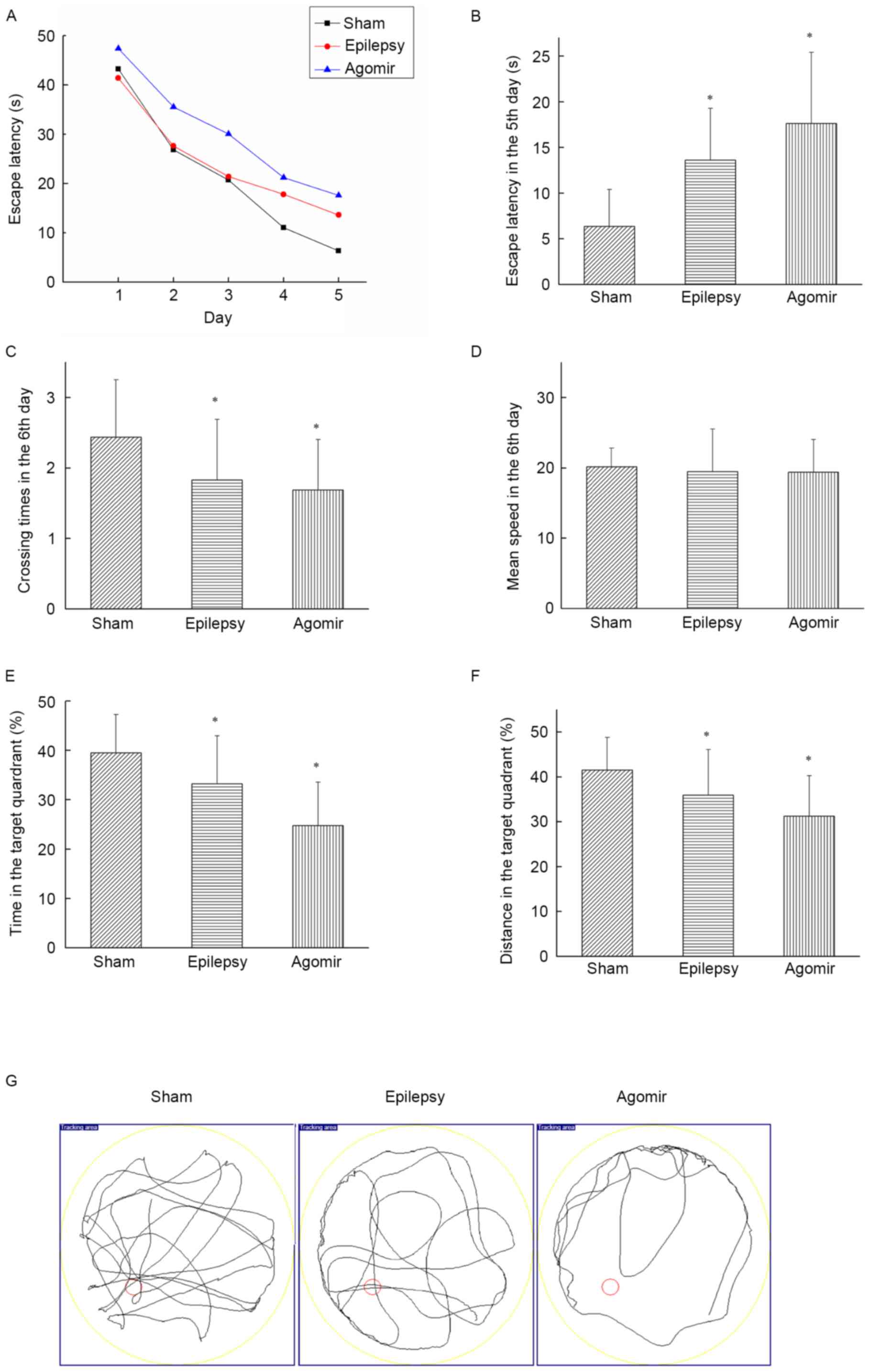
The place navigation test may be used to analyze
learning ability in animals. The results of the present study
demonstrated that the escape latency of each group decreased as the
number of training days increased. The miR-34c agomir group
exhibited increased escape latencies at each time point compared
with the sham and epilepsy groups (Fig. 1A). As presented in Fig. 1B, compared with the sham group, the
escape latency on the 5th day was significantly increased in the
epilepsy and miR-34c agomir groups (both P<0.05), indicating a
decreased learning ability following epileptic seizures.
Additionally, rats in the miR-34c agomir group exhibited an
impairment in studying ability.
On the 6th day, the platform was removed to perform
the spatial probe test. As presented in Fig. 1C, the epilepsy and miR-34c agomir
groups exhibited significantly decreased crossing times compared
with the sham group (both P<0.05); however, no significant
differences were observed between the epilepsy and agomir groups.
As presented in Fig. 1D, no
differences were observed in mean speed among the three groups on
the 6th day. The time and distance for which the rats remained in
the target quadrant in the epilepsy and miR-34c agomir groups were
decreased compared with the sham group (all P<0.05; Fig. 1E and F). In the miR-34c agomir
group, the time and distance for which the rats remained in the
target quadrant were significantly decreased compared with the
epilepsy group (both P<0.05). The results of the present study
demonstrated that treatment with the miR-34c agomir led to memory
impairment. The track plot from the 6th day is presented in
Fig. 1G.
miR-34c is upregulated following
epilepsy induced by PTZ
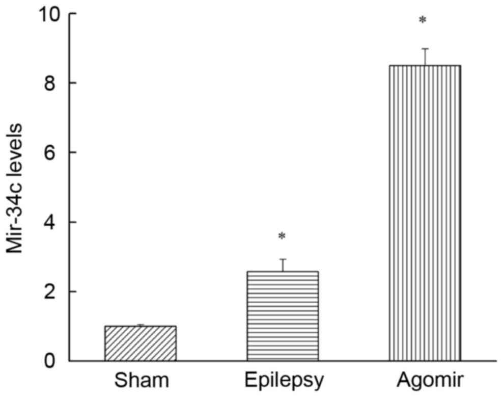
As presented in Fig.
2, compared with the sham group, the miR-34c expression in the
epilepsy group was significantly upregulated (P<0.05),
demonstrating that miR-34c was increased following epilepsy induced
by PTZ. In addition, the expression of miR-34c in the miR-34c
agomir group was also significantly increased compared with the
sham group (P<0.05).
Functional analysis of miR-34c
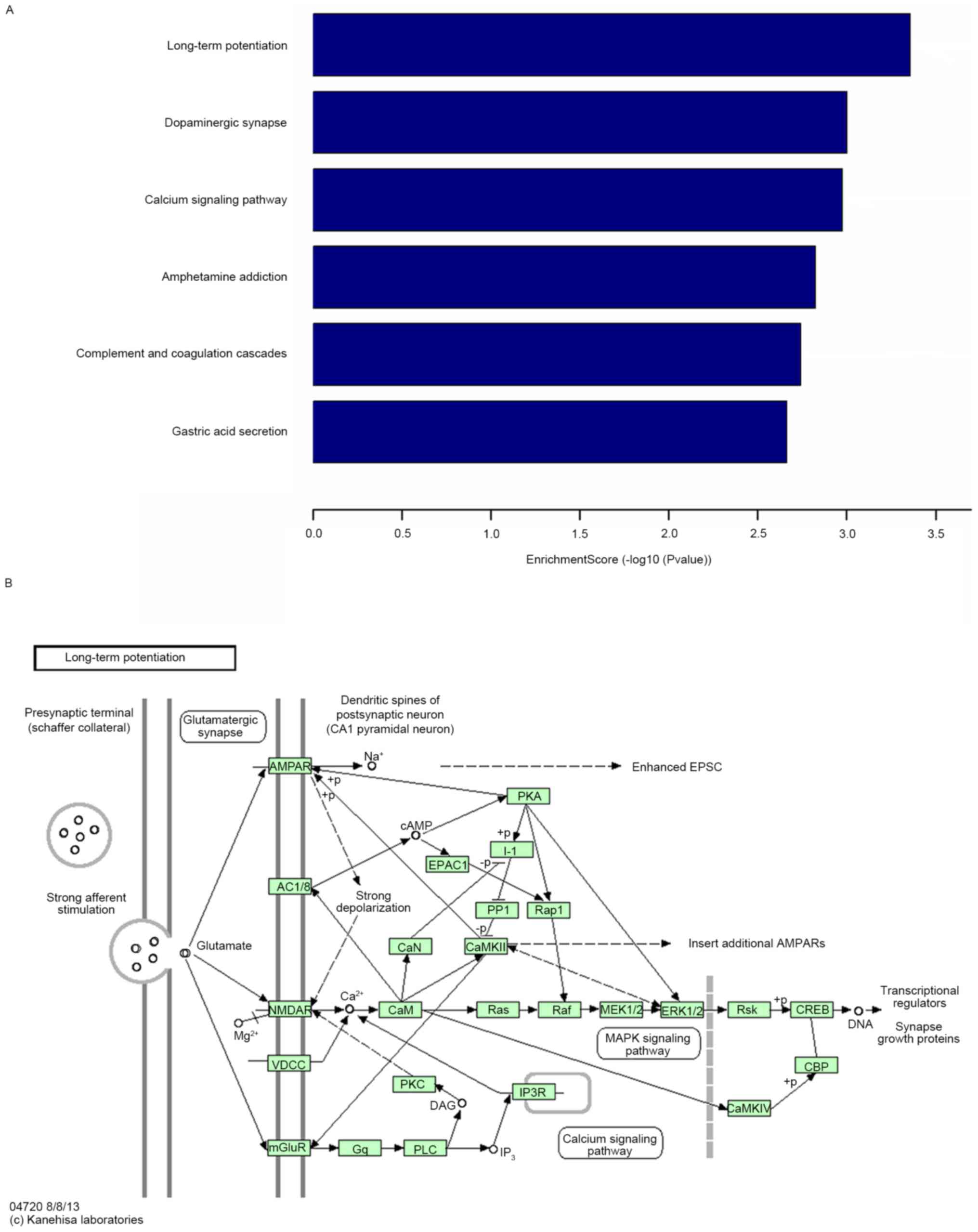
Following KEGG pathway enrichment analysis, the
predicted target genes of miR-34c were primarily enriched in six
KEGG pathways, including LTP, dopaminergic synapse, calcium
signaling pathway and amphetamine addiction. It is known that LTP
serves an important role in cognitive function (Fig. 3).
Expression of NR2B in the cortex
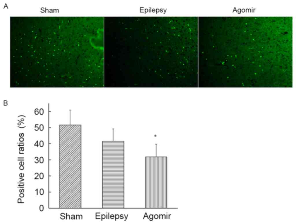
As demonstrated by counting positive cells in the
cortex, rats in the epilepsy and miR-34c agomir groups exhibited
decreased NR2B expression compared with the sham group, and the
number of NR2B-positive cells in miR-34c agomir group was decreased
compared with the sham group (Fig.
4).
Expression of NR2B, p-GluR1 and p-NR1
in the hippocampus
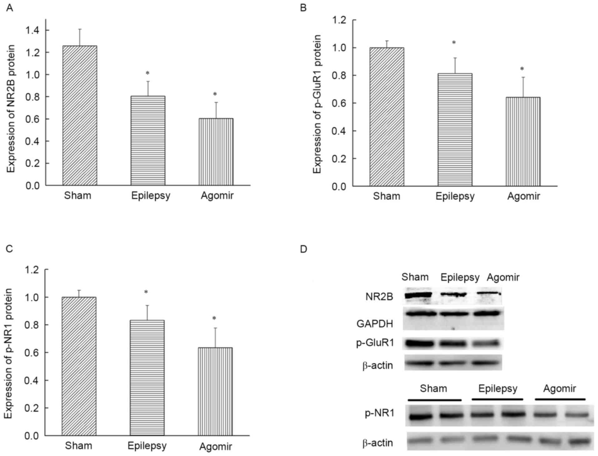
As presented in Fig.
5A, the expression of NR2B in the epilepsy group was
significantly downregulated compared with the sham group
(P<0.05), indicating that NR2B expression in the hippocampus was
decreased following epilepsy. The expression of NR2B protein in the
miR-34c agomir group was also decreased compared with the sham
group (P<0.05), which demonstrated that the levels of NR2B in
the hippocampus were consistent with those in the cortex.
Compared with the sham group, the levels of p-GluR1
and p-NR1 in the PSD of the hippocampus in the epilepsy group were
significantly decreased (P<0.05). In addition, the expression of
p-GluR1 and p-NR1 in the PSD of the hippocampus in the miR-34c
agomir group were also decreased compared with the sham group
(P<0.05) (Fig. 5B and C). The
results of the western blot analysis are presented in Fig. 5D.
Discussion
In the clinical manifestation of epilepsy,
particularly TLE, 30–40% of patients develop memory impairment,
attention dispersion and other cognitive disorders (16). PTZ-induced epileptic rats are
similar to humans with temporal lobe epilepsy. PTZ-induced rats are
frequently used to study the mechanisms of cognitive impairment in
epilepsy. Cognitive impairment due to TLE has been demonstrated to
be associated with lesions of the limbic system, including the
hippocampus. Studies have demonstrated that the synaptic plasticity
of the hippocampus, represented by LTP (17) and long term depression (18,19),
is associated with cognitive function.
The chronic epilepsy modal generated in the present
study began with an injection of PTZ (60 mg/kg) as a
preconditioning treatment, followed by a total of 14 injections of
PTZ (35 mg/kg), instead of the standard 15 injections, to mimic a
prominent feature of clinical patients with epilepsy. miRNA agomir
is a chemically-modified antisense strand containing 2
phosphorothioates at the 5′ end, 4 phosphorothioates and 4
cholesterol groups at the 3′ end, and a full-length nucleotide
2′-methoxy modification. Agomirs are able to mimic mature
endogenous miRNAs and stimulate miRNA activity following transfer
into cells in vivo. Agomirs and antagomirs have been
widely-used in vivo to investigate the role of miRNAs
(20).
The results of the present study demonstrated that
LTP is one of the principal pathways of miR-34c, via KEGG pathway
enrichment analysis. LTP in the hippocampus is a cellular mechanism
hypothesized to underlie memory formation, and previous studies
have demonstrated that stress may acutely and chronically impair
memory acquisition and LTP induction (21,22).
When glutamate activates NMDARs on the postsynaptic membrane, the
influx of Ca2+ is increased, causing a series of cascade
reactions and leading to LTP. NMDARs are voltage-dependent
ligand-gated ionotropic glutamate receptors and exhibit an
increased permeability to calcium ions. NMDARs, including NR1,
NR2A-D and NMDA receptor subunit 3, are expressed in the cerebral
cortex and hippocampus. NR2A-D act as regulatory subunits. NR2B is
the primary regulatory subunit of NMDAR, causing the change in
Ca2+ permeability and serving an important role in
learning and memory. It is frequently termed a ‘smart gene’
(23). Functional NMDARs consist
of 1–2 constitutive glycine-binding NR1 subunits and 1–2 NR2
glutamate-binding subunits (24,25).
A previous study demonstrated that the specific deletion of the NR1
subunit of NMDAR may lead to hippocampus-specific manipulation of
LTP and an increase in a behavioral phenotype which impairs spatial
working memory (26). Previous
studies have observed that when rats were injected with an NMDA
receptor antagonist, spatial working memory was impaired (27,28),
while learning ability was improved by increasing the level of NR2B
(29). Genetic deletion of the
NR2B subunit in the hippocampus or forebrain may lead to memory
deficits and impaired LTP (30).
Similar results demonstrated that acute administration of ghrelin
increased NR2B protein levels in the hippocampus and may result in
increased LTP generation and long-term memory (31). The results of the present study
indicated that, when treated with miR-34c agomir, cognitive
function in epileptic rats was impaired and the expression of NR2B
in the cortex, and p-NR1 and NR2B in the hippocampus, were
decreased. It was hypothesized that miR-34c may exert an effect on
cognitive dysfunction in epileptic rats induced by PTZ, which may
account for the impaired LTP.
GluR1 (also termed GluR-A or GluA1) acts as an AMPAR
subunit. GluR1-containing AMPARs serve a role in
hippocampus-dependent forms of learning and memory (32). Previous studies have demonstrated
that GluR1 deletion may impair spatial working memory. The influx
of Ca2+, activated by NMDARs, stimulates
calcium/calmodulin-dependent protein kinase type II subunit γ
(CaMKII), which phosphorylates the GluR1 subunit of the AMPARs
(24). The upregulation of NR2B
subunits may increase the influx of Ca2+ and activation
of CaMKII (31). NMDAR activation
and downstream signaling events induced by Ca2+ influx
result in the phosphorylation of GluR1-containing AMPARs (33). It was hypothesized that
downregulation of NR2B subunits may cause a decrease in
Ca2+ influx and activation of CaMKII, leading to p-GluR1
expression at the PSD of the hippocampus and resulting in LTP
impairment.
In conclusion, the present study used a PTZ-induced
epileptic rat model and caused miR-34c overexpression using
treatment with miR-34c agomir. The results of the present study
demonstrated that, following epileptic seizures, the levels of
miR-34c were upregulated and cognitive function was impaired,
possibly due to the increased expression of miR-34c. It was
hypothesized that miR-34c may serve a negative role in cognitive
function in a PTZ-induced epileptic model, by reducing the
expression of NR2B, p-NR1 and p-GluR1 proteins associated with LTP,
in pathways including the NMDARs and AMPARs, which may elucidate a
potential anabolic strategy for treating cognitive dysfunction in
epilepsy.
Acknowledgements
The present study was supported by the National
Natural Science Foundation of China (grant nos. 81360201 and
81160167).
References
|
1
|
Fineberg SK, Kosik KS and Davidson BL:
MicroRNAs potentiate neural development. Neuron. 64:303–309. 2009.
View Article : Google Scholar : PubMed/NCBI
|
|
2
|
Smalheiser NR and Lugli G: microRNA
regulation of synaptic plasticity. Neuromolecular Med. 11:133–140.
2009. View Article : Google Scholar : PubMed/NCBI
|
|
3
|
Ouellet DL, Perron MP, Gobeil LA, Plante P
and Provost P: MicroRNAs in gene regulation: When the smallest
governs it all. J Biomed Biotechnol. 2006:696162006. View Article : Google Scholar : PubMed/NCBI
|
|
4
|
Parsons MJ, Grimm CH, Paya-Cano JL, Sugden
K, Nietfeld W, Lehrach H and Schalkwyk LC: Using hippocampal
microRNA expression differences between mouse inbred strains to
characterise miRNA function. Mamm Genome. 19:552–560. 2008.
View Article : Google Scholar : PubMed/NCBI
|
|
5
|
Haramati S, Navon I, Issler O, Ezra-Nevo
G, Gil S, Zwang R, Hornstein E and Chen A: MicroRNA as repressors
of stress-induced anxiety: The case of amygdalar miR-34. J
Neurosci. 31:14191–14203. 2011. View Article : Google Scholar : PubMed/NCBI
|
|
6
|
Zovoilis A, Agbemenyah HY, Agis-Balboa RC,
Stilling RM, Edbauer D, Rao P, Farinelli L, Delalle I, Schmitt A,
Falkai P, et al: microRNA-34c is a novel target to treat dementias.
EMBO J. 30:4299–4308. 2011. View Article : Google Scholar : PubMed/NCBI
|
|
7
|
Cao SE, Tian J, Chen S, Zhang X and Zhang
Y: Role of miR-34c in ketamine-induced neurotoxicity in neonatal
mice hippocampus. Cell Biol Int. 39:164–168. 2015. View Article : Google Scholar : PubMed/NCBI
|
|
8
|
Ryan B, Joilin G and Williams JM:
Plasticity-related microRNA and their potential contribution to the
maintenance of long-term potentiation. Front Mol Neurosci. 8:42015.
View Article : Google Scholar : PubMed/NCBI
|
|
9
|
Stepan J, Dine J and Eder M: Functional
optical probing of the hippocampal trisynaptic circuit in vitro:
Network dynamics, filter properties, and polysynaptic induction of
CA1 LTP. Front Neurosci. 9:1602015. View Article : Google Scholar : PubMed/NCBI
|
|
10
|
Bowden JB, Abraham WC and Harris KM:
Differential effects of strain, circadian cycle, and stimulation
pattern on LTP and concurrent LTD in the dentate gyrus of freely
moving rats. Hippocampus. 22:1363–1370. 2012. View Article : Google Scholar : PubMed/NCBI
|
|
11
|
Joilin G, Guévremont D, Ryan B, Claudianos
C, Cristino AS, Abraham WC and Williams JM: Rapid regulation of
microRNA following induction of long-term potentiation in vivo.
Front Mol Neurosci. 7:982014. View Article : Google Scholar : PubMed/NCBI
|
|
12
|
Cercato MC, Colettis N, Snitcofsky M,
Aguirre AI, Kornisiuk EE, Baez MV and Jerusalinsky DA: Hippocampal
NMDA receptors and the previous experience effect on memory. J
Physiol Paris. 108:263–269. 2014. View Article : Google Scholar : PubMed/NCBI
|
|
13
|
Liu X, Wu Y, Huang Q, Zou D, Qin W and
Chen Z: Grouping pentylenetetrazol-induced epileptic rats according
to memory impairment and MicroRNA expression profiles in the
hippocampus. PLoS One. 10:e01261232015. View Article : Google Scholar : PubMed/NCBI
|
|
14
|
Paxinos G, Watson CR and Emson PC:
AChE-stained horizontal sections of the rat brain in stereotaxic
coordinates. J Neurosci Methods. 3:129–149. 1980. View Article : Google Scholar : PubMed/NCBI
|
|
15
|
Livak KJ and Schmittgen TD: Analysis of
relative gene expression data using real-time quantitative PCR and
the 2(-Delta Delta C(T)) method. Methods. 25:402–408. 2001.
View Article : Google Scholar : PubMed/NCBI
|
|
16
|
Sayin U, Sutula TP and Stafstrom CE:
Seizures in the developing brain cause adverse long-term effects on
spatial learning and anxiety. Epilepsia. 45:1539–1548. 2004.
View Article : Google Scholar : PubMed/NCBI
|
|
17
|
Kerchner GA and Nicoll RA: Silent synapses
and the emergence of a postsynaptic mechanism for LTP. Nat Rev
Neurosci. 9:813–825. 2008. View
Article : Google Scholar : PubMed/NCBI
|
|
18
|
Pierrefiche O: Long term depression in rat
hippocampus and the effect of ethanol during fetal life. Brain Sci.
7:pii: E1572017. View Article : Google Scholar
|
|
19
|
Ramachandran B, Ahmed S and Dean C:
Long-term depression is differentially expressed in distinct lamina
of hippocampal CA1 dendrites. Front Cell Neurosci. 9:232015.
View Article : Google Scholar : PubMed/NCBI
|
|
20
|
Izumi Y, O'Dell KA and Zorumski CF:
Corticosterone enhances the potency of ethanol against hippocampal
long-term potentiation via local neurosteroid synthesis. Front Cell
Neurosci. 9:2542015. View Article : Google Scholar : PubMed/NCBI
|
|
21
|
Liao Y, Huang Y, Liu X, Luo C, Zou D, Wei
X, Huang Q and Wu Y: MicroRNA-328a regulates water maze performance
in PTZ-kindled rats. Brain Res Bull. 125:205–210. 2016. View Article : Google Scholar : PubMed/NCBI
|
|
22
|
Tabassum H and Frey JU: The effect of
acute swim stress and training in the water maze on hippocampal
synaptic activity as well as plasticity in the dentate gyrus of
freely moving rats: Revisiting swim-induced LTP reinforcement.
Hippocampus. 23:1291–1298. 2013. View Article : Google Scholar : PubMed/NCBI
|
|
23
|
Cui Y, Jin J, Zhang X, Xu H, Yang L, Du D,
Zeng Q, Tsien JZ, Yu H and Cao X: Forebrain NR2B overexpression
facilitating the prefrontal cortex long-term potentiation and
enhancing working memory function in mice. PLoS One. 6:e203122011.
View Article : Google Scholar : PubMed/NCBI
|
|
24
|
Szczurowska E and Mareš P: NMDA and AMPA
receptors: Development and status epilepticus. Physiol Res. 62
Suppl 1:S21–S38. 2013.PubMed/NCBI
|
|
25
|
Traynelis SF, Wollmuth LP, McBain CJ,
Menniti FS, Vance KM, Ogden KK, Hansen KB, Yuan H, Myers SJ and
Dingledine R: Glutamate receptor ion channels: Structure,
regulation, and function. Pharmacol Rev. 62:405–496. 2010.
View Article : Google Scholar : PubMed/NCBI
|
|
26
|
Niewoehner B, Single FN, Hvalby Ø, Jensen
V, Meyer zum Alten Borgloh S, Seeburg PH, Rawlins JN, Sprengel R
and Bannerman DM: Impaired spatial working memory but spared
spatial reference memory following functional loss of NMDA
receptors in the dentate gyrus. Eur J Neurosci. 25:837–846. 2007.
View Article : Google Scholar : PubMed/NCBI
|
|
27
|
Watson DJ, Herbert MR and Stanton ME: NMDA
receptor involvement in spatial delayed alternation in developing
rats. Behav Neurosci. 123:44–53. 2009. View
Article : Google Scholar : PubMed/NCBI
|
|
28
|
Watson DJ and Stanton ME: Intrahippocampal
administration of an NMDA-receptor antagonist impairs spatial
discrimination reversal learning in weanling rats. Neurobiol Learn
Mem. 92:89–98. 2009. View Article : Google Scholar : PubMed/NCBI
|
|
29
|
Hawasli AH, Benavides DR, Nguyen C, Kansy
JW, Hayashi K, Chambon P, Greengard P, Powell CM, Cooper DC and
Bibb JA: Cyclin-dependent kinase 5 governs learning and synaptic
plasticity via control of NMDAR degradation. Nat Neurosci.
10:880–886. 2007. View
Article : Google Scholar : PubMed/NCBI
|
|
30
|
von Engelhardt J, Doganci B, Jensen V,
Hvalby Ø, Göngrich C, Taylor A, Barkus C, Sanderson DJ, Rawlins JN,
Seeburg PH, et al: Contribution of hippocampal and
extra-hippocampal NR2B-containing NMDA receptors to performance on
spatial learning tasks. Neuron. 60:846–860. 2008. View Article : Google Scholar : PubMed/NCBI
|
|
31
|
Ghersi MS, Gabach LA, Buteler F, Vilcaes
AA, Schiöth HB, Perez MF and de Barioglio SR: Ghrelin increases
memory consolidation through hippocampal mechanisms dependent on
glutamate release and NR2B-subunits of the NMDA receptor.
Psychopharmacology (Berl). 232:1843–1857. 2015. View Article : Google Scholar : PubMed/NCBI
|
|
32
|
Sanderson DJ, Good MA, Seeburg PH,
Sprengel R, Rawlins JN and Bannerman DM: The role of the GluR-A
(GluR1) AMPA receptor subunit in learning and memory. Prog Brain
Res. 169:159–178. 2008. View Article : Google Scholar : PubMed/NCBI
|
|
33
|
Balu DT and Coyle JT: Glutamate receptor
composition of the post-synaptic density is altered in genetic
mouse models of NMDA receptor hypo- and hyperfunction. Brain Res.
1392:1–7. 2011. View Article : Google Scholar : PubMed/NCBI
|


