Introduction
Anesthesia has an important role in invasive
medicinal procedures, as it allows for various surgeries to be
performed, which may otherwise be impossible to complete due to
intolerable pain (1,2). The anesthetic procedures have been
further classified based upon the need of medical application, such
as general or local anesthesia (3,4). In
general anesthesia, the activity of the central nervous system is
suppressed, which leads to an overall lack of sensation and
unconsciousness, whereas in local anesthesia, the transmission of
nerve impulses is blocked between the central nervous system and
the part of the body which is targeted (5–9).
Among the various anesthetic agents used today to sedate patients,
etomidate has been identified as advantageous due to most favorable
therapeutic index for single bolus administration (10,11).
Etomidate also provides advantages for induction of anesthesia in
hemorrhagic shock conditions. It was administered intravenously in
order to actasageneral anesthetic without affecting blood pressure
and avoid any cardiovascular side effects (12–14).
Etomidate has been previously identified to exert protection
against cerebral ischemia, cause minimal ventilation suppression
and reduce the liberation of histamines (15,16).
Previous studies have investigated the cytotoxic effect of
etomidate (17–19); however, to the best of our
knowledge no previous studies have investigated the underlying
mechanism. The present study determined that etomidate induced
apoptosis in the N2a neuroblastoma cell line.
Materials and methods
Chemicals and reagents
The reagents and chemicals such as DMEM medium, MTT,
dimethyl sulfoxide (DMSO), penicillinG, streptomycin, sodium
bicarbonate, 4-(2-hydroxyethyl)-1-piperazineethanesulfonic acid
(HEPES) and sodium pyruvate were obtained from Sigma-Aldrich; Merck
Millipore, (Darmstadt, Germany). Fetal bovine serum (FBS) was
purchased from Gibco (Thermo Fisher Scientific, Inc., Waltham, MA,
USA) and Annex in V-FITC/PI Apoptosis Detection kit (cat. no.
556570) from BD Biosciences (San Jose, CA, USA).
Cell culture
N2a neuroblastoma cell line was obtained from the
American Type Culture Collection (Manassas, VA, USA) and strictly
grown as per the manufacturer's protocol. Dulbecco's modified
Eagle's medium was supplemented with 10% fetal bovine serum (FBS),
penicillin G (70 mg/l), streptomycin (100 mg/l) and
NaHCO3 (3.7 g/l). Cells were maintained at 37°C in a
humidified CO2 incubator with 95% humidity and 5%
CO2.
Viability assay
The effect of etomidate on cell viability was
investigated using an MTT assay. Briefly, in a 96-well plate, the
cells were seeded at a density of 2 ×10⁶ cells/well and
allowed to grow for 24 h. After 24 h, the cells were treated with
increasing concentrations of etomidate and were maintained for an
additional 48 h. The 2.5 mg/ml MTT was added 4 h prior to the
termination of experiment. After 4 h, the media was removed and
formazan crystals were dissolved by adding 150 µl DMSO per well.
The absorbance was quantified using the Synergy MX microplate
reader at 570 nm.
Mitochondrial membrane potential
Cells at the density of 2×10⁶ were seeded
in 60 mm dish and maintained for 24 h after the 0, 1, 5 and 10 µM
etomidate treatment was administered for 48 h. Rhodamine-123 was
added to the cells at 37°C for 30 min prior to the termination of
the experiment. Finally, the cells were collected in respective
tubes, centrifuged for 5 min at 400 × g at 37°C and then washed
three times with PBS. In flow tubes, the samples were finally
transferred and resuspended in 500 µl PBS. The inverted fluorescent
microscope was used to visualize the mitochondrial depolarization
level, the mitochondrial depolarization level was quantitatively
analyzed by imaging software Image J (version 1.49; National
Institutes of Health, Bethesda, MD, USA).
Colony formation assay
In a 6-well plate, N2a cells were seeded at
2.0×10⁶ cells/well cultured for 24 h followed by
treatment with 0, 1, 5 and 10 µM etomidate for 48 h at 37°C.
Subsequently the cells were trypsinized and replated in a 6-well
plate with 500 cells/well. The cells were subsequently cultured for
21 days at 37°C. During termination of the experiment, the cells
were washed 3 times with PBS and were subsequently fixed for 10–12
min in 4% paraformaldehyde. Crystal violet (0.06%) was used to
stain (30–60 min at 37°C) the live cells and the number of colonies
(>50 cells/colony) was counted under an inverted microscope
using a camera (Olympus Corporation, Tokyo, Japan).
Reactive oxygen species (ROS)
assay
In order to determine the effect of etomidate on the
generation of ROS, N2a cells at a cell count of 2.0×10⁶
were seeded in 6-well plates and maintained for 24 h. Subsequently,
the cells were treated with 0.5, 1, 5 and 10 µM etomidate for 48 h.
Finally, cells were collected in tubes, washed three times with PBS
and then resuspended in 500 µl PBS to which 10 µM DCFH-DA was added
and cells were incubated for 30 min in dark at 37°C. ROS-induced
green fluorescence of DCF-DA was imaged using 488-nm laser
excitation. The laser power was set to 1–3%. This power setting
allowed complete discrimination of DCF fluorescence and the
autofluorescence originating from the oxidized form of
mitochondrial flavoproteins. The 515–530 nm emission range was used
to monitor an increase in dichlorofluorescein, the oxidized product
of DCF-DA.
Detection of apoptosis via Annexin V
PI
Cells (1×10⁶) were seeded in 50-ml dishes
and incubated for 24 h at 37°C. Subsequently etomidate at different
concentration was added directly to the dishes and incubated for
additional 24, 48 and 72 h. Cells were collected, washed with PBS
and resuspended in PBS. Apoptotic cell death was identified by
double supravital staining with recombinant FITC-conjugated Annexin
V and PI, using the Annexin V-FITC Apoptosis Detection kit (BD
Biosciences) according to the manufacturer's protocol. Flow
cytometry analysis was performed immediately following supravital
staining. Data acquisition and analysis were performed in a
Becton-Dickinson FACS Calibur flow cytometer using Cell Quest
software (version 5.1; BD Biosciences).
Western blot analysis
In the 60 mm dishes, the N2a cells (at a density
2×10⁶) were seeded and maintained for 24 h after which
they were treated with 0.5, 1, 5 and 10 µM etomidate for 48 h.
Cells were trypsinized and lysed using a radioimmunoprecipitation
assay buffer (Abcam, Cambridge, MA, USA). Bradford method (Thermo
Fisher Scientific, Inc.) was used for protein quantification, after
which proteins were separated (10 µg/sample) by 12% SDS-PAGE and
were then transferred at 100 V for 2 h onto a PVDF membrane.
Blocking of the membrane was performed with 5% skimmed milk for 1 h
to avoid non-specific binding of the antibodies. Membranes were
incubated with primary antibodies: Caspase-3 (procaspase-3 and
active caspase-3; cat. no. 9665; 1:1,000), caspase-9 (procaspase-9
and active caspase-9; cat. no. 9502; 1:1,000), poly ADP-ribose
polymerase (PARP; cleaved PARP p89; cat. no. 9542; 1:1,000),
β-actin (cat. no. 4967; 1:1,000) all from Cell Signaling
Technology, Inc., (Beverly, MA, USA) at 4°C for 12–14 h after which
membrane was washed twice with TBST for 5 min each. The secondary
antibody, horseradish peroxidase-conjugated goat anti-mouse
immunoglobulin G (IgG; cat. no. K0211589; 1:3,000; KOMA Biotech,
Seoul, South Korea) was added at room temperature for 1 h. The
blots were analyzed with enhanced chemiluminescence detection
system (GE Healthcare, Chicago, IL, USA) and visualized using a LAS
4000 (GE Healthcare, Chicago, IL, USA) imaging system and images
were processed using ImageJ software (version 1.49; National
Institutes of Health).
Statistical analysis
All experiments were performed in triplicate and
data were presented as the mean ± standard error of the mean.
One-way analysis of variance or the independent samples
Kruskal-Wallis H test, as appropriate was used to compare
differences among normally distributed variables. For statistically
significant differences, post hoc pairwise comparisons were
performed by using Tukey's honestly significant difference test or
Dunn's test. P<0.05 was considered to indicate a statistically
significant difference.
Results
Inhibition of cell proliferation by
etomidate in N2a cells
N2a cells were first treated with a single
concentration of etomidate (20 µM) for 48 h and the cell viability
was reduced up to 80% in 48 h (data not shown). For the
IC50 value determination, the cells were treated with
increasing concentrations of etomidate for 48 h in a 96-well plate.
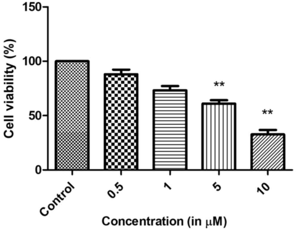
The cell viability was reduced with increased etomidate
concentration (0.5, 1, 5 and 10 µM). These findings demonstrated
that N2a cells responded to etomidate in a dose-dependent manner.
The IC50 was determined as 5 µM (Fig. 1). These findings collectively
suggested that etomidate reduced the viability of N2a neuroblastoma
cells.
Effect on colony formation
When N2a cells were treated with different
concentrations of etomidate, the clonogenic potential of the cells
reduced with the increase in etomidate concentration. This led to
an inhibition of the colony formation potential of N2a cells in a
dose-dependent manner as presented in Fig. 2.
Detection of apoptosis in N2a cells by
Annexin V/PI staining
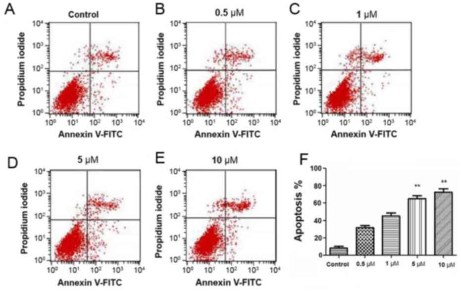
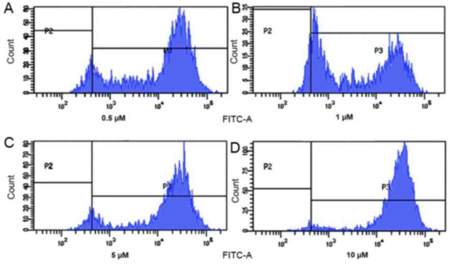
In order to confirm that etomidate induces apoptosis
in N2a cells, Annexin V/PI assay was performed. The current study
determined that etomidate increased the apoptotic population in a
dose-dependent manner as presented in in Fig. 3. These finding sclearly indicated
that etomidate induced concentration dependent apoptosis in N2a
cells.
Effect of etomidateon generation of
reactive oxygen species (ROS) in N2a cells
As a fore mentioned etomidate induced apoptosis in
N2a cells; therefore, the current study investigated the effect of
etomidate on the generation of reactive oxygen species using
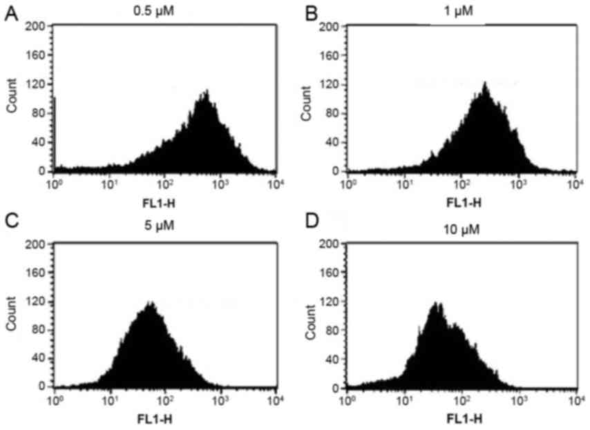
DCFH2-DA dye. Cells were treated with different concentrations of
etomidate for 48 h and it was evident that etomidate treatment had
a considerable impact on generation of reactive oxygen species in
N2a cells as presented in Fig. 4.
Therefore, these findings clearly indicated that etomidate induced
generation of reactive oxygen species that led to apoptosis of N2a
cells.
Mitochondrial membrane potential loss
is induced by etomidatein N2a cells
The current study investigated whether etomidate
treatment of N2a cells had any influence on mitochondrial membrane
potential using Rodamine-123 dye. It was evident that etomidate
treatment of N2a cells lead to loss of their mitochondrial membrane
potential in a dose-dependent manner which led to quenching of the
fluorescence when compared with the untreated cells which retained
fluorescence. These findings confirmed that etomidate induced
apoptosis in N2a cells by reducing the mitochondrial membrane
potential of these cells (Fig.
5).
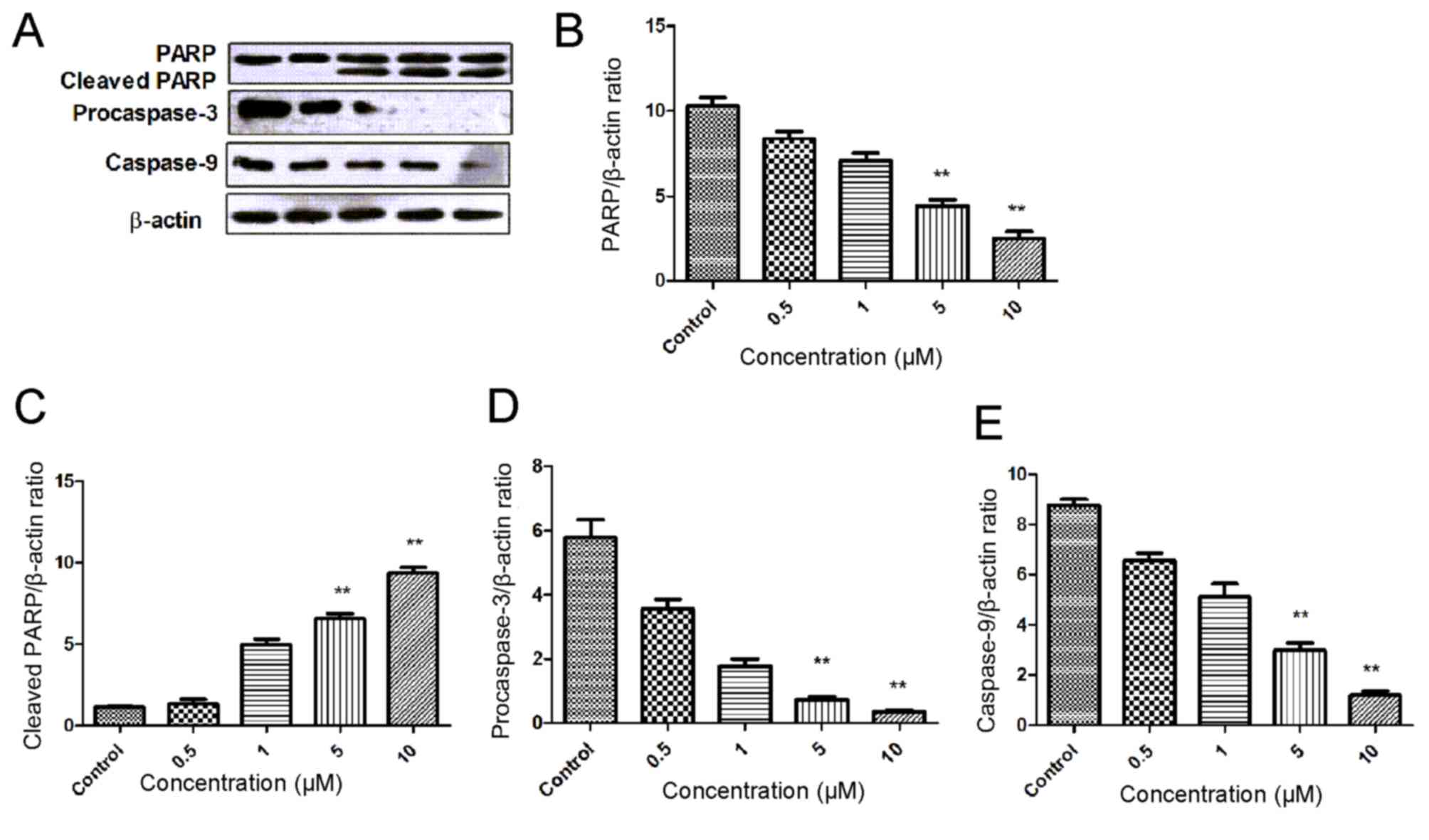
Etomidate induces PARP cleavage in N2a
cells
In the present study, the effect of etomidate on the
caspase activation and cleaving of PARP was investigated using
western blotting. The N2a cells were treated with different
concentrations of etomidate for 48 h. An increase in the expression
of cleaved PARP was observed in a dose dependent manner. There was
also a decrease in the expression level of the initiator caspase
(caspase-9), along with a decrease in the expression of
procaspase-3 in a dose-dependent manner. These findings suggested
that etomidate induces apoptosis in a dose-dependent manner
(Fig. 6).
Discussion
Surgery is one of the most common procedures used to
treat patients in various diseases, including cancer (20,21).
However, to lessen the pain for the patients, sedatives or
anesthetics are administered prior the medical procedures being
performed (22,23). Anesthesia is the induction of
temporary loss of sensation or awareness (24). It is important as it aids in the
execution of surgeries that may be otherwise impossible to be
performed due to intolerable pain (24). The type of anesthesia administered
to a patient depends upon the type of medical procedure that is to
be performed on the patient during toothache, local anesthesia is
administered that helps by blocking the transmission of the nerve
impulse between the central nervous system and the tooth that is to
be treated. During surgeries involving the removal of tumor mass
from a patient with cancer, general anesthesia is administered,
which leads to total suppression of central nervous system
resulting in total lack of sensation and unconsciousness (9,25).
Etomidate is a drug that is administered as general anesthetic to
patients. Etomidate is widely used due to easy dosing pattern and
because it has no effect on the blood pressure of the patient
(26,27). Additionally, it also provides
myocardial and cerebral ischemia protection, minimal suppression of
ventilation and decrease in liberation of histamines (15,28).
To the best of our knowledge, whilst the way etomidate functions
has been previously established, how etomidate exerts some of its
cytotoxic effects remains to be determined. The present study
revealed that anesthetic agent etomidate induces apoptosis in the
N2a neuroblastoma cell line. The cells responded to etomidate in a
concentration-dependent manner with increased cell death. In
previous study, etomidate was identified to exert cytotoxic effect
on RAW264.7 murine leukemia macrophage cell line (29). It has been determined that
etomidate may lead to enhancement of apoptotic cell morphological
changes and reduced cell viability in RAW264.7 cells. It also leads
to an increase in expression of cytochrome c,
apoptosis-inducing factor (AIF), endonuclease G (Endo G),
caspase-9, caspase-3 active form and BCL2 associated X (Bax)
proteins; however, it inhibited the expression of B cell
leukemia/lymphoma 2 extra-large (Bcl-xL), leading to apoptosis
(29). This would appear to be the
case with etomidate viaa mitochondria-dependent pathway based on
the change of the ratio of Bax/Bcl-xL which led to cytochrome c,
AIF and Endo G, release from mitochondria (30–32).
In present study, western blot analysis of etomidate-treated N2a
cells revealed an decrease in the expression of pro-apoptotic
proteins, such as initiator caspase-9 and procaspase-3. It was
evident that there was an increase in the expression of cleaved
PARP in a dose-dependent manner. Additionally, etomidate treatment
led to generation of reactive oxygen species in N2a cells.
Etomidate treatment also led to loss of mitochondrial membrane
potential in N2a cells.
In conclusion, the current study revealed that
etomidate is an anesthetic drug that induces apoptosis in the N2a
neuroblastoma cell line. However, further studies in vitro
and in vivo are required in order to confirm the cytotoxic
effects of etomidate.
Acknowledgements
Authors would like to thank the Affiliated Foshan
Hospital of Sun Yat-sen University (Guangdong, China) for providing
the laboratory facility for the present study.
Funding
No funding was received.
Availability of data and materials
The datasets used and/or analyzed during the current
study are available from the corresponding author on reasonable
request.
Authors' contributions
JZ and ZYY conceived and designed the experiments
and wrote the manuscript. HTC, YLF, MX, XQH and CL performed the
experiments. BJL, LXF, XQH and LX analyzed the data.
Ethics approval and consent to
participate
Not applicable.
Consent for publication
Not applicable.
Competing interests
The authors declare that they have no competing
interests.
References
|
1
|
Ghoneim MM, Block RI, Haffarnan M and
Mathews MJ: Awareness during anesthesia: Risk factors, causes and
sequelae: A review of reported cases in the literature. Anesth
Analg. 108:527–535. 2009. View Article : Google Scholar : PubMed/NCBI
|
|
2
|
Hsu GL, Hsieh CH, Chen HS, Ling PY, Wen
HS, Liu LJ, Chen CW and Chua C: The advancement of pure local
anesthesia for penile surgeries: Can an outpatient basis be
sustainable? J Androl. 28:200–205. 2007. View Article : Google Scholar : PubMed/NCBI
|
|
3
|
Lee JS, Hayanga AJ, Kubus JJ, Makepeace H,
Hutton M, Campbell DA Jr and Englesbe MJ: Local anesthesia: A
strategy for reducing surgical site infections? World J Surg.
35:2596–2602. 2011. View Article : Google Scholar : PubMed/NCBI
|
|
4
|
Breen P and Park KW: General anesthesia
versus regional anesthesia. Int Anesthesiol Clin. 40:61–71. 2002.
View Article : Google Scholar : PubMed/NCBI
|
|
5
|
Craig AD: Interoception: The sense of the
physiological condition of the body. Curr Opin Neurobiol.
13:500–505. 2003. View Article : Google Scholar : PubMed/NCBI
|
|
6
|
Bery A, Cardona A, Martinez P and
Hartenstein V: Structure of the central nervous system of a
juvenile acoel, Symsagittifera roscoffensis. Dev Genes Evol.
220:61–76. 2010. View Article : Google Scholar : PubMed/NCBI
|
|
7
|
Koizumi O: Nerve ring of the hypostome in
hydra: Is it an origin of the central nervous system of bilaterian
animals? Brain Behav Evol. 69:151–159. 2007. View Article : Google Scholar : PubMed/NCBI
|
|
8
|
Mace SE: Central nervous system infections
as a cause of an altered mental status? What is the pathogen
growing in your central nervous system? Emerg Med Clin North Am.
28:535–570. 2010. View Article : Google Scholar : PubMed/NCBI
|
|
9
|
Uhrig L, Dehaene S and Jarraya B: Cerebral
mechanisms of general anesthesia. Ann Fr Anesth Reanim. 33:72–82.
2014. View Article : Google Scholar : PubMed/NCBI
|
|
10
|
Bajwa S and Kulshrestha A:
Dexmedetomidine: An adjuvant making large inroads into clinical
practice. Ann Med Health Sci Res. 3:475–483. 2013. View Article : Google Scholar : PubMed/NCBI
|
|
11
|
Chohan AS: Anesthetic considerations in
orthopedic patients with or without trauma. Top Companion Anim Med.
25:107–119. 2010. View Article : Google Scholar : PubMed/NCBI
|
|
12
|
Sarkiss M: Anesthesia for bronchoscopy and
interventional pulmonology: From moderate sedation to jet
ventilation. Curr Opin Pulm Med. 17:274–278. 2011. View Article : Google Scholar : PubMed/NCBI
|
|
13
|
Lü F, Lin J and Benditt DG: Conscious
sedation and anesthesia in the cardiac electrophysiology
laboratory. J Cardiovasc Electrophysiol. 24:237–245. 2013.
View Article : Google Scholar : PubMed/NCBI
|
|
14
|
Hansson L, Zanchetti A, Carruthers SG,
Dahlöf B, Elmfeldt D, Julius S, Ménard J, Rahn KH, Wedel H and
Westerling S: Effects of intensive blood-pressure lowering and
low-dose aspirin in patients with hypertension: Principal results
of the Hypertension Optimal Treatment (HOT) randomised trial. HOT
study Group. Lancet. 351:1755–1762. 1998. View Article : Google Scholar : PubMed/NCBI
|
|
15
|
El-Khatib MF and Bou-Khalil P: Clinical
review: Liberation from mechanical ventilation. Crit Care.
12:2212008. View
Article : Google Scholar : PubMed/NCBI
|
|
16
|
Nedergaard M and Dirnagl U: Role of glial
cells in cerebral ischemia. Glia. 50:281–286. 2005. View Article : Google Scholar : PubMed/NCBI
|
|
17
|
Tavare AN, Perry NJ, Benzonana LL, Takata
M and Ma D: Cancer recurrence after surgery: Direct and indirect
effects of anesthetic agents. Int J Cancer. 130:1237–1250. 2012.
View Article : Google Scholar : PubMed/NCBI
|
|
18
|
Drlica K: Mechanism of fluoroquinolone
action. Curr Opin Microbiol. 2:504–508. 1999. View Article : Google Scholar : PubMed/NCBI
|
|
19
|
Alonso-Castro AJ, Domínguez F and
García-Carrancá A: Rutin exerts antitumor effects on nude mice
bearing SW480 tumor. Arch Med Res. 44:346–351. 2013. View Article : Google Scholar : PubMed/NCBI
|
|
20
|
Duffy MJ: The war on cancer: Are we
winning? Tumor Biol. 34:1275–1284. 2013. View Article : Google Scholar
|
|
21
|
De La Puerta B and Baines S: Surgical
diseases of the genital tract in male dogs 1. Scrotum, testes and
epididymides. In Pract. 34:58–65. 2012. View Article : Google Scholar
|
|
22
|
Hansen TG: Sedative medications outside
the operating room and the pharmacology of sedatives. Curr Opin
Anaesthesiol. 28:446–452. 2015. View Article : Google Scholar : PubMed/NCBI
|
|
23
|
Anderson SL, Duke-Novakovski T and Singh
B: The immune response to anesthesia: Part 2 sedatives, opioids,
and injectable anesthetic agents. Vet Anaesth Analg. 41:553–566.
2014. View Article : Google Scholar : PubMed/NCBI
|
|
24
|
Chamisha Y, Shamir MH, Merbl Y and Chai O:
Reversible paralysis and loss of deep pain sensation after topical
intrathecal morphine administration following durotomy. Vet Surg.
44:41–45. 2015.PubMed/NCBI
|
|
25
|
Dierdorf SF: Awareness during anesthesia.
Anesthesiol Clin. 14:369–384. 1996. View Article : Google Scholar
|
|
26
|
de la Grandville B, Arroyo D and Walder B:
Etomidate for critically ill patients. Con: Do you really want to
weaken the frail? Eur J Anaesthesiol. 29:511–514. 2012. View Article : Google Scholar : PubMed/NCBI
|
|
27
|
Janouschek H, Nickl-Jockschat T, Haeck M,
Gillmann B and Grözinger M: Comparison of methohexital and
etomidate as anesthetic agents for electroconvulsive therapy in
affective and psychotic disorders. J Psychiatr Res. 47:686–693.
2013. View Article : Google Scholar : PubMed/NCBI
|
|
28
|
Mendez-Tellez PA and Needham DM: Early
physical rehabilitation in the ICU and ventilator liberation.
Respir Care. 57:1663–1669. 2012. View Article : Google Scholar : PubMed/NCBI
|
|
29
|
Wu RS, Wu KC, Yang JS, Chiou SM, Yu CS,
Chang SJ, Chueh FS and Chung JG: Etomidate induces cytotoxic
effects and gene expression in a murine leukemia macrophage cell
line (RAW264.7). Anticancer Res. 31:2203–2208. 2011.PubMed/NCBI
|
|
30
|
Chiang JH, Yang JS, Ma CY, Yang MD, Huang
HY, Hsia TC, Kuo HM, Wu PP, Lee TH and Chung JG: Danthron, an
anthraquinone derivative, induces DNA damage and caspase
cascades-mediated apoptosis in SNU-1 human gastric cancer cells
through mitochondrial permeability transition pores and
Bax-triggered pathways. Chem Res Toxicol. 24:20–29. 2011.
View Article : Google Scholar : PubMed/NCBI
|
|
31
|
Yu FS, Yang JS, Yu CS, Lu CC, Chiang JH,
Lin CW and Chung JG: Safrole induces apoptosis in human oral cancer
HSC-3 cells. J Dent Res. 90:168–174. 2011. View Article : Google Scholar : PubMed/NCBI
|
|
32
|
Chen JC, Lu KW, Lee JH, Yeh CC and Chung
JG: Gypenosides induced apoptosis in human colon cancer cells
through the mitochondria-dependent pathways and activation of
caspase-3. Anticancer Res. 26:4313–4326. 2006.PubMed/NCBI
|


