Percutaneous ultrasound-guided irreversible electroporation: A goat liver study

  • Authors:
    • Ying Liu
    • Zhengai Xiong
    • Wei Zhou
    • Yuanyuan Hua
    • Chengxiang Li
    • Chenguo Yao
  • View Affiliations

  • Published online on: June 29, 2012     https://doi.org/10.3892/ol.2012.781
  • Pages: 450-454
Metrics: Total Views: 0 (Spandidos Publications: | PMC Statistics: )
Total PDF Downloads: 0 (Spandidos Publications: | PMC Statistics: )


Abstract

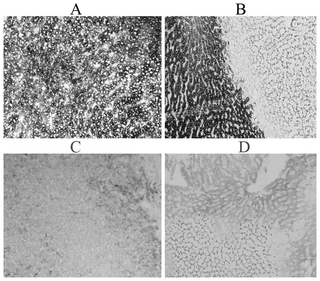
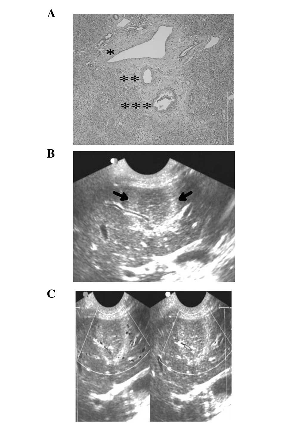
Irreversible electroporation (IRE) is a new tumor ablation technique. Pulsed electric fields (PEFs) with permanent duration (100 µsec) permanently permeabilize the cell membrane, causing the formation of innumerable permanent nanopores in the cell membrane and leading to cell death. In this study, percutaneous IRE was performed on 24 goat livers under the guidance of ultrasonography (US). IRE-exposed tissues were pathologically examined and glucose-6-phosphatase (G-6-P) and succinodehydrogenase (SDH) staining were used to detect the activity and function of the endoplasmic reticulum and mitochondria of liver tissues at 0 and 24 h after IRE ablation. Tissue ablation responses were monitored in real-time with US in the percutaneous IRE group; the largest diameter of the ablation zones was measured immediately (D1) and after 24 h (D2). Following D2 measurement, the animals were sacrificed and the gross sections (D3) were measured. The pathological examination results showed complete tissue necrosis after 24 h instead of immediately following IRE. The largest long diameters measured by intraprocedural US immediately after IRE (D1, 39.58±2.13 mm) were larger than those measured by US after 24 h (D2, 37.07±3.51 mm) and in gross section measurements (D3, 36.44±2.04 mm; P<0.05). D1 showed a good linear correlation with D3 (r=0.949). We conclude from these studies that IRE is not an acute ablating effect which leads to cell death. If US-guided percutaneous IRE focused on the target liver areas accurately, physicians would be able to assess the extent of necrosis through the regression equation during the IRE ablation procedure, and evaluate whether sufficient electric field energy had been applied to the desired tissue. Assisted with US guiding and monitoring, the minimally invasive IRE procedure in intraperitoneal lesions may become an important tumor ablation technique.
View Figures
View References

Related Articles

Journal Cover

September 2012
Volume 4 Issue 3

Print ISSN: 1792-1074
Online ISSN:1792-1082

Sign up for eToc alerts

Recommend to Library

Copy and paste a formatted citation
x
Spandidos Publications style
Liu Y, Xiong Z, Zhou W, Hua Y, Li C and Yao C: Percutaneous ultrasound-guided irreversible electroporation: A goat liver study. Oncol Lett 4: 450-454, 2012.
APA
Liu, Y., Xiong, Z., Zhou, W., Hua, Y., Li, C., & Yao, C. (2012). Percutaneous ultrasound-guided irreversible electroporation: A goat liver study. Oncology Letters, 4, 450-454. https://doi.org/10.3892/ol.2012.781
MLA
Liu, Y., Xiong, Z., Zhou, W., Hua, Y., Li, C., Yao, C."Percutaneous ultrasound-guided irreversible electroporation: A goat liver study". Oncology Letters 4.3 (2012): 450-454.
Chicago
Liu, Y., Xiong, Z., Zhou, W., Hua, Y., Li, C., Yao, C."Percutaneous ultrasound-guided irreversible electroporation: A goat liver study". Oncology Letters 4, no. 3 (2012): 450-454. https://doi.org/10.3892/ol.2012.781