Biomarker expression in normal fimbriae: Comparison of high- and low-grade serous ovarian carcinoma
- Authors:
- Zhang Xuyin
- Ding Jingxin
- Tao Xiang
- Jia Luoqi
- Hua Keqin
View Affiliations
Affiliations: Department of Obstetrics and Gynecology, Obstetrics and Gynecology Hospital, Fudan University, Shanghai 200090, P.R. China
- Published online on: August 23, 2012 https://doi.org/10.3892/ol.2012.877
-
Pages:
1008-1012
Metrics: Total
Views: 0 (Spandidos Publications: | PMC Statistics: )
Metrics: Total PDF Downloads: 0 (Spandidos Publications: | PMC Statistics: )
This article is mentioned in:
Abstract
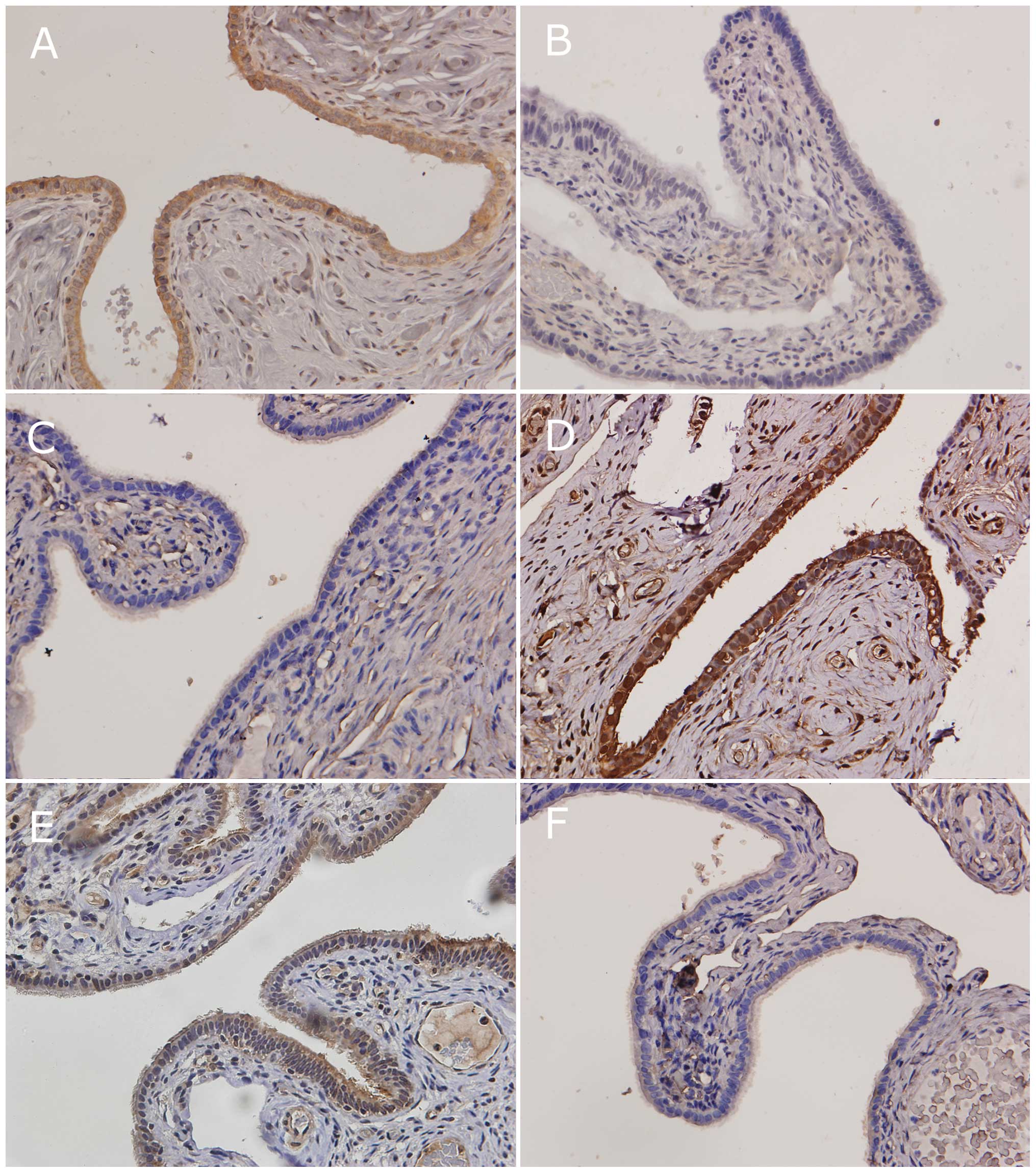
The objective of this study was to assess the difference in fimbriae of high- and low-grade ovarian serous carcinoma (OSC). The fimbriae of normal appearance [without serous tubal intraepithelial carcinoma (STIC)] from 28 patients with high-grade OSCs and 24 patients with low-grade OSCs were assessed for the expression of 6 markers [E-cadherin, matrix metalloproteinase-2 (MMP-2), phospho‑AKT (pAKT), cyclooxygenase-2 (COX-2), vascular endothelial growth factor (VEGF) and p53] using immunohistochemistry. Sectioning and extensively examining the fimbria (SEE-FIM) was performed to exclude fimbrial involvement for all the cases. The immunostaining levels of pAKT and COX-2 were significantly higher in the fimbriae of normal appearance from high-grade OSCs compared with low-grade OSCs (61 vs. 8% and 71 vs. 21%; P=0.005 and 0.007, respectively). The immunostaining of E-cadherin was significantly higher in the fimbriae of low‑grade OSCs compared with high-grade OSCs (83 vs. 21%; P=0.003). The remaining 3 markers (MMP-2, VEGF and p53) had similar expression in low- and high-grade OSCs (21 vs. 13%; 25 vs. 21%; and 14 vs. 8%; P=0.78, 0.86 and 0.82, respectively). Our results suggest marked biological differences in the behavior of the fimbriae in high- and low-grade OSCs and indicate that proliferation, cell adhesion and the inflammatory microenvironment of fimbriae in high-grade OSCs without STIC had changed prior to p53 mutation.
View References
1.
|
A MalpicaMT DeaversK LuGrading ovarian
serous carcinoma using a two-tier systemAm J Surg
Pathol28496504200410.1097/00000478-200404000-0000915087669
|
2.
|
RT BurksME ShermanRJ KurmanMicropapillary
serous carcinoma of the ovary. A distinctive low-grade carcinoma
related to serous borderline tumorsAm J Surg
Pathol2013191330199610.1097/00000478-199611000-000038898836
|
3.
|
RJ KurmanJD SeidmanIM ShihSerous
borderline tumours of the
ovaryHistopathology47310315200510.1111/j.1365-2559.2005.02186.x16115232
|
4.
|
DD BowtellThe genesis and evolution of
high-grade serous ovarian cancerNat Rev
Cancer10803808201010.1038/nrc294620944665
|
5.
|
Y LeeA MironR DrapkinA candidate precursor
to serous carcinoma that originates in the distal fallopian tubeJ
Pathol2112635200710.1002/path.209117117391
|
6.
|
DW KindelbergerY LeeA MironIntraepithelial
carcinoma of the fimbria and pelvic serous carcinoma: Evidence for
a causal relationshipAm J Surg
Pathol31161169200710.1097/01.pas.0000213335.40358.4717255760
|
7.
|
S VaughanJI CowardRC Bast JrRethinking
ovarian cancer: recommendations for improving outcomesNat Rev
Cancer10719725201110.1038/nrc314421941283
|
8.
|
RJ KurmanIe-M ShihMolecular pathogenesis
and extraovarian origin of epithelial ovarian cancer - shifting the
paradigmHum
Pathol42918931201110.1016/j.humpath.2011.03.00321683865
|
9.
|
V ShtilbansM WuDE BursteinCurrent overview
of the role of Akt in cancer studies via applied
immunohistochemistryAnn Diagn
Pathol12153160200810.1016/j.anndiagpath.2007.12.00118325479
|
10.
|
T FujiokaY TakebayashiT KihanaExpression
of E-cadherin and beta-catenin in primary and peritoneal metastatic
ovarian carcinomaOncol Rep8249255200111182035
|
11.
|
K VleminckxL Vakaet JrM MareelGenetic
manipulation of E-cadherin expression by epithelial tumor cells
reveals an invasion suppressor
roleCell66107119199110.1016/0092-8674(91)90143-M2070412
|
12.
|
A MunkarahR Ali-FehmiCOX-2: a protein with
an active role in gynecological cancersCurr Opin Obstet
Gynecol174953200510.1097/00001703-200502000-0000915711411
|
13.
|
J KaernM AghmeshehJM NeslandPrognostic
factors in ovarian carcinoma stage III patients. Can biomarkers
improve the prediction of short- and long-term survivors?Int J
Gynecol
Cancer1510141022200510.1111/j.1525-1438.2005.00185.x16343177
|
14.
|
HA KennyE LengyelMMP-2 functions as an
early response protein in ovarian cancer metastasisCell
Cycle8683688200910.4161/cc.8.5.770319221481
|
15.
|
B SchmalfeldtD PrechtelK HärtkingIncreased
expression of matrix metalloproteinases (MMP)-2, MMP-9, and the
urokinase-type plasminogen activator is associated with progression
from benign to advanced ovarian cancerClin Cancer
Res723962404200111489818
|
16.
|
P PaleyK StaskusK GebhardVascular
endothelial growth factor expression in early stage ovarian
carcinomaCancer8098106199710.1002/(SICI)1097-0142(19970701)80:1%3C98::AID-CNCR13%3E3.0.CO;2-A9210714
|
17.
|
BC CooperJM RitchieCL
BroghammerPreoperative serum vascular endothelial growth factor
levels: significance in ovarian cancerClin Cancer
Res831933197200212374688
|
18.
|
L HeflerR ZeillingerC GrimmPreoperative
serum vascular endothelial growth factor as a prognostic parameter
in ovarian cancerGynecol
Oncol103512517200610.1016/j.ygyno.2006.03.05816750560
|
19.
|
IeM ShihRJ KurmanOvarian tumorigenesis: a
proposed model based on morphological and molecular genetic
analysisAm J
Pathol16415111518200410.1016/S0002-9440(10)63708-X15111296
|