Intradermal melanocytic nevus with lymphatic nevus cell embolus: A case report
- Authors:
- Hyun‑Soo Kim
- Sang Hwa Lee
- Hyung‑Sik Moon
- Youn Wha Kim
View Affiliations
Affiliations: Department of Experimental Analysis, Aerospace Medical Center, Republic of Korea Air Force, Cheongju, Chungcheongbuk‑do 363-849, Republic of Korea, Department of Pathology, Aerospace Medical Center, Republic of Korea Air Force, Cheongju, Chungcheongbuk‑do 363-849, Republic of Korea, Department of Dermatology, Aerospace Medical Center, Republic of Korea Air Force, Cheongju, Chungcheongbuk‑do 363-849, Republic of Korea, Department of Pathology, School of Medicine, Kyung Hee University, Seoul 130‑701, Republic of Korea
- Published online on: November 25, 2013 https://doi.org/10.3892/ol.2013.1704
-
Pages:
331-333
Metrics: Total
Views: 0 (Spandidos Publications: | PMC Statistics: )
Metrics: Total PDF Downloads: 0 (Spandidos Publications: | PMC Statistics: )
This article is mentioned in:
Abstract
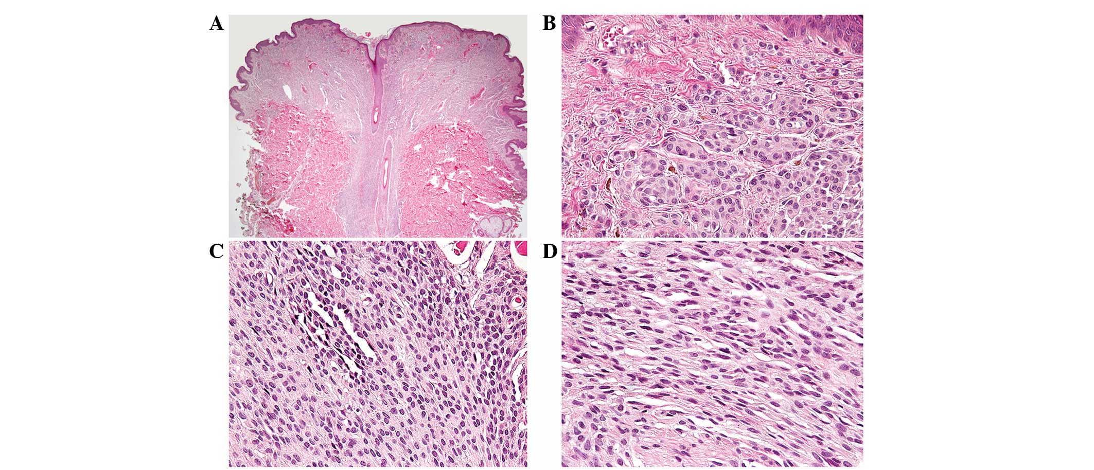
The current study presents a rare case of intradermal melanocytic nevus with lymphatic nevus cell embolus. A 26‑year‑old male presented with a slowly enlarging, pigmented nodule on the back, measuring 1 cm in diameter. Histological observations of the lesion were typical of an intradermal melanocytic nevus. The most notable feature of this nevus, however, was an aggregate of nevus cells within a lymphatic vessel of the upper dermis. The nevus cells observed within the lymphatic lumen demonstrated characteristic morphological features of type A nevus cells. The cells were round‑to‑cuboidal, exhibited abundant cytoplasm with well‑defined cell borders and formed nests. In addition, the nevus cell aggregate was lined by flattened endothelial cells. Nevus cell aggregates occur in the collagenous framework of lymph nodes, however, the mechanism by which nevus cells are deposited in lymph nodes has been a source of interest and controversy. The histological observation presented may be regarded as support for the mechanical transport or benign metastasis theories, which posit transfer of nevus cell emboli, via lymphatics, from a cutaneous nevus to the draining regional lymph node. Due to its rarity, a lymphatic nevus cell embolus creates diagnostic and management issues for pathologists and clinicians. This observation must not be interpreted as evidence of malignancy, but must be assessed in context with the associated histological features of the lesion.
View References
1
|
Katsumata M, Matsunaga T, Maruyama R and
Ezoe K: Lymphatic invasion of nevus cells observed in intradermal
nevus. J Dermatol. 17:264–265. 1990.PubMed/NCBI
|
2
|
Subramony C and Lewin JR: Nevus cells
within lymph nodes: possible metastases from a benign intradermal
nevus. Am J Clin Pathol. 84:220–223. 1985.
|
3
|
Stewart FW and Copeland MM: Neurogenic
sarcoma. Am J Cancer. 15:12351931.
|
4
|
Erlandson RA and Rosen PP: Electron
microscopy of a nevus cell aggregate associated with axillary lymph
node. Cancer. 49:269–272. 1982. View Article : Google Scholar
|
5
|
Hart WR: Primary nevus of lymph node. Am J
Clin Pathol. 55:88–92. 1971.
|
6
|
McCarthy SW, Palmer AA, Bale PM and Herst
E: Nevus cells in lymph nodes. Pathology. 6:351–358. 1974.
View Article : Google Scholar : PubMed/NCBI
|
7
|
Ridolfi RL, Rosen PP and Thaler H: Nevus
cell aggregates associated with lymph nodes: estimated frequency
and clinical significance. Cancer. 39:164–171. 1977. View Article : Google Scholar : PubMed/NCBI
|
8
|
Johnson WT and Helwig EB: Benign nevus
cells in the capsule of lymph nodes. Cancer. 23:747–753. 1969.
View Article : Google Scholar : PubMed/NCBI
|
9
|
Carson KF, Wen DR, Li PX, et al: Nodal
nevi and cutaneous melanomas. Am J Surg Pathol. 20:834–840. 1996.
View Article : Google Scholar : PubMed/NCBI
|
10
|
Patterson JW: Nevus cell aggregates in
lymph nodes. Am J Clin Pathol. 121:13–15. 2004. View Article : Google Scholar : PubMed/NCBI
|
11
|
Fontaine D, Parkhill W, Greer W and Walsh
N: Nevus cells in lymph nodes. Am J Dermatopathol. 24:1–5. 2002.
View Article : Google Scholar
|
12
|
Bautista NC, Cohen S and Anders KH: Benign
melanocytic nevus cells in axillary lymph nodes: a prospective
incidence and immunohistochemical study with literature review. Am
J Clin Pathol. 102:102–108. 1994.
|
13
|
Azzopardi JG, Ross CM and Frizzera G: Blue
naevi of lymph node capsule. Histopathology. 1:451–461. 1977.
View Article : Google Scholar : PubMed/NCBI
|
14
|
Bell ME, Hill DP and Bhargava MK:
Lymphatic invasion in pigmented nevi. Am J Clin Pathol. 72:97–100.
1979.PubMed/NCBI
|