Manifestations and pathological features of solitary thin‑walled cavity lung cancer observed by CT and PET/CT imaging

  • Authors:
    • Yuangang Qi
    • Qing Zhang
    • Yong Huang
    • Daoqing Wang
  • View Affiliations

  • Published online on: April 15, 2014     https://doi.org/10.3892/ol.2014.2065
  • Pages: 285-290
Metrics: Total Views: 0 (Spandidos Publications: | PMC Statistics: )
Total PDF Downloads: 0 (Spandidos Publications: | PMC Statistics: )


Abstract

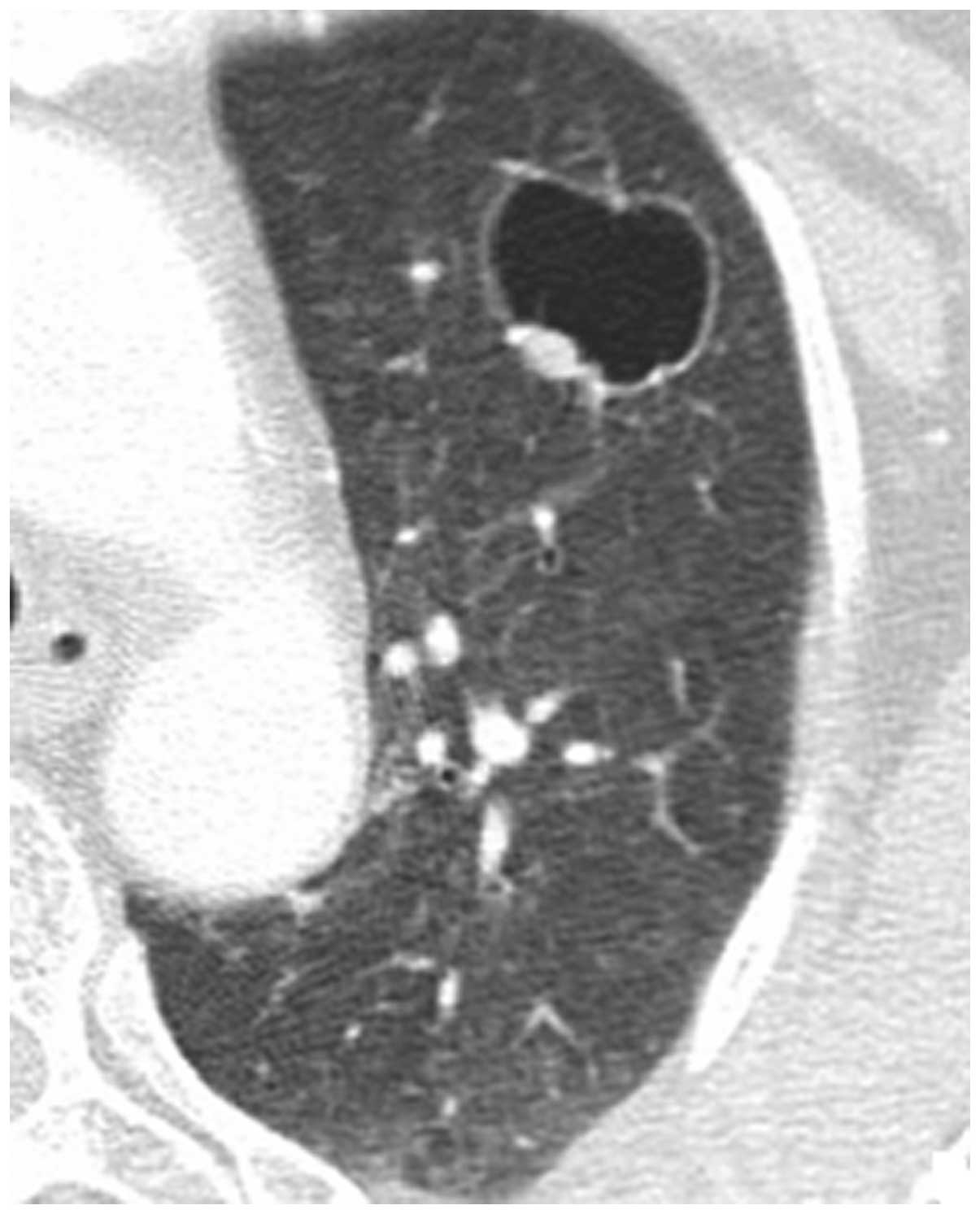
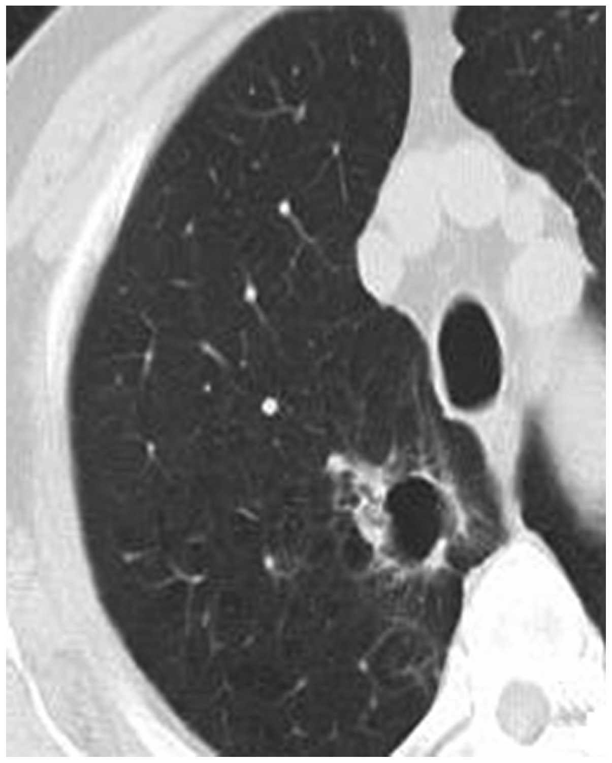
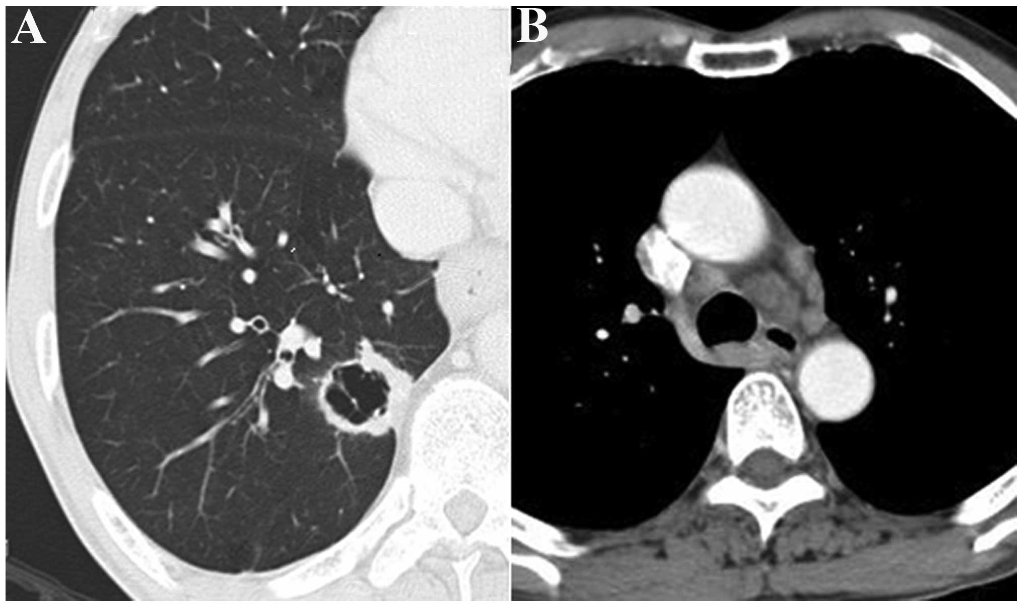
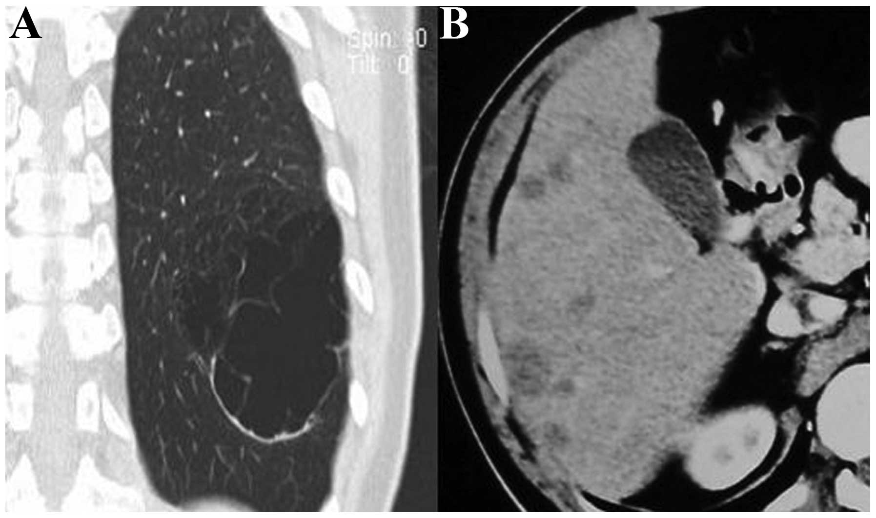
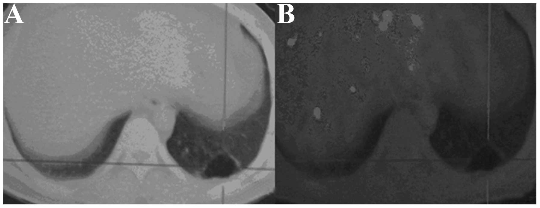
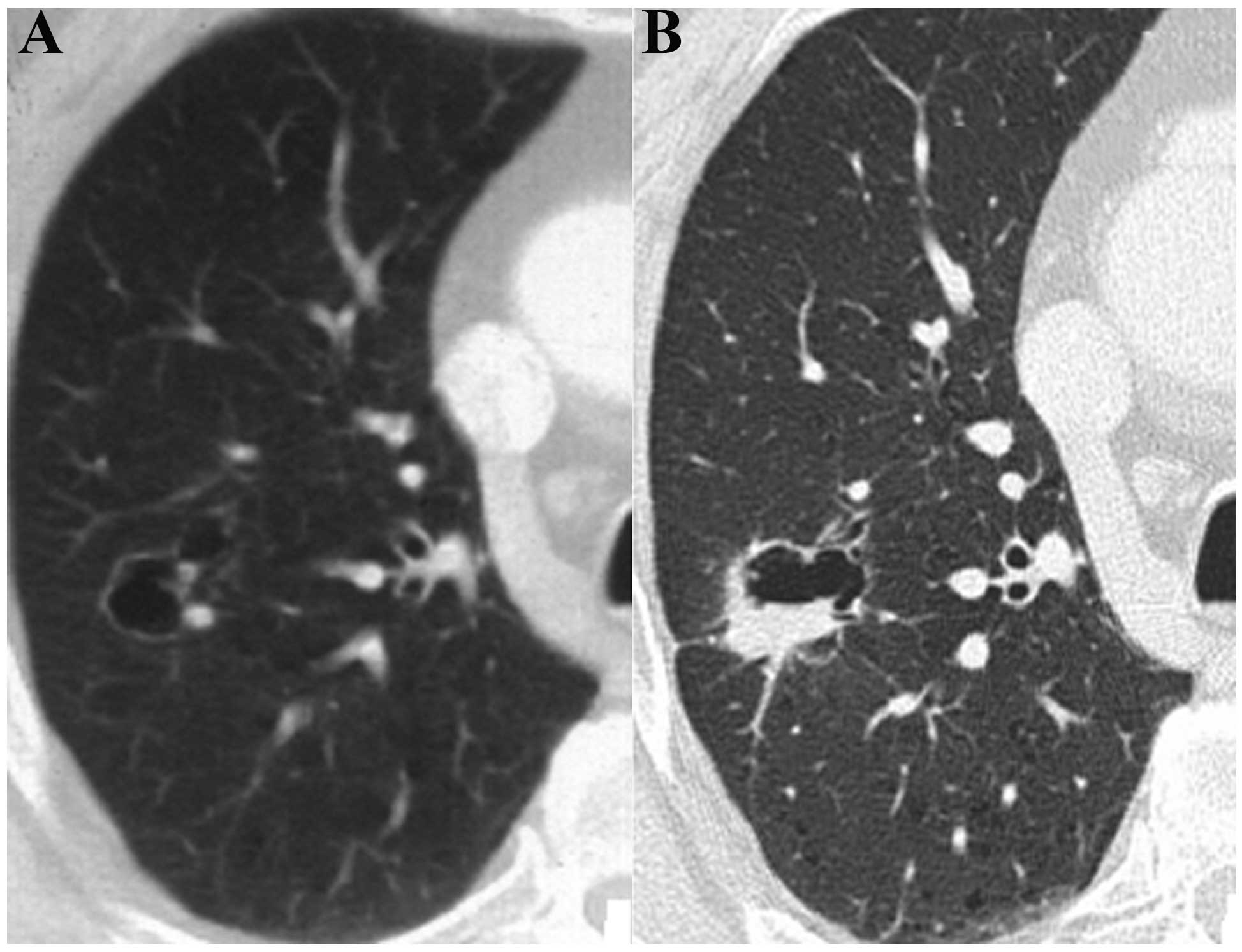
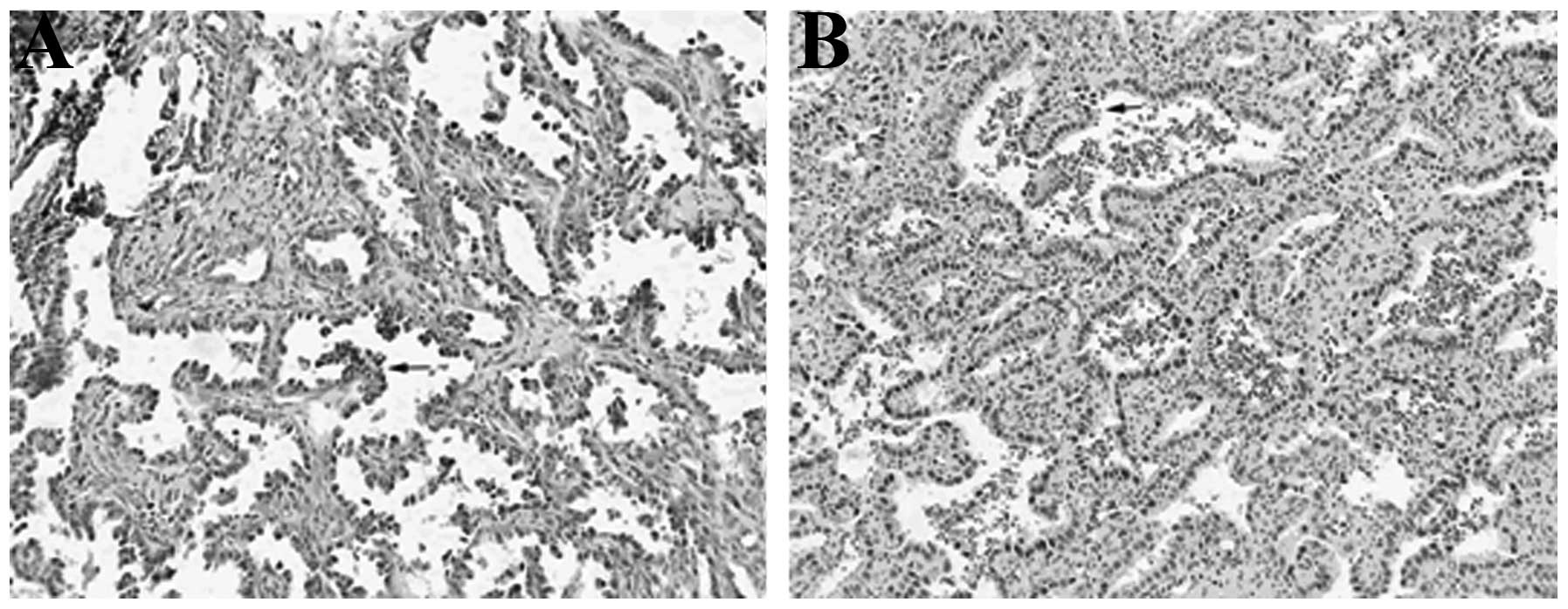
The aim of the present study was to analyze and improve the understanding of computed tomography (CT) and positron emission tomography (PET)/CT imaging and the pathological features of solitary thin‑walled cavity lung cancer. A total of 16 patients with pathologically confirmed solitary thin‑walled cavity lung cancer were included in the present study. All of the patients received CT scans. Among these, two patients underwent an additional PET/CT examination. The CT and PET/CT images were analyzed and a cross‑check analysis of the pathological results was conducted. In total, 16 cases of lesions demonstrated thin‑walled cavities on the CT images. Among these cases, three presented with an uneven thickening of the cavity walls, 10 cases exhibited wall nodules and three cases presented with compartments in the cavity. The standard uptake value (SUV) of the cavity wall increased in two patients who underwent PET/CT examinations. The 16 cases of lesions were pathologically confirmed as adenocarcinomas. Light microscopy revealed that the tumor cells, which were observed in 12 cases of lesions, had diffused along the inner cavity wall and the tumor cells of four cases had invaded the bronchial wall. Images of the chest that demonstrated a single thin‑walled cavity accompanied by uneven thickening of the cavity wall or wall nodules, in addition to an increase in the SUV and compartments in the cavity, indicated potential lung cancer. Valves formed as a result of bronchial wall damage may have led to the cavity.
View Figures
View References

Related Articles

Journal Cover

July-2014
Volume 8 Issue 1

Print ISSN: 1792-1074
Online ISSN:1792-1082

Sign up for eToc alerts

Recommend to Library

Copy and paste a formatted citation
x
Spandidos Publications style
Qi Y, Zhang Q, Huang Y and Wang D: Manifestations and pathological features of solitary thin‑walled cavity lung cancer observed by CT and PET/CT imaging. Oncol Lett 8: 285-290, 2014.
APA
Qi, Y., Zhang, Q., Huang, Y., & Wang, D. (2014). Manifestations and pathological features of solitary thin‑walled cavity lung cancer observed by CT and PET/CT imaging. Oncology Letters, 8, 285-290. https://doi.org/10.3892/ol.2014.2065
MLA
Qi, Y., Zhang, Q., Huang, Y., Wang, D."Manifestations and pathological features of solitary thin‑walled cavity lung cancer observed by CT and PET/CT imaging". Oncology Letters 8.1 (2014): 285-290.
Chicago
Qi, Y., Zhang, Q., Huang, Y., Wang, D."Manifestations and pathological features of solitary thin‑walled cavity lung cancer observed by CT and PET/CT imaging". Oncology Letters 8, no. 1 (2014): 285-290. https://doi.org/10.3892/ol.2014.2065