A novel cyclic pentapeptide, H‑10, inhibits B16 cancer cell growth and induces cell apoptosis
- Authors:
- Geng Zhang
- Shouxin Liu
- Yunjiang Liu
- Feifei Wang
- Jingwen Ren
- Jifeng Gu
- Kaixuan Zhou
- Baoen Shan
View Affiliations
Affiliations: Research Center, The Fourth Hospital of Hebei Medical University, Shijiazhuang, Hebei 050011, P.R. China, State Key Laboratory Breeding Base‑Hebei Province Key Laboratory of Molecular Chemistry for Drug, Hebei University of Science and Technology, Shijiazhuang, Hebei 050018, P.R. China, Breast Center, The Fourth Hospital of Hebei Medical University, Shijiazhuang, Hebei 050011, P.R. China
- Published online on: May 8, 2014 https://doi.org/10.3892/ol.2014.2121
-
Pages:
248-252
Metrics: Total
Views: 0 (Spandidos Publications: | PMC Statistics: )
Metrics: Total PDF Downloads: 0 (Spandidos Publications: | PMC Statistics: )
This article is mentioned in:
Abstract
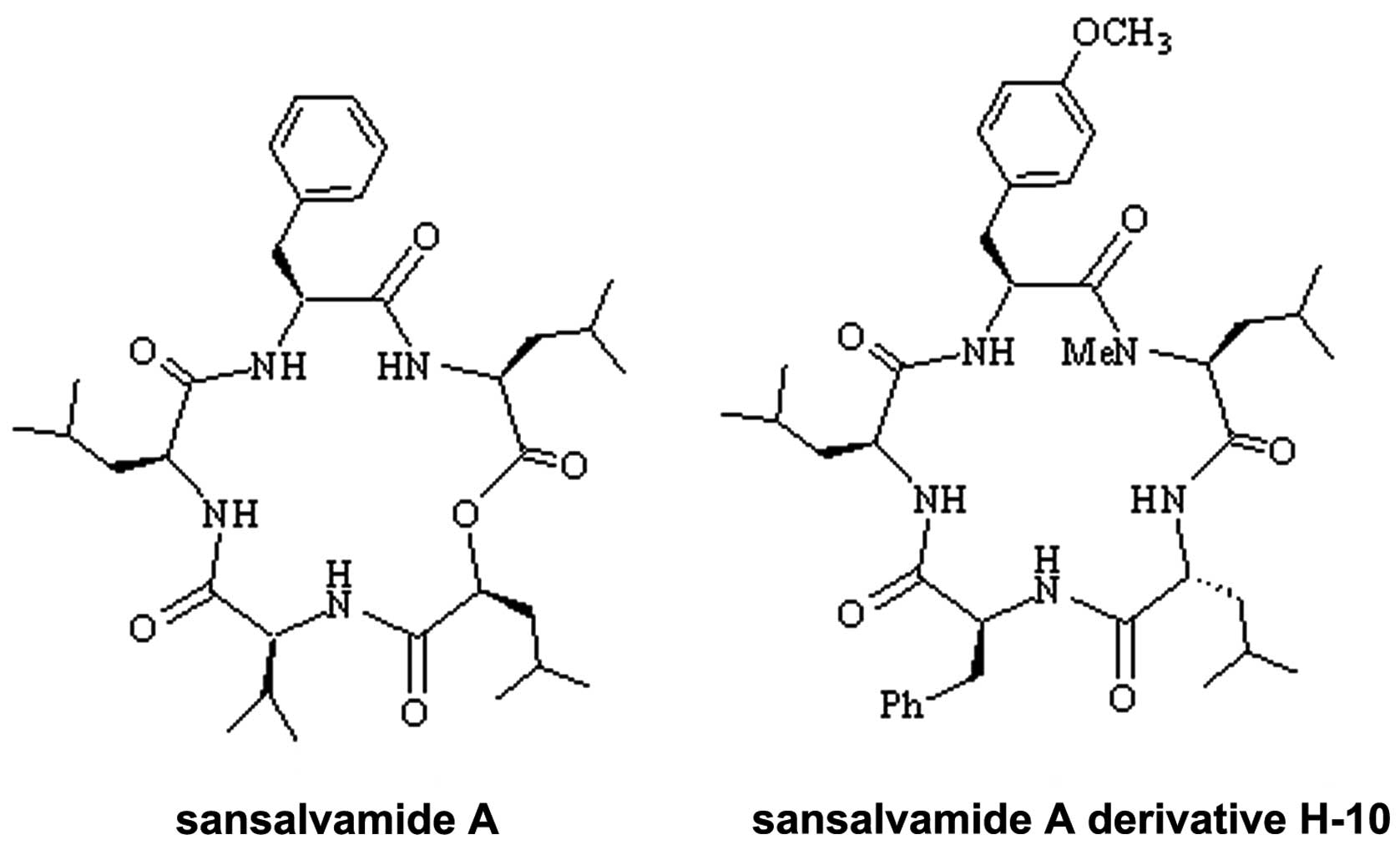
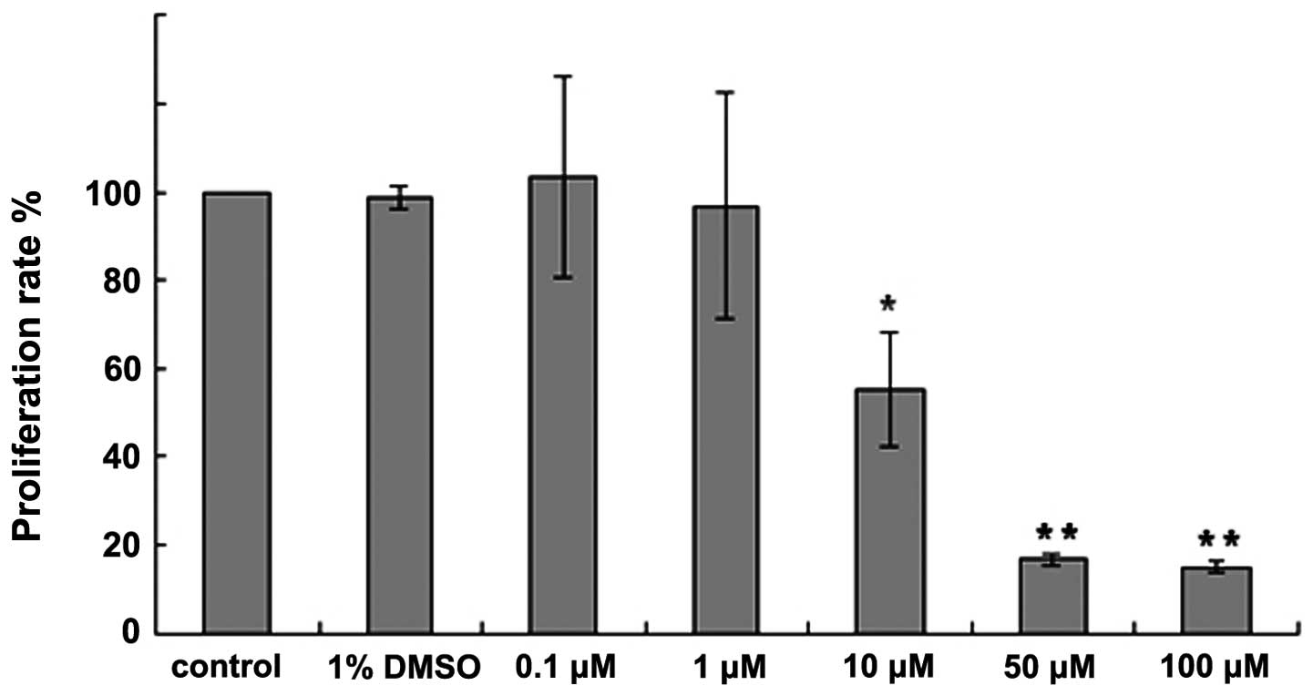
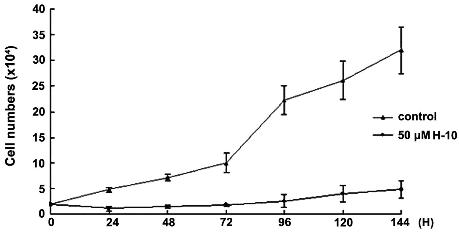
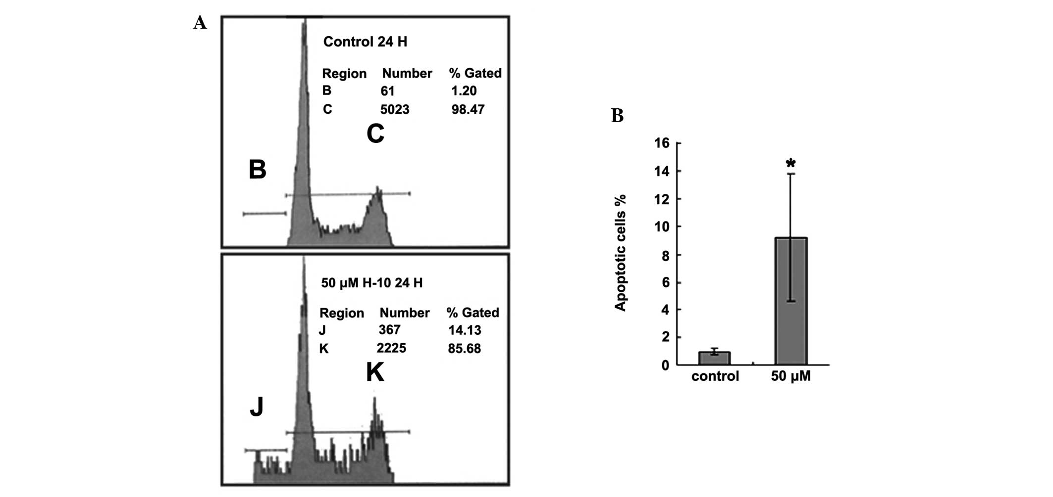
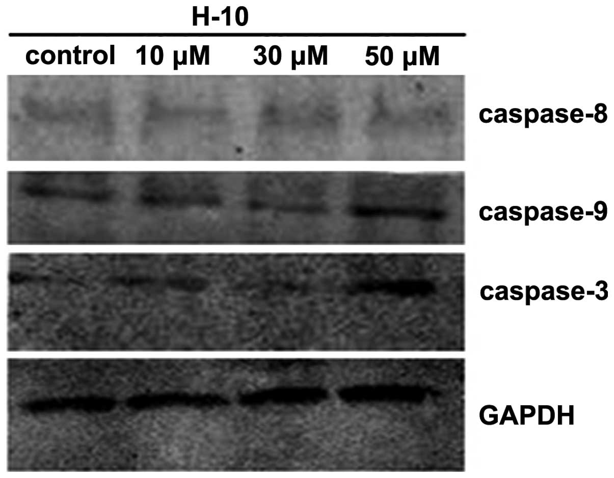
Sansalvamide A, a cyclic depsipeptide isolated from a marine fungus of the Fusarium genus, exhibits significant antitumor activity. In the present study, H‑10 (molecular formula, C38H55N5O6; molecular weight, 677.8732), a novel sansalvamide A derivative, demonstrated an inhibitory effect on the proliferation of murine melanoma B16 cells. It was confirmed that H‑10 induced the apoptosis of the B16 cells. The inhibitory rate of various concentrations of H‑10 on the B16 cells was measured by sulforhodamine B colorimetric assay, and the results revealed that the inhibitory effect of H‑10 on the B16 cells occurred in a concentration‑dependent manner. In addition, a growth curve model of the B16 cells treated with 50 µM H‑10 revealed that the effect of H‑10 also occurred in a time‑dependent manner. The apoptotic morphology of the B16 cells was observed using an optical microscope. Following the treatment of the cells with 50 µM H‑10 for 24 h, the cell apoptosis rate was analyzed using flow cytometry. The expression levels of caspase‑3, ‑8 and ‑9 were analyzed by western blot analysis, and the results indicated that H‑10 may induce the apoptosis of B16 cells.
View Figures |
Figure 1
|
|
Figure 2
|
|
Figure 3
|
|
Figure 4
|
|
Figure 5
|
|
Figure 6
|
View References
1
|
Liu S, Gu W, Lo D, Ding XZ, Ujiki M,
Adrian TE, Soff GA and Silverman RB: N-methylsansalvamide a peptide
analogues. Potent new antitumor agents. J Med Chem. 48:3630–3638.
2005.
|
2
|
Belofsky GN, Jensen PR and Fenical W:
Sansalvamide: a new cytotoxic cyclic depsipeptide produced by a
marine fungus of the genus Fusarium. Tetrahedron Lett.
40:2913–2916. 1999.
|
3
|
Pan PS, Vasko RC, Lapera SA, Johnson VA,
Seller RP, Lin CC, Pan CM, Davis MR, Ardi VC and McAlpine SR: A
comprehensive study of Sansalvamide A derivatives: The
structure-activity relationships of 78 derivatives in two
pancreatic cancer cell lines. Bioorg Med Chem. 17:5806–5825.
2009.
|
4
|
Styers TJ, Kekec A, Rodriguez R, Brown JD,
Cajica J, Pan PS, Parry E, Carroll CL, Medina I, Corral R, et al:
Synthesis of Sansalvamide A derivatives and their cytotoxicity in
the MSS colon cancer cell line HT-29. Bioorg Med Chem.
14:5625–5631. 2006.
|
5
|
Rodriguez RA, Pan PS, Pan CM, Ravula S,
Lapera S, Singh EK, Styers TJ, Brown JD, Cajica J, Parry E, et al:
Synthesis of second-generation sansalvamide A derivatives: novel
templates as potential antitumor agents. J Org Chem. 72:1980–2002.
2007.
|
6
|
Otrubova K, Lushington G, Vander Velde D,
McGuire KL and McAlpine SR: Comprehensive study of sansalvamide A
derivatives and their structure-activity relationships against
drug-resistant colon cancer cell lines. J Med Chem. 51:530–544.
2008.
|
7
|
Ujiki MB, Milam B, Ding XZ, Roginsky AB,
Salabat MR, Talamonti MS, Bell RH, Gu W, Silverman RB and Adrian
TE: A novel peptide sansalvamide analogue inhibits pancreatic
cancer cell growth through G0/G1 cell-cycle arrest. Biochem Biophys
Res Commun. 340:1224–1228. 2006.
|
8
|
Kunicki JB, Petersen MN, Alexander LD, et
al: Synthesis and evaluation of biotinylated sansalvamide A analogs
and their modulation of Hsp90. Bioorg Med Chem Lett. 21:4716–4719.
2011.
|
9
|
Poter AG and Jänicke RU: Emerging roles of
caspase-3 in apoptosis. Cell Death Differ. 6:99–104. 1999.
|
10
|
Li H, Zhu H, Xu CJ and Yuan J: Cleavage of
BID by caspase 8 mediates the mitochondrial damage in the Fas Path
of apoptosis. Cell. 94:491–501. 1998.
|