Prostate cancer with cyst formation detected by whole body positron emission tomography/computed tomography: A case report
- Authors:
- Hakushi Kim
- Sunao Shoji
- Tetsuro Tomonaga
- Masanori Shima
- Toshiro Terachi
- Toyoaki Uchida
View Affiliations
Affiliations: Department of Urology, Tokai University Hachioji Hospital, Hachioji, Tokyo 192‑0032, Japan, Department of Urology, Tokai University School of Medicine, Isehara, Kanagawa 259-1193, Japan
- Published online on: September 8, 2014 https://doi.org/10.3892/ol.2014.2510
-
Pages:
2037-2039
Metrics: Total
Views: 0 (Spandidos Publications: | PMC Statistics: )
Metrics: Total PDF Downloads: 0 (Spandidos Publications: | PMC Statistics: )
This article is mentioned in:
Abstract
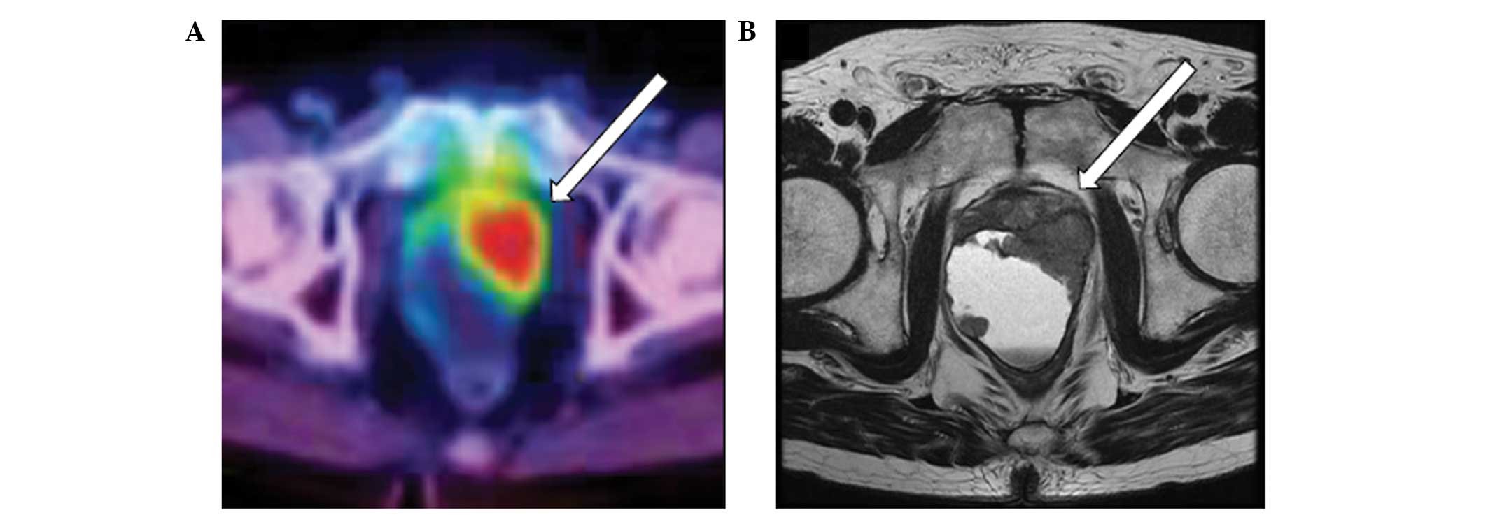
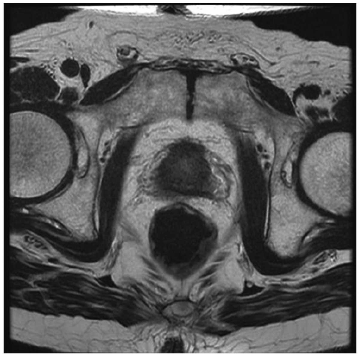
The present study reports a case of prostate adenocarcinoma with cyst formation. A 72‑year‑old male diagnosed with multiple lung metastases at a local clinic was referred to Tokai University Hachioji Hospital (Tokyo, Japan) for detection of a primary lesion. Whole body positron emission tomography/computed tomography showed strong accumulation of [18F]‑fluoro‑deoxy‑2‑glucose (FDG) in the small pelvis, and pelvic magnetic resonance imaging revealed a 60x40‑mm cystic lesion, with an irregular thickened wall, behind the prostate. The serum prostate‑specific antigen (PSA) level was elevated to 211.99 ng/ml, therefore, the patient underwent a transperineal prostate needle biopsy, and was diagnosed with prostate adenocarcinoma with cyst formation. Androgen deprivation therapy was administered for 8 months following the diagnosis of prostate cancer; consequently, the cyst reduced in size and the serum PSA level decreased to 0.14 ng/ml. To the best of our knowledge, this is the first report of a malignant prostatic cyst detected by FDG‑positron emission tomography/computed tomography.
View References
1
|
Emmett JL and Braasch WF: Cysts of the
prostate gland. J Urol. 36:236–249. 1936.
|
2
|
Kojima K, Uehara H, Naruo S, Kanayama H
and Kagawa S: Papillary cystadenocarcinoma of the prostate. Int J
Urol. 3:511–513. 1996.
|
3
|
Itami Y, Nagai Y, Kobayashi Y, Shimizu N,
Yamamoto Y, Minami T, Hayashi T, Nozawa M, Yoshimura K, Ishii T and
Uemura H: A case of prostatic cancer with a low PSA level
accompanied with cystic formation requiring differentiation from
adenocarcinoma of the seminal vesicle. Hinyokika Kiyo. 58:349–353.
2012.(In Japanese).
|
4
|
Naoe M, Ogawa Y, Fuji K, Fukagai T, Inoue
K and Yoshida H: Papillary cystadenocarcinoma of the prostate. Int
J Urol. 11:1036–1038. 2004.
|
5
|
Kim SC1, Fujimoto K, Matsumoto Y, et al: A
case of prostate cancer with cyst formation. Hinyokika Kiyo.
47:653–656. 2001.(In Japanese).
|
6
|
Niimi K, Hashimoto Y, Kurokawa S, Okada A,
Tozawa K and Kohri K: Embryonal rhabdomyosarcoma of the prostate.
Int J Clin Oncol. 15:93–96. 2010.
|
7
|
Chung HC, Lee HS, Kim TI, Kim DI, Park KH
and Song JM: A large cystic phyllodes tumor of the prostate. Yonsei
Med J. 50:174–176. 2009.
|
8
|
Epstein JI, Allsbrook WC Jr, Amin MB, et
al: The 2005 International Society of Urological Pathology (ISUP)
Consensus Conference on Gleason Grading of prostatic carcinoma. Am
J Surg Pathol. 29:1228–1242. 2005.
|
9
|
Hinev A, Chaushev B and Klisarova A: FDG
PET/CT in prostate cancer: A valuable method to detect the primary
and metastatic tumor sites and to monitor cancer response to
hormonal therapy. Nephrourol Mon. 4:644–645. 2012.
|
10
|
Han EJ, JH O, Choi WH, Yoo IR and Chung
SK: Significance of incidental focal uptake in prostate on
18-fluoro-2-deoxyglucose positron emission tomography CT images. Br
J Radiol. 83:915–920. 2010.
|
11
|
Minamimoto R, Uemura H, Sano F, Terao H,
Nagashima Y, Yamanaka S, Shizukuishi K, Tateishi U, Kubota Y and
Inoue T: The potential of FDG-PET/CT for detecting prostate cancer
in patients with an elevated serum PSA level. Ann Nucl Med.
25:21–27. 2011.
|