Solitary plasmacytoma of the sternum with a spiculated periosteal reaction: A case report
- Authors:
- Jingpin Zhao
- Yuqing Li
- Wenjuan Wu
- Zekun Zhang
- Yang Ding
View Affiliations
Affiliations: Department of Radiology, Third Hospital of Hebei Medical University, Hebei Province Biomechanical Key Laboratory of Orthopedics, Shijiazhuang, Hebei 050051, P.R. China
- Published online on: October 24, 2014 https://doi.org/10.3892/ol.2014.2636
-
Pages:
191-194
Metrics: Total
Views: 0 (Spandidos Publications: | PMC Statistics: )
Metrics: Total PDF Downloads: 0 (Spandidos Publications: | PMC Statistics: )
This article is mentioned in:
Abstract
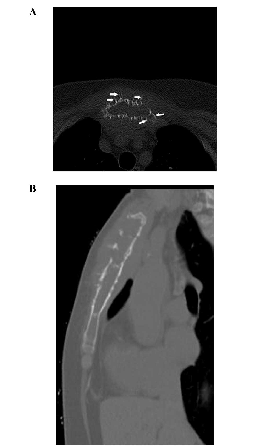
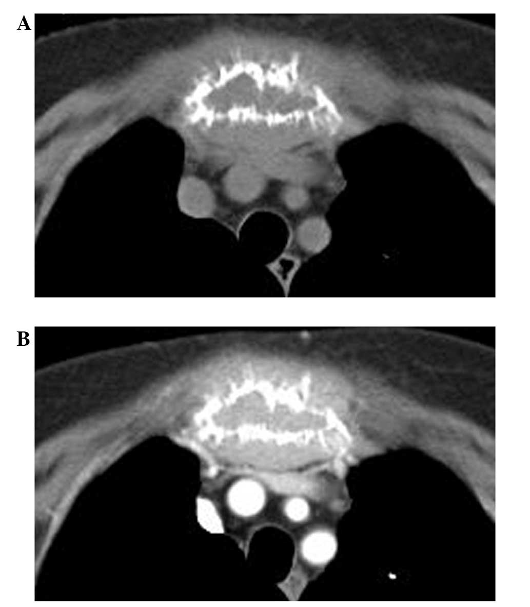
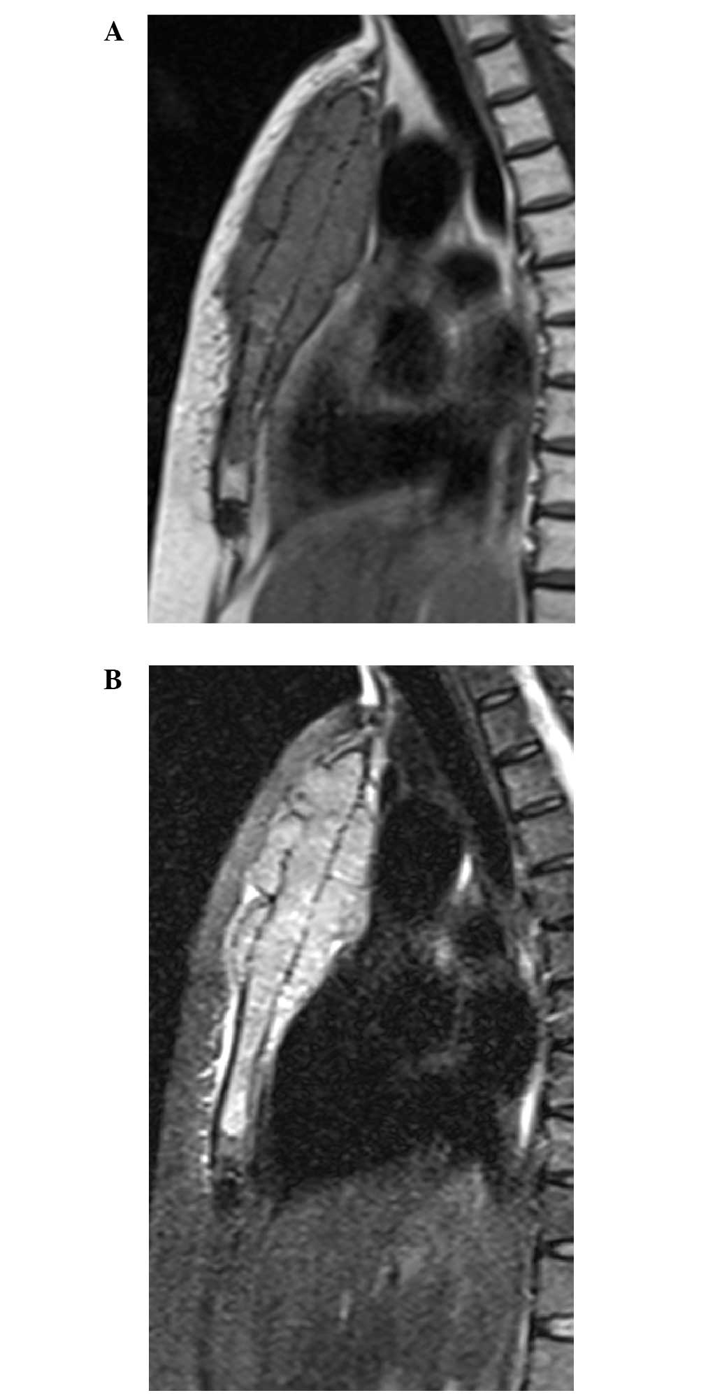
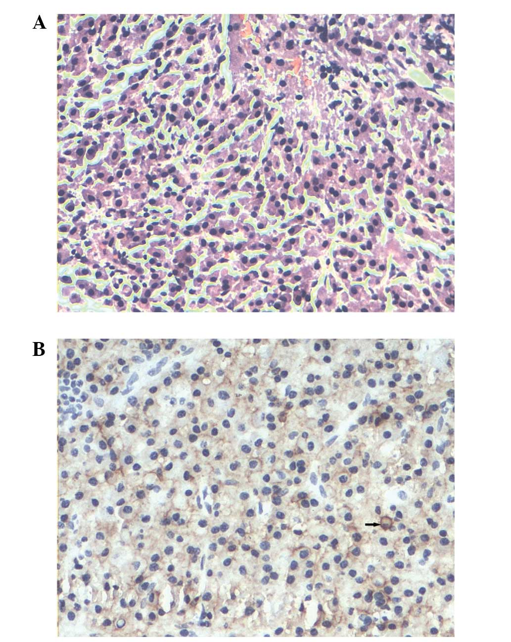
Solitary plasmacytomas (SPs) represent ≤5% of all plasma cell neoplasms and mostly occur in the spine, pelvis, ribs and pectoral girdle, while rarely occurring in the sternum. The tumors typically appear as osteolytic lesions. In rare cases, SPs can manifest as bony spicules on the surface of the bone. The present study reports the case of a 74 year‑old female with an osteolytic tumor localized in the sternum. The tumor displayed extensive bony destruction, with a large quantity of thick straight spicules on the surface of the bone, resembling a sunray in appearance. The imaging, laboratory and pathological examinations of the patient met the diagnostic criteria of SP. The patient was initially treated with radiotherapy at a dose of 45 Gy. Six months later, chemotherapy consisting of vindesine, Adriamycin and dexamethasone was administered. Vindesine and Adriamycin were administered at a dose of 2 and 15 mg/day, respectively on days 1-4 in a 20-day cycle. Dexamethasone was administered at a dose of 20 mg/day on days 1-4, 9-12 and 17-20 in the 20-day cycle. In total, the patient underwent 6 cycles of chemotherapy, with a total duration of 7 months. The patient was followed‑up for two years after beginning therapy. At present, the patient is well, without any evidence of progressive disease or multiple myeloma. To the best of our knowledge, this is the first case in the English literature of SP in the sternum, with an unusual sunray periosteal reaction on radiological imaging. The sites of bony spiculation in the lesions that have previously been described in the literature are the mandible, orbit, vertebral body and skull vault. To the best of our knowledge, the current study presents the first case of a SP of the sternum with a unusual spiculated periosteal reaction on radiological imaging to be reported in the English literature.
View References
1
|
Reed V, Shah J, Medeiros LJ, et al:
Solitary plasmacytomas: outcome and prognostic factors after
definitive radiation therapy. Cancer. 117:4468–4474. 2011.
View Article : Google Scholar : PubMed/NCBI
|
2
|
Nakanishi K, Kashiwagi N, Hamada K, et al:
Solitary plasmacytoma of the sternum detected incidentally by MR
imaging of the cervical spine. Magn Reson Med Sci. 9:227–231. 2010.
View Article : Google Scholar : PubMed/NCBI
|
3
|
Woodruff RK, Malpas JS and White FE:
Solitary plasmacytoma. II: Solitary plasmacytoma of bone. Cancer.
43:2344–2347. 1979. View Article : Google Scholar : PubMed/NCBI
|
4
|
Woodring JH, Umer MA and Bernardy MO:
Solitary plasmacytoma of the sternum: diagnosis by computed
tomography. J Comput Tomogr. 9:17–19. 1985. View Article : Google Scholar : PubMed/NCBI
|
5
|
Ghosh S, Wadhwa P, Kumar A, et al:
Abnormal radiological features in a multiple myeloma patient: a
case report and radiological review of myelomas. Dentomaxillofac
Radiol. 40:513–518. 2011. View Article : Google Scholar : PubMed/NCBI
|
6
|
Mohammadi A, Ilkhanizadeh B and
Ghasemi-Rad M: Mandibular plasmocytoma with sun-ray periosteal
reaction: A unique presentation. Int J Surg Case Rep. 3:296–298.
2012. View Article : Google Scholar : PubMed/NCBI
|
7
|
Ramon Y, Oberman M, Horowitz I, et al: A
large mandibular tumor with a distinct radiological ‘sun-ray
effect’ as the primary manifestation of multiple myeloma. J Oral
Surg. 36:52–54. 1978.PubMed/NCBI
|
8
|
Baykul T, Aydin U and O Carroll MK:
Unusual combination of presenting features in multiple myeloma.
Dentomaxillofac Radiol. 33:413–419. 2004. View Article : Google Scholar
|
9
|
Porter EC: Osteogenesis in multiple
myeloma. Report of a case Radiology. 76:457–458. 1961.
|
10
|
Shozushima M, Suzuki M, Ito S, et al:
Multiple myeloma with a distinct ‘sun-ray appearance’ occurring in
the jaw. Oral Radiol. 3:121–126. 1987. View Article : Google Scholar
|
11
|
Lipper S, Kahn LB and Hesselson N:
Localised myeloma with osteogenesis and Russell body formation. S
Afr Med J. 49:2041–2045. 1975.PubMed/NCBI
|
12
|
Grover SB and Dhar A: Imaging spectrum in
sclerotic myelomas: an experience of three cases. Eur Radiol.
10:1828–1831. 2000. View Article : Google Scholar : PubMed/NCBI
|
13
|
Bataille R and Sany J: Solitary myeloma:
clinical and prognostic features of a review of 114 cases. Cancer.
48:845–851. 1981. View Article : Google Scholar : PubMed/NCBI
|
14
|
Jiang ZH: Radiologic diagnosis of tumors
of the sternum (analysis of 30 cases). Zhonghua Fang She Xue Za
Zhi. 22:286–289. 1988.(In Chinese). PubMed/NCBI
|
15
|
Restrepo CS, Martinez S, Lemos DF, et al:
Imaging appearances of the sternum and sternoclavicular joints.
Radiographics. 29:839–859. 2009. View Article : Google Scholar : PubMed/NCBI
|
16
|
Jeung MY, Gangi A, Gasser B, et al:
Imaging of chest wall disorders. Radiographics. 19:617–637. 1999.
View Article : Google Scholar : PubMed/NCBI
|