TP53-induced glycolysis and apoptosis regulator promotes proliferation and invasiveness of nasopharyngeal carcinoma cells
- Authors:
- Published online on: December 12, 2014 https://doi.org/10.3892/ol.2014.2797
- Pages: 569-574
-
Copyright: © Wong et al. This is an open access article distributed under the terms of Creative Commons Attribution License.
Metrics: Total
Views: 0 (Spandidos Publications: | PMC Statistics: )
Total PDF Downloads: 0 (Spandidos Publications: | PMC Statistics: )
Abstract
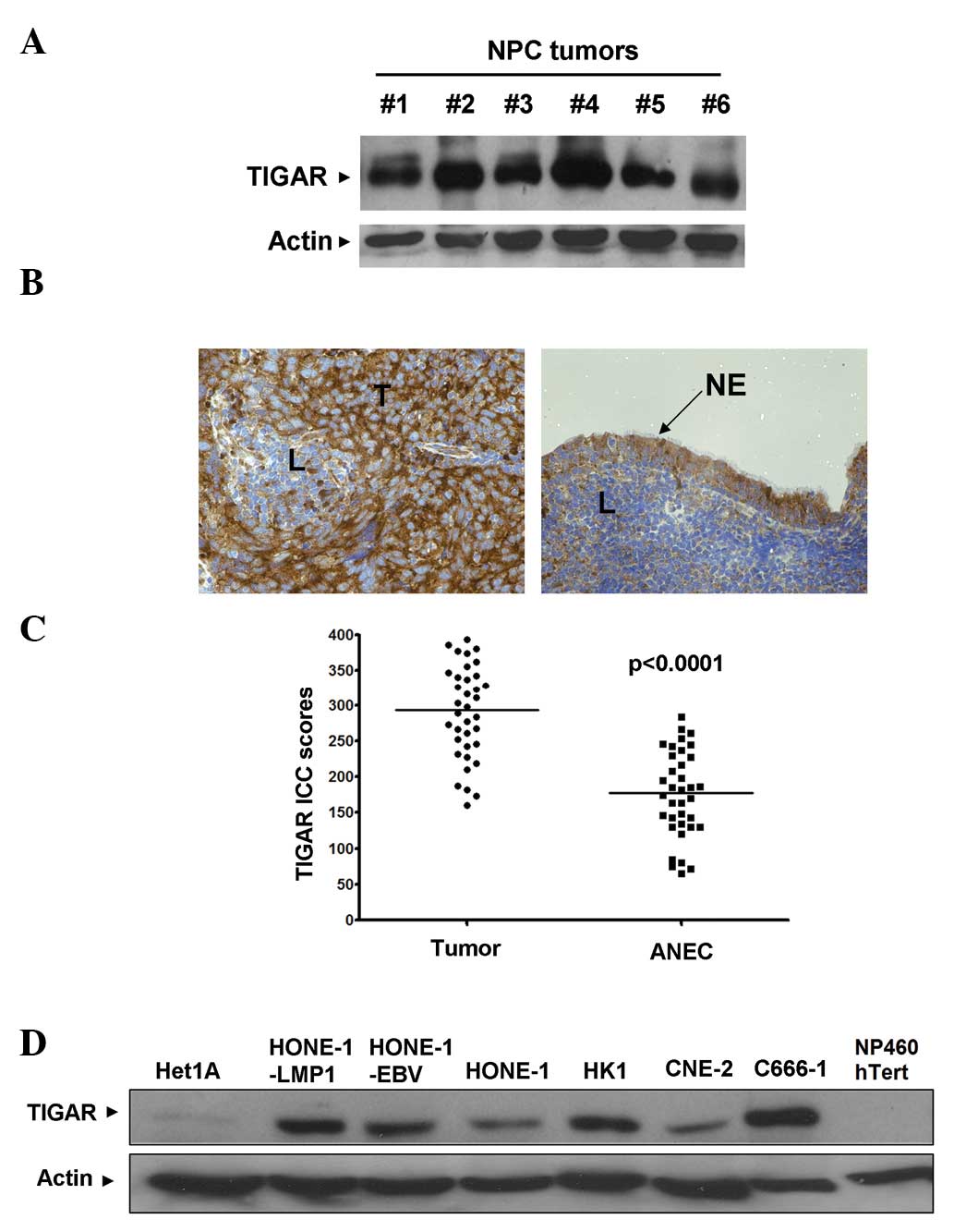
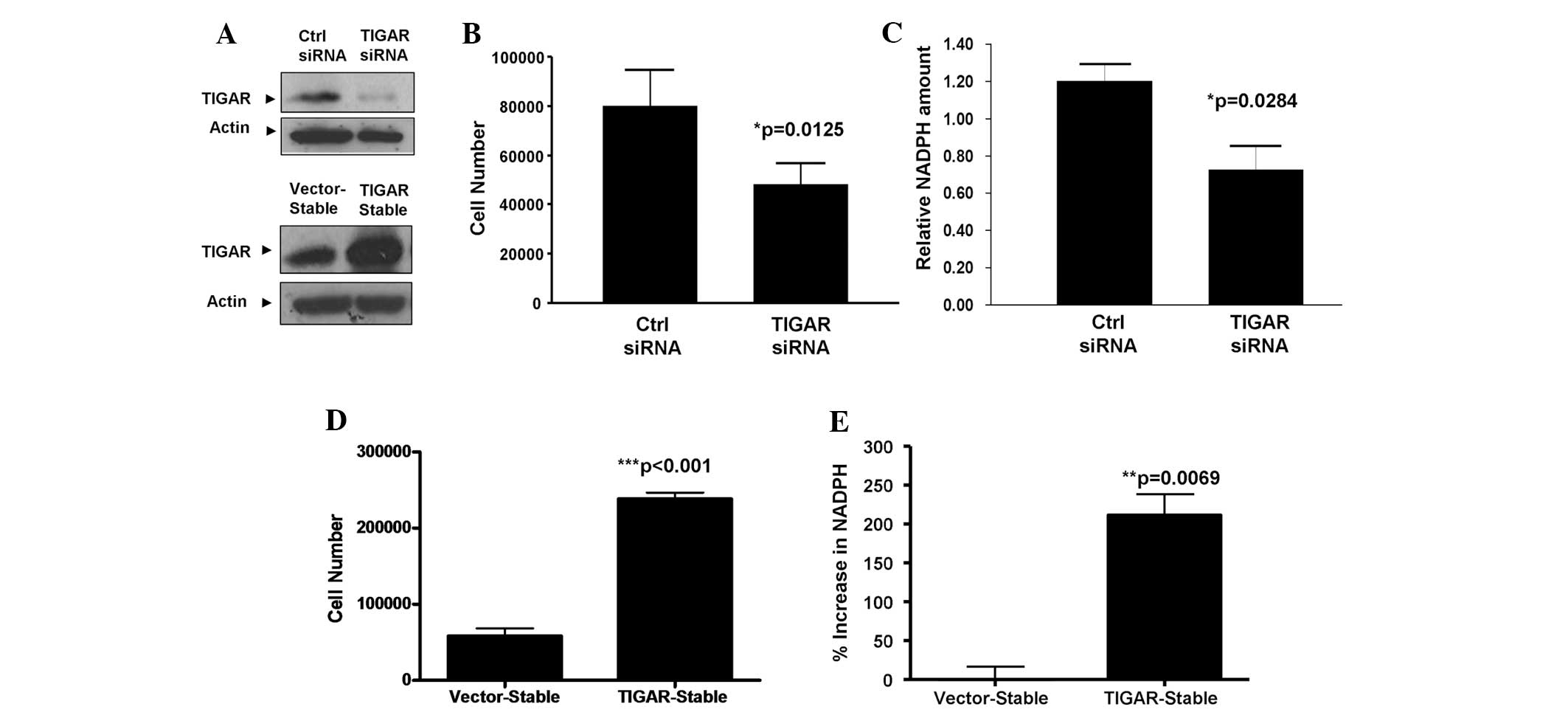
The TP53‑induced glycolysis and apoptosis regulator (TIGAR) is the protein product of the p53 target gene, C12orf5. TIGAR blocks glycolysis and promotes cellular metabolism via the pentose phosphate pathway; it promotes the production of cellular nicotinamide adenine dinucleotide phosphate (NADPH), which leads to enhanced scavenging of intracellular reactive oxygen species, and inhibition of oxidative stress‑induced apoptosis in normal cells. Our previous study identified a novel nucleoside analog that inhibited cellular growth and induced apoptosis in nasopharyngeal carcinoma (NPC) cell lines via downregulation of TIGAR expression. Furthermore, the growth inhibitory effects of c‑Met tyrosine kinase inhibitors were ameliorated by the overexpression of TIGAR in the NPC cell lines. These results indicate a significant role for TIGAR expression in the survival of NPCs. The present study aimed to further define the function of TIGAR expression in NPC cells. In total, 36 formalin‑fixed, paraffin‑embedded NPC tissue samples were obtained for the immunohistochemical determination of TIGAR expression. The effects of TIGAR expression on cell proliferation, NADPH production and cellular invasiveness were also assessed in NPC cell lines. Overall, TIGAR was overexpressed in 27/36 (75%) of the NPC tissues compared with the adjacent non‑cancer epithelial cells. Similarly, TIGAR overexpression was also observed in a panel of six NPC cell lines compared with normal NP460 hTert and Het1A cell lines. TIGAR overexpression led to increased cellular growth, NADPH production and invasiveness of the NPC cell lines, whereas a knockdown of TIGAR expression resulted in significant inhibition of cellular growth and invasiveness. The expression of the two mesenchymal markers, fibronectin and vimentin, was increased by TIGAR overexpression, but reduced following TIGAR‑knockdown. The present study revealed that TIGAR overexpression led to increased cellular growth, NADPH production and invasiveness, and the maintenance of a mesenchymal phenotype, in NPC tissues.