Symptomatic schwannoma of the abdominal wall: A case report and review of the literature
- Authors:
- Ruben Balzarotti
- Fabio Rondelli
- Jessica Barizzi
- Roberto Cartolari
View Affiliations
Affiliations: Surgical Unit, San Giovanni Hospital ‑ Ente Ospedaliero Cantonale, Bellinzona, Ticino 6500, Switzerland, Surgical Pathology, Istituto Cantonale di Patologia, Locarno, Ticino 6601, Switzerland, Radiological Unit, San Giovanni Hospital ‑ Ente Ospedaliero Cantonale, Bellinzona, Ticino 6500, Switzerland
- Published online on: January 12, 2015 https://doi.org/10.3892/ol.2015.2866
-
Pages:
1095-1098
-
Copyright: © Balzarotti
et al. This is an open access article distributed under the
terms of Creative
Commons Attribution License.
Metrics: Total
Views: 0 (Spandidos Publications: | PMC Statistics: )
Metrics: Total PDF Downloads: 0 (Spandidos Publications: | PMC Statistics: )
This article is mentioned in:
Abstract
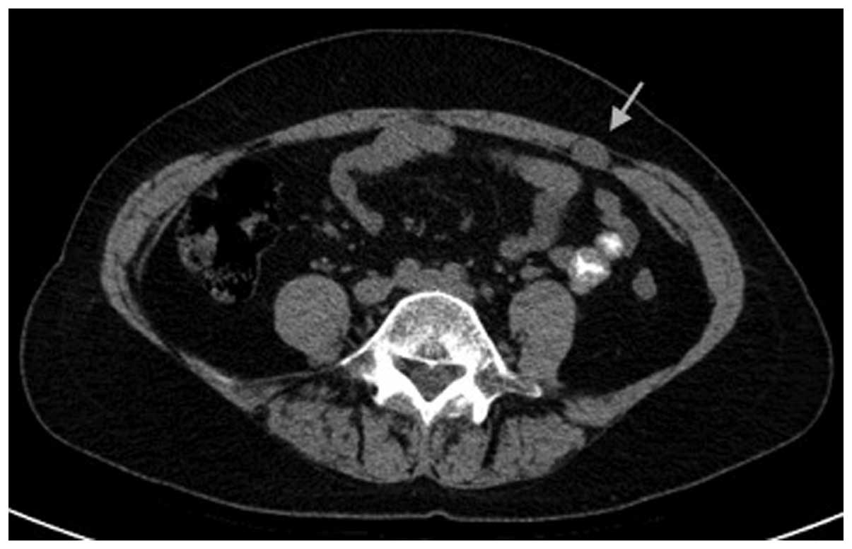
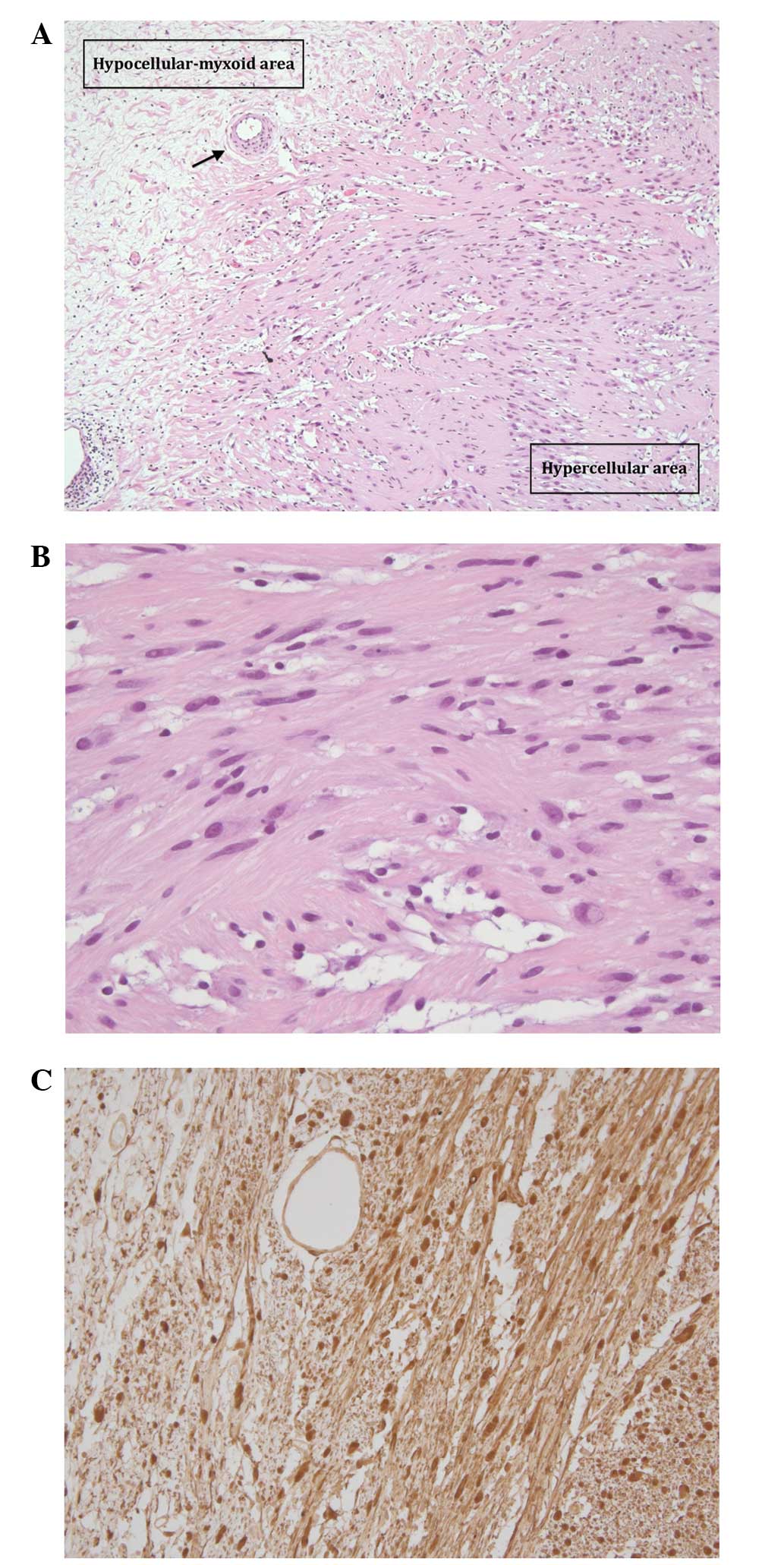
Schwannoma is a rare, benign tumor that arises from the nerve sheath. This tumor usually involves the extremities, but can also be found in the head and neck, trunk, pelvis, retroperitoneum, mediastinum and gastrointestinal tract. In numerous cases, the tumors are asymptomatic and are identified incidentally on physical examination or imaging. Occasionally, schwannoma is symptomatic due to compression of surrounding large nerves. In the present study, a 57‑year‑old female presented to the surgical outpatient's department due to a well‑localized parietal pain in the left lower quadrant. The onset of the pain occurred three years prior to presentation, without apparent cause and in the absence of other symptoms. Ultrasound and a computed tomography scan revealed a small solid tumor in the anterior abdominal wall, which was dimensionally stable over time, but was not noted in a preliminary analysis by a radiologist. The lesion was surgically removed using an anterior surgical approach. Histopathology revealed the tumor to be benign schwannoma. The painful symptoms completely disappeared. To the best of our knowledge, this is the third case of an abdominal wall benign schwannoma in the medical literature, and the first symptomatic case.
View Figures |
Figure 1
|
|
Figure 2
|
|
Figure 3
|
|
Figure 4
|
|
Figure 5
|
|
Figure 6
|
View References
1
|
Forthman CL and Blazar PE: Nerve tumors of
the hand and upper extremity. Hand Clin. 20:233–242. 2004.
View Article : Google Scholar : PubMed/NCBI
|
2
|
Abernathey CD, Onofrio BM, Scheithauer B,
et al: Surgical management of giant sacral schwannomas. J
Neurosurg. 65:286–295. 1986. View Article : Google Scholar : PubMed/NCBI
|
3
|
Li Q, Gao C, Juzi JT and Hao X: Analysis
of 82 cases of retroperitoneal schwannoma. ANZ J Surg. 77:237–240.
2007. View Article : Google Scholar : PubMed/NCBI
|
4
|
Bhatia RK, Banerjea A, Ram M and Lovett
BE: Benign ancient schwannoma of the abdominal wall: an unwanted
birthday present. BMC Surg. 10:12010. View Article : Google Scholar : PubMed/NCBI
|
5
|
Mishra A, Hamadto M, Azzabi M and Elfagieh
M: Abdominal wall schwannoma: case report and review of the
literature. Case Rep Radiol. 2013:4568632013.PubMed/NCBI
|
6
|
Hayashi M, Takeshita A, Yamamoto K and
Tanigawa N: Primary hepatic benign schwannoma. World J Gastrointest
Surg. 4:73–78. 2012. View Article : Google Scholar : PubMed/NCBI
|
7
|
White W, Shiu MH, Rosenblum MK, Erlandson
RA and Woodruff JM: Cellular schwannoma. A clinicopathologic study
of 57 patients and 58 tumors. Cancer. 66:1266–1275. 1990.
View Article : Google Scholar : PubMed/NCBI
|
8
|
Theodosopoulos T, Stafyla VK, Tsiantoula
P, et al: Special problems encountering surgical management of
large retroperitoneal schwannomas. World J Surg Oncol. 6:1072008.
View Article : Google Scholar : PubMed/NCBI
|
9
|
Hide IG, Baudouin CJ, Murray SA and
Malcolm AJ: Giant ancient schwannoma of the pelvis. Skeletal
Radiol. 29:538–542. 2000. View Article : Google Scholar : PubMed/NCBI
|
10
|
Dodd LG, Marom EM, Dash RC, Matthews MR
and McLendon RE: Fine-needle aspiration cytology of “ancient”
schwannoma. Diagn Cytopathol. 20:307–311. 1999. View Article : Google Scholar : PubMed/NCBI
|
11
|
Argenyi ZB, Balogh K and Abraham AA:
Degenerative (“ancient”) changes in benign cutaneous schwannoma. A
light microscopic, histochemical and immunohistochemical study. J
Cutan Pathol. 20:148–153. 1993. View Article : Google Scholar : PubMed/NCBI
|
12
|
Beaman FD, Kransdorf MJ and Menke DM:
Schwannoma: radiologic-pathologic correlation. Radiographics.
24:1477–1481. 2004. View Article : Google Scholar : PubMed/NCBI
|
13
|
Tsai WC, Chiou HJ, Chou YH, et al:
Differentiation between schwannomas and neurofibromas in the
extremities and superficial body: the role of high-resolution and
color Doppler ultrasonography. J Ultrasound Med. 27:161–166.
2008.PubMed/NCBI
|
14
|
Eizinger FM and Weiss SW: Benign tumors of
peripheral nerves. Soft Tissue Tumors. Third edition. Mosby; St.
Louis, MO: pp. 829–842. 1995
|
15
|
Akin M, Bozkirli B, Leventoglu S, et al:
Liver schwannoma incidentally discovered in a patient with breast
cancer. Bratisl Lek Listy. 110:298–300. 2009.PubMed/NCBI
|