Poorly differentiated cecal adenocarcinoma showing prominent rhabdoid feature combined with appendiceal mucinous cystadenoma: A case report and review of the literature
- Authors:
- In‑Ju Cho
- Sung‑Soo Kim
- Young‑Don Min
- Mun-Whan Noh
- Ran Hong
View Affiliations
Affiliations: Department of Pathology, College of Medicine, Chosun University, Gwangju 501‑759, Republic of Korea, Department of Surgery, College of Medicine, Chosun University, Gwangju 501‑759, Republic of Korea, Department of Complementary and Alternative Medicine, Chosun University Graduate School, Gwangju 501‑759, Republic of Korea
- Published online on: January 27, 2015 https://doi.org/10.3892/ol.2015.2905
-
Pages:
1527-1530
-
Copyright: © Cho
et al. This is an open access article distributed under the
terms of Creative
Commons Attribution License.
Metrics: Total
Views: 0 (Spandidos Publications: | PMC Statistics: )
Metrics: Total PDF Downloads: 0 (Spandidos Publications: | PMC Statistics: )
This article is mentioned in:
Abstract
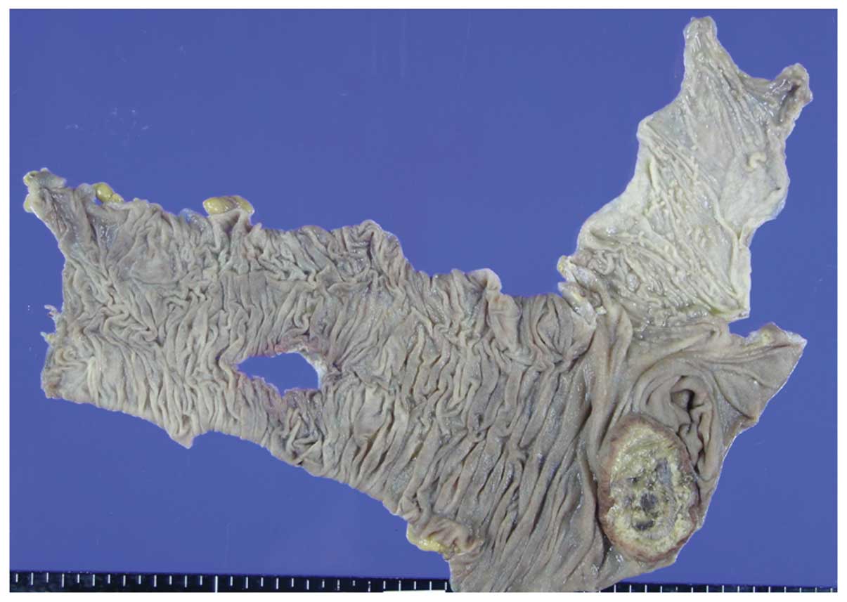
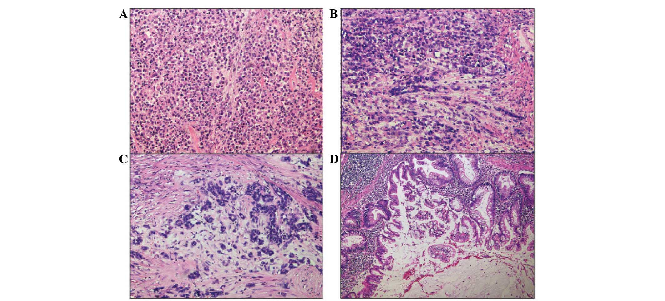
Extrarenal rhabdoid tumors (ERRTs) are extremely rare neoplasms; of these, colorectal ERRTs are the most rare, and only nine cases have been previously described in the English language literature. The current study reports the pathological features of a case of poorly differentiated cecal adenocarcinoma with prominent rhabdoid feature, which was combined with mucinous cystadenoma of the appendix in a 73‑year‑old male, and additionally reviews the previously reported cases. Microscopically, the majority of tumor cells were non‑cohesive or loosely cohesive, with a polygonal morphology and prominent rhabdoid feature, showing eccentric vesicular nuclei, prominent nucleoli and abundant eosinophilic cytoplasm. Immunohistochemically, the tumor cells were positive for cytokeratin (CK) and vimentin, but negative for CK20, CK7, desmin and smooth muscle actin. This indicated a diagnosis of poorly differentiated adenocarcinoma with prominent rhabdoid features, combined with appendiceal mucinous cystadenoma. At two months following surgery the patient succumbed to peritoneal seeding and metastasis of liver and bone The emergence of the rhabdoid phenotype is invariably associated with an aggressive and almost always fatal clinical course. The present case is the 10th example of such a tumor in the colon, and to the best of our knowledge, this is the first case of colonic rhabdoid tumor coinciding with appendiceal benign mucinous neoplasm.
View References
1
|
Beckwith JB and Palmer NF: Histopathology
and prognosis of Wilms tumors: results from the First National
Wilms Tumor Study. Cancer. 41:1937–1948. 1978. View Article : Google Scholar : PubMed/NCBI
|
2
|
Pancione M, Di Blasi A, Sabatino L, et al:
A novel case of rhabdoid colon carcinoma associated with a positive
CpG island methylator phenotype and BRAF mutation. Hum Pathol.
42:1047–1052. 2011. View Article : Google Scholar : PubMed/NCBI
|
3
|
Yang AH, Chen WY and Chiang H: Malignant
rhabdoid tumour of colon. Histopathology. 24:89–91. 1994.
View Article : Google Scholar : PubMed/NCBI
|
4
|
Marcus VA, Viloria J, Owen D and Tsao MS:
Malignant rhabdoid tumor of the colon. Report of a case with
molecular analysis. Dis Colon Rectum. 39:1322–1326. 1996.
View Article : Google Scholar : PubMed/NCBI
|
5
|
Nakamura I, Nakano K, Nakayama K, et al:
Malignant rhabdoid tumor of the colon: report of a case. Surg
Today. 29:1083–1087. 1999. View Article : Google Scholar : PubMed/NCBI
|
6
|
Kono T, Imai Y, Imura J, et al: Cecal
adenocarcinoma with prominent rhabdoid feature: report of a case
with immunohistochemical, ultrastructural, and molecular analyses.
Int J Surg Pathol. 15:414–420. 2007. View Article : Google Scholar : PubMed/NCBI
|
7
|
Remo A, Zanella C, Molinari E, et al:
Rhabdoid carcinoma of the colon: a distinct entity with a very
aggressive behavior: a case report associated with a polyposis coli
and review of the literature. Int J Surg Pathol. 20:185–190. 2012.
View Article : Google Scholar
|
8
|
Chetty R and Bhathal PS: Caecal
adenocarcinoma with rhabdoid phenotype: an immunohistochemical and
ultrastructural analysis. Virchows Arch A Pathol Anat Histopathol.
422:179–182. 1993. View Article : Google Scholar : PubMed/NCBI
|
9
|
Lee SH, Seol H, Kim WY, et al: Rhabdoid
colorectal carcinomas: reports of two cases. Korean J Pathol.
47:372–377. 2013. View Article : Google Scholar : PubMed/NCBI
|
10
|
Frierson HF Jr, Mills SE and Innes DJ Jr:
Malignant rhabdoid tumor of the pelvis. Cancer. 55:1963–1967. 1985.
View Article : Google Scholar : PubMed/NCBI
|
11
|
Kodet R, Newton WA Jr, Sachs N, et al:
Rhabdoid tumors of soft tissues: a clinicopathologic study of 26
cases enrolled on the Intergroup Rhabdomyosarcoma Study. Hum
Pathol. 22:674–684. 1991. View Article : Google Scholar : PubMed/NCBI
|
12
|
Wick MR, Ritter JH and Dehner LP:
Malignant rhabdoid tumors: a clinicopathologic review and
conceptual discussion. Semin Diagn Pathol. 12:233–248.
1995.PubMed/NCBI
|
13
|
Berry PJ and Vujanic GM: Malignant
rhabdoid tumour. Histopathology. 20:189–193. 1992. View Article : Google Scholar : PubMed/NCBI
|
14
|
Tsuneyoshi M, Daimaru Y, Hashimoto H, et
al: Malignant soft tissue neoplasms with the histologic features of
renal rhabdoid tumors: an ultrastructural and immunohistochemical
study. Hum Pathol. 16:1235–1242. 1985. View Article : Google Scholar : PubMed/NCBI
|
15
|
Haas JE, Palmer NF, Weinberg AG and
Beckwith JB: Ultrastructure of malignant rhabdoid tumor of the
kidney. A distinctive renal tumor of children. Hum Pathol.
12:646–657. 1981. View Article : Google Scholar : PubMed/NCBI
|
16
|
Flatcher C, Unni K and Mertens F:
Pathology and Genetics of Tumours of Soft tissue and Bond. IARC
Press; Lyon: 2002
|