Intrapulmonary myelolipoma and its computed tomography features: A case report and literature review
- Authors:
- Published online on: January 28, 2015 https://doi.org/10.3892/ol.2015.2913
- Pages: 1677-1680
Metrics: Total
Views: 0 (Spandidos Publications: | PMC Statistics: )
Total PDF Downloads: 0 (Spandidos Publications: | PMC Statistics: )
Abstract
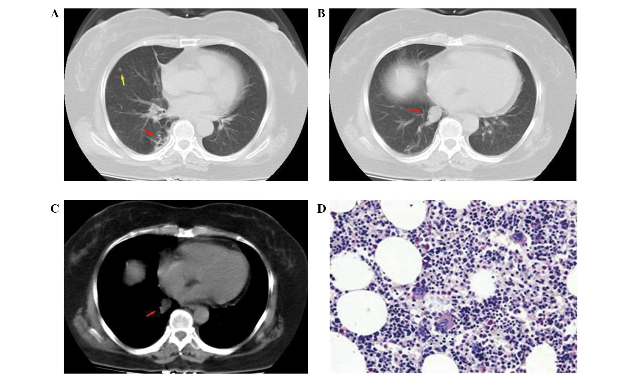
Intrapulmonary myelolipoma is a rare, benign tumor composed of mature adipose tissue and normal hematopoietic cells. To the best of our knowledge, 10 cases of intrapulmonary myelolipoma, including the present case, have been reported to date, and the majority have focused on the pathological diagnosis of the disease. The radiological features of intrapulmonary myelolipoma have not been studied. Therefore, the present study reports a case of primary myelolipoma in the lung, and examines its computed tomography features and pathology. Furthermore, other potential diagnoses are discussed in the context of the relevant literature. The present report describes the case of a 57-year-old female who experienced chills, but no coughing or expectoration, with an intermittent fever of 38.6˚C that had been apparent for 13 days. Chest CT scan revealed a benign nodule and bronchiectasis in the lower lobe of the right lung. The patient then underwent a lobectomy of the lower right lung by thoracoscopy. The histological analysis of the excised specimen identifid a myelolipoma consisting of mature adipose tissue and hematopoietic cells. There was no recurrence after 513 days of follow-up, as shown by CT.