Imaging features of primary hepatic leiomyosarcoma: A case report and review of literature
- Authors:
- Wei‑Fu Lv
- Jian‑Kui Han
- De‑Lei Cheng
- Wen‑Jing Tang
- Dong Lu
View Affiliations
Affiliations: Positron Emission Tomography/Computed Tomography Center, Qilu Hospital, First Affiliated Hospital of Shandong University, Jinan, Shandong 250012, P.R. China, Department of Radiology, Affiliated Anhui Provincial Hospital of Anhui Medical University, Hefei, Anhui 230001, P.R. China
- Published online on: March 3, 2015 https://doi.org/10.3892/ol.2015.3006
-
Pages:
2256-2260
Metrics: Total
Views: 0 (Spandidos Publications: | PMC Statistics: )
Metrics: Total PDF Downloads: 0 (Spandidos Publications: | PMC Statistics: )
This article is mentioned in:
Abstract
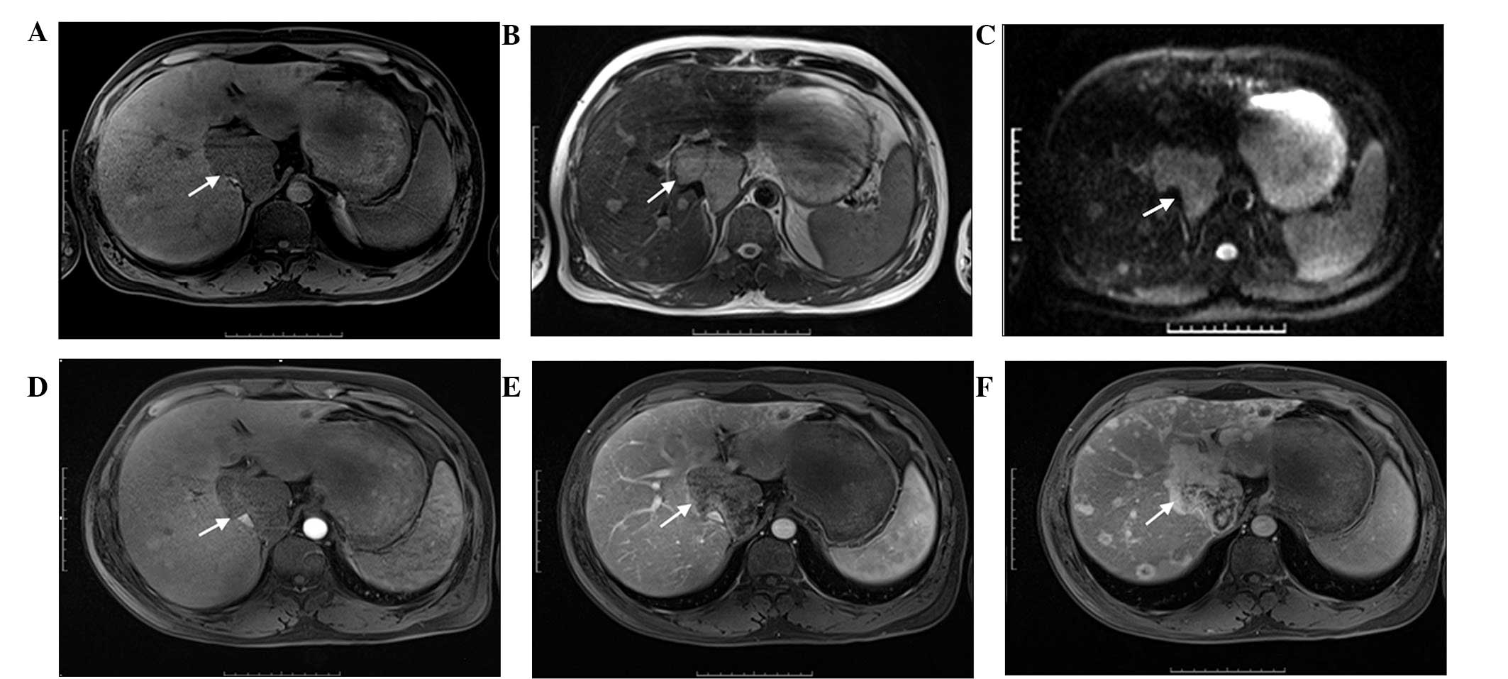
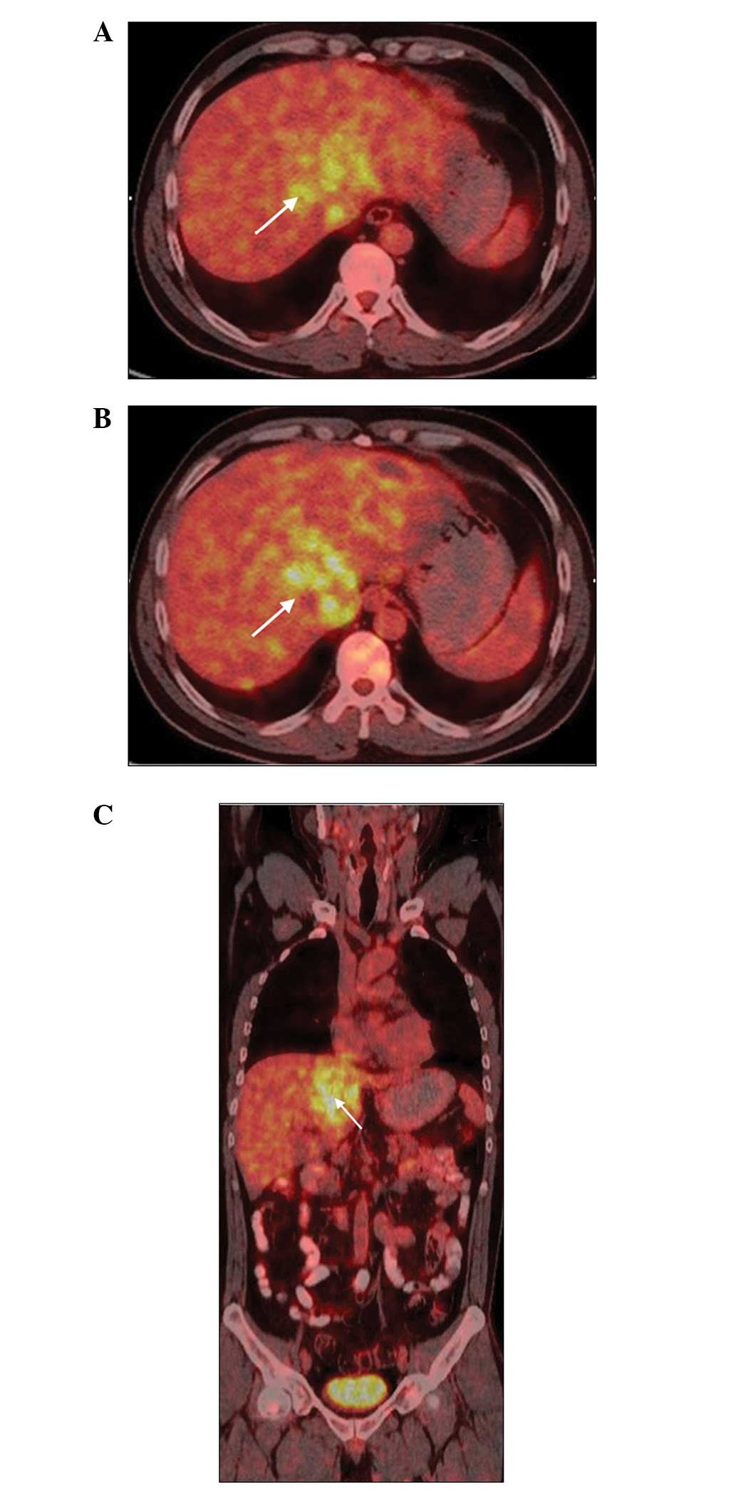
Primary hepatic leiomyosarcoma (PHL) is an extremely rare tumour. This tumour is difficult to diagnose by imaging examinations due to its rarity, and non‑specific conventional imaging manifestations and clinical presentation. The present study reports the case of a 42‑year‑old male with PHL that was confirmed by histopathological and immunohistochemical examinations. Multimodal imaging examinations, including ultrasound, computed tomography (CT), magnetic resonance imaging (MRI), positron emission tomography‑CT and digital subtraction angiography, were performed. The imaging manifestations were analysed and the associated literature was reviewed. The results found that no characteristic imaging appearance was present on ultrasound or plain CT scan. However, on unenhanced MRI, the tumours presented with a heterogeneous low signal density on T1‑weighted imaging (WI) and a high signal density on T2WI and diffusion‑WI. On gadopentetate dimeglumine enhanced MRI, the lesions were not enhanced during the arterial and portal venous phases; by contrast, these lesions were evidently enhanced during the 5‑min delayed phase. Therefore, the delayed imaging of enhanced MRI is likely to be used to differentiate PHL from other hepatic tumours.
View Figures |
Figure 1
|
|
Figure 2
|
|
Figure 3
|
|
Figure 4
|
|
Figure 5
|
|
Figure 6
|
View References
1
|
Ferrozzi F, Bova D, Zangrandi A and
Garlaschi G: Primary liver leiomyosarcoma: CT appearance. Abdom
Imaging. 21:157–160. 1996. View Article : Google Scholar : PubMed/NCBI
|
2
|
Shamseddine A, Faraj W, Mukherji D, et al:
Unusually young age distribution of primary hepatic leiomyosarcoma:
case series and review of the adult literature. World J Surg Oncol.
8:562010. View Article : Google Scholar : PubMed/NCBI
|
3
|
Civardi G, Cavanna L, Iovine E, et al:
Diagnostic imaging of primary hepatic leiomyosarcoma: a case
report. Ital J Gastroenterol. 28:98–101. 1996.PubMed/NCBI
|
4
|
Gandhi MR, Wong DC and Wood DJ: Ultrasound
and computed tomography appearances of primary hepatic
leiomyosarcoma. Australas Radiol. 39:289–291. 1995. View Article : Google Scholar : PubMed/NCBI
|
5
|
Soyer P, Blanc F, Vissuzaine C, et al:
Primary leiomyosarcoma of the liver MR findings. Clin Imaging.
20:273–275. 1996. View Article : Google Scholar : PubMed/NCBI
|
6
|
Chi M, Dudek AZ and Wind KP: Primary
hepatic leiomyosarcoma in adults: analysis of prognostic factors.
Onkologie. 35:210–214. 2012. View Article : Google Scholar : PubMed/NCBI
|
7
|
Tsai PS, Yeh TC and Shih SL: Primary
hepatic leiomyosarcoma in a 5-month-old female infant. Acta Radiol
Short Rep. 2:20479816134987222013.PubMed/NCBI
|
8
|
Lencioni R and Llovet JM: Modified RECIST
(mRECIST) assessment for hepatocellular carcinoma. Semin Liver Dis.
30:52–60. 2010. View Article : Google Scholar : PubMed/NCBI
|
9
|
Holloway H, Walsh CB, Thomas R and
Fielding J: Primary hepatic leiomyosarcoma. J Clin Gastroenterol.
23:131–133. 1996. View Article : Google Scholar : PubMed/NCBI
|
10
|
Rosas JS, Del Rosario A, Bui HX, et al:
Primary hepatic leiomyosarcoma in a child with the acquired
immunodeficiency syndrome. Hum Pathol. 23:69–72. 1992. View Article : Google Scholar : PubMed/NCBI
|
11
|
Brichard B, Smets F, Sokal E, et al:
Unusual evolution of an Epstein-Barr virus-associated
leiomyosarcoma occurring after liver transplantation. Pediatr
Transplant. 5:365–369. 2001. View Article : Google Scholar : PubMed/NCBI
|
12
|
Jeong TY, Kim YS, Park KJ, et al: A case
of primary leiomyosarcoma of the liver presenting with acute
bleeding. Korean J Gastroenterol. 51:194–198. 2008.(Korean).
PubMed/NCBI
|
13
|
Gates LK Jr, Cameron AJ, Nagorney DM, et
al: Primary leiomyosarcoma of the liver mimicking liver abscess. Am
J Gastroenterol. 90:649–652. 1995.PubMed/NCBI
|
14
|
Yu RS, Chen Y, Jiang B, et al: Primary
hepatic sarcomas: CT findings. Eur Radiol. 18:2196–2205. 2008.
View Article : Google Scholar : PubMed/NCBI
|
15
|
Smith MB, Silverman JF, Raab SS, et al:
Fine-needle aspiration cytology of hepatic leiomyosarcoma. Diagn
Cytopathol. 11:321–327. 1994. View Article : Google Scholar : PubMed/NCBI
|
16
|
Sprogøe-Jakobsen S and Hølund B:
Immunohistochemistry (Ki-67 and p53) as a tool in determining
malignancy in smooth muscle neoplasms (exemplified by a myxoid
leiomyosarcoma of the uterus). APMIS. 104:705–708. 1996. View Article : Google Scholar : PubMed/NCBI
|