First adult case of sporadic localized glomerulocystic kidney mimicking a tumor
- Authors:
- Fumiyoshi Kojima
- Mitsuaki Ishida
- Yuichi Tsujimoto
- Masahiro Hosomi
- Kinouchi Toshiaki
- Hidetoshi Okabe
-
View Affiliations
Affiliations: Department of Diagnostic Pathology, Saiseikai Senri Hospital, Suita, Osaka 565‑0862, Japan, Department of Clinical Laboratory Medicine and Division of Diagnostic Pathology, Shiga University of Medical Science, Otsu, Shiga 520‑2192, Japan, Department of Urology, Saiseikai Senri Hospital, Suita, Osaka 565‑0862, Japan
- Published online on: March 20, 2015 https://doi.org/10.3892/ol.2015.3060
-
Pages:
2368-2370
Metrics:
Total
Views: 0 (Spandidos Publications: | PMC Statistics:
)
Metrics:
Total PDF Downloads: 0 (Spandidos Publications: | PMC Statistics:
)
This article is mentioned in:
Abstract
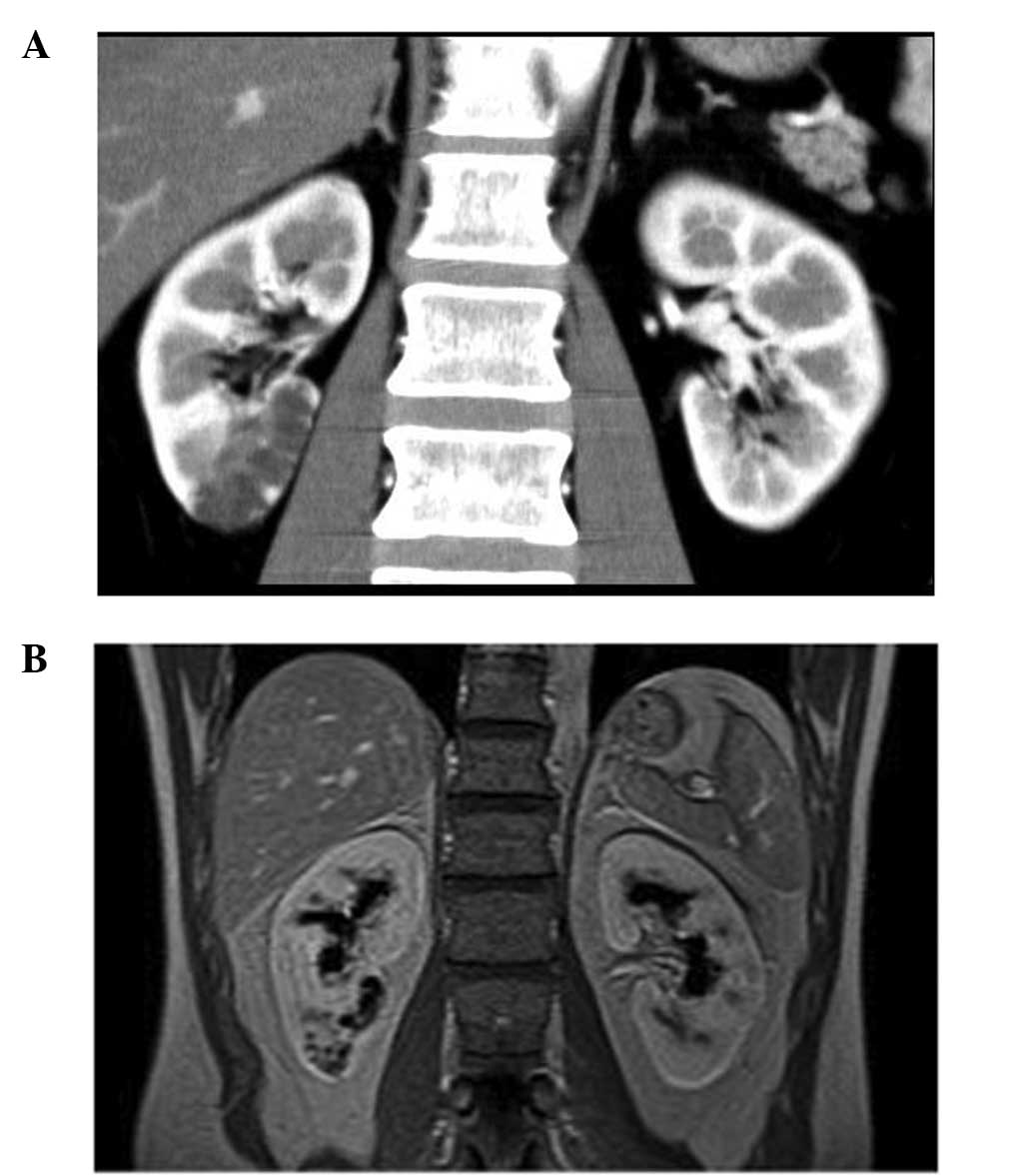
Glomerulocystic kidneys (GCKs) are mainly observed in infants and young children, and are characterized by the cystic dilatation of Bowman's space to form glomerular cysts (GCs). GCKs are associated with various conditions. Additionally, the cystogenesis of GCKs remains controversial. The present study describes a rare adult case of a sporadic localized GCK that radiologically mimicked a multilocular cystic tumor, and analyses the features of GC. A 42‑year‑old male with hematuria underwent a right partial nephrectomy for a cystic mass. The majority of the cyst was distributed in the cortex and contained a single collapsed glomerulus. Using serial sections, narrow and serpiginous proximal tubules that continued to the GCs were detected. These findings suggested that obliteration at the glomerulotubular junction was not the primary cause of GC in this case. To the best of our knowledge, this is the first adult case of a sporadic localized GCK mimicking a tumor. Unnecessary surgical procedures may be avoided by careful evaluation of computed tomography scans and magnetic resonance imaging, although localized GCKs are quite rare.
View References
1
|
Lennerz JK, Spence DC, Iskandar SS, Dehner
LP and Liapis H: Glomerulocystic kidney: one hundred-year
perspective. Arch Pathol Lab Med. 134:583–605. 2010.PubMed/NCBI
|
2
|
Obata Y, Furuse A, Miyazaki M, Nishino T,
Kawazu T, Kanamoto Y, Nishikido M, Taguchi T and Kohno S:
Glomerulocystic kidney disease in an adult with enlarged kidneys: a
case report and review of the literature. Clin Nephrol. 75:158–164.
2011.PubMed/NCBI
|
3
|
Liu JS, Ishikawa I, Saito Y, Nakazawa T,
Tomosugi N and Ishikawa Y: Digital glomerular reconstruction in a
patient with a sporadic adult form of glomerulocystic kidney
disease. Am J Kidney Dis. 35:216–220. 2000. View Article : Google Scholar : PubMed/NCBI
|
4
|
Bosniak MA: The Bosniak renal cyst
classification: 25 years later. Radiology. 262:781–785. 2012.
View Article : Google Scholar : PubMed/NCBI
|
5
|
Hotta O, Sato M, Furuta T and Taguma Y:
Pathogenic role of glomerulo-tubular junction stenosis in
glomerulocystic disease. Clin Nephrol. 51:177–180. 1999.PubMed/NCBI
|
6
|
Liu JS, Ishikawa I, Saito Y, Nakazawa T,
Tomosugi N and Ishikawa Y: Digital glomerular reconstruction in a
patient with a sporadic adult form of glomerulocystic kidney
disease. Am J Kidney Dis. 35:216–220. 2000. View Article : Google Scholar : PubMed/NCBI
|
7
|
Kanouzawa K, Tamura H, Matsumura O,
Nagasawa R, Mitarai T, Isoda K and Yamanaka N: An adult case of
glomerulocystic kidney disease. Nihon Jinzo Gakkai Shi. 36:762–768.
1994.[(In Japanese)]. PubMed/NCBI
|