Primary glioblastoma of the cerebellar vermis: A case report
- Authors:
- Xiaolin Jing
- Guohua Shen
- Minggang Su
- Rong Tian
- Zhiyun Jia
View Affiliations
Affiliations: Department of Nuclear Medicine, West China Hospital, Sichuan University, Chengdu, Sichuan 610041, P.R. China
- Published online on: May 6, 2015 https://doi.org/10.3892/ol.2015.3188
-
Pages:
402-404
Metrics: Total
Views: 0 (Spandidos Publications: | PMC Statistics: )
Metrics: Total PDF Downloads: 0 (Spandidos Publications: | PMC Statistics: )
This article is mentioned in:
Abstract
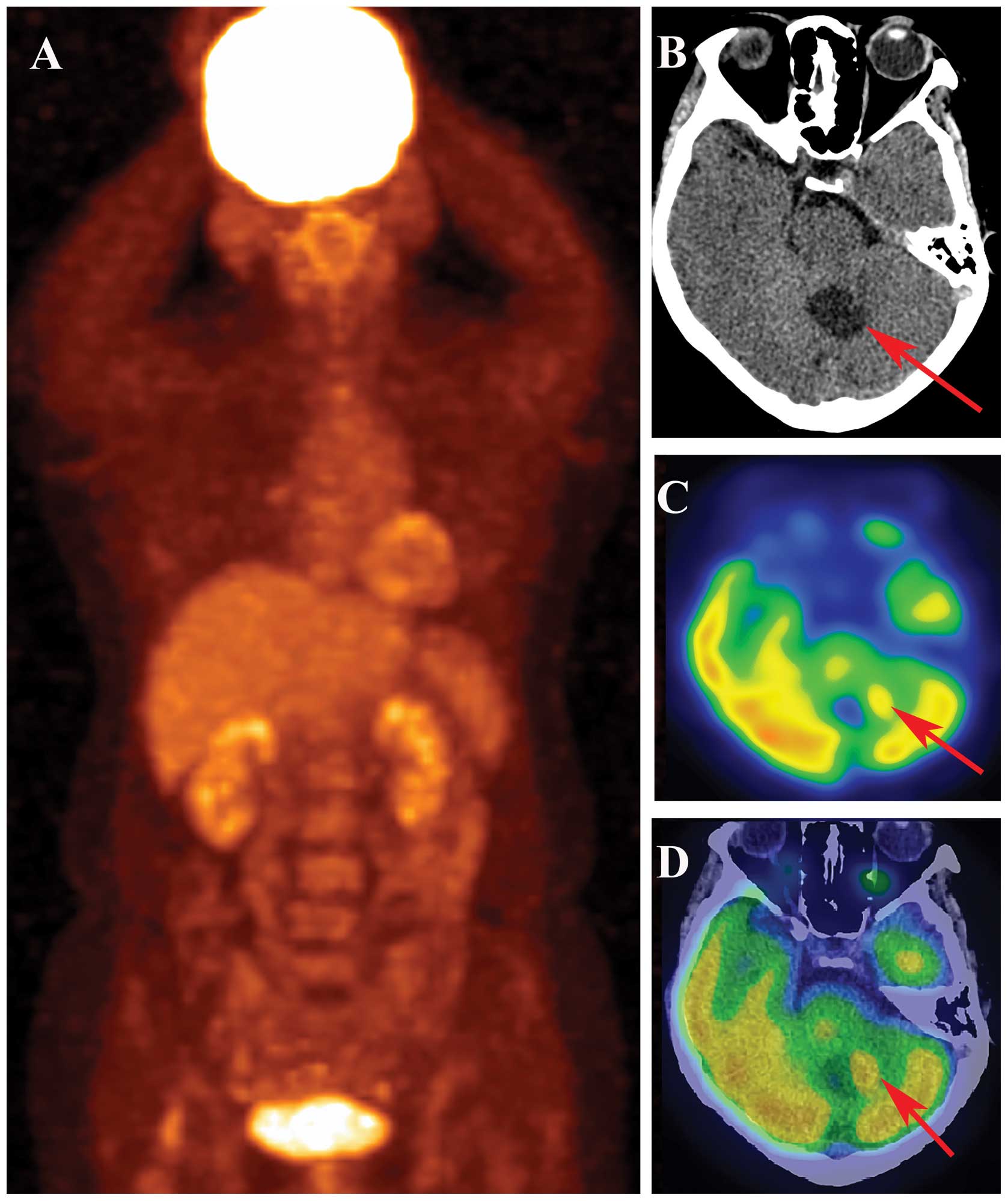
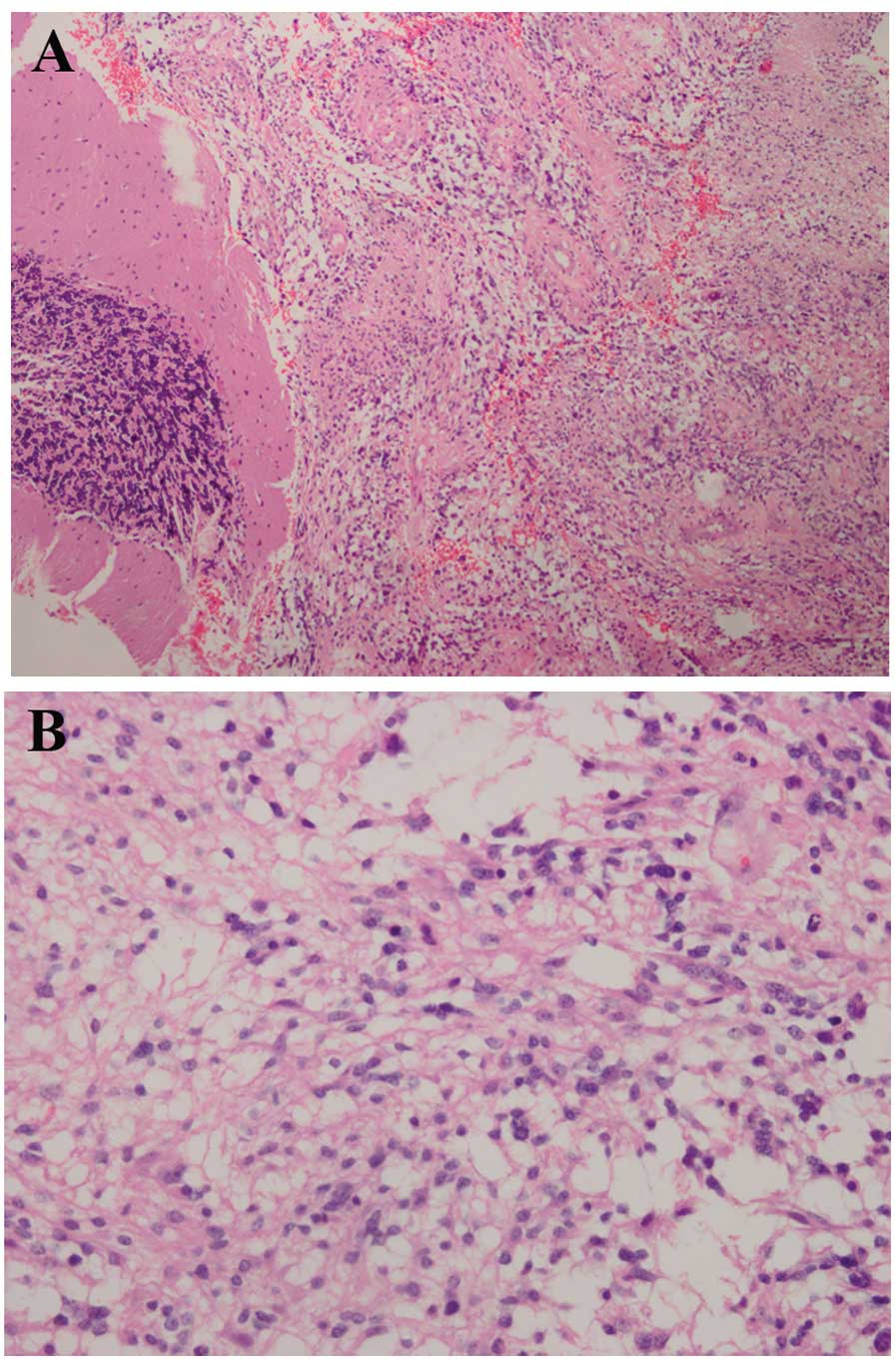
Cerebellar glioblastoma is a rare adult tumor. The accurate diagnosis of cerebellar glioblastoma is important for establishing a suitable therapeutic schedule. However, it is occasionally difficult to diagnosis these tumors. Clinical presentation, computed tomography (CT) and magnetic resonance imaging can provide useful information, but they may not lead to a definitive diagnosis. Positron emission tomography/computed tomography (PET/CT) may provide a novel way of forming a differential diagnoses. The lesions of glioblastoma multiforme (GBM) rarely occur in the cerebellum, with prior studies reporting that only 0.4‑3.4% of all GBM tumors occur here. In the current study, a case of primary cerebellar glioblastoma is presented and the physiopathology, clinical presentation, diagnosis, differential diagnosis, treatment and general outcome of this disease are discussed. A 61‑year‑old female presented with nausea, vomiting, balance problems and cerebellar signs. Cranial magnetic resonance imaging (MRI) and positron emission tomography/computed tomography (PET/CT) examination demonstrated one regular contour of a mass lesion in the cerebellar vermis. Following surgery, glioblastoma was histologically confirmed. The outcome of the patient was favorable after 18 months of follow‑up. Cerebellar GBM should be considered in the differential diagnosis of a cerebellar mass lesion, and PET/CT may provide a novel identification method for different cerebellar mass lesions.
View References
1
|
El El Maaqili MR, Hossini A, El Fatemi N,
et al: Primary glioblastoma of the cerebellum in a 19-year-old
woman: a case report. J Med Case Rep. 6:3292012. View Article : Google Scholar : PubMed/NCBI
|
2
|
Grahovac G, Tomac D, Lambasa S, et al:
Cerebellar glioblastomas: Pathophysiology, clinical presentation
and management. Acta Neurochir. 151:653–657. 2009. View Article : Google Scholar : PubMed/NCBI
|
3
|
Babu R, Sharma R, Karikari IO, et al:
Outcome and prognostic factors in adult cerebellar glioblastoma. J
Clin Neurosci. 20:1117–1121. 2013. View Article : Google Scholar : PubMed/NCBI
|
4
|
Kuroiwa T, Numaguchi Y, Rothman MI, et al:
Posterior fossa glioblastoma multiforme: MR findings. AJNR Am J
Neuroradiol. 16:583–589. 1995.PubMed/NCBI
|
5
|
Roth JG and Elvidge AR: Glioblastoma
multiforme: A clinical survey. J Neurosurg. 17:736–750. 1960.
View Article : Google Scholar : PubMed/NCBI
|
6
|
Mattos JP, Marenco HA, Campos JM, et al:
Cerebellar glioblastoma multiforme in an adult. Arq Neuropsiquiatr.
64:132–135. 2006. View Article : Google Scholar : PubMed/NCBI
|
7
|
Louis DN, Ohgaki H, Wiestler OD, et al:
The 2007 WHO classification of tumours of the central nervous
system. Acta Neuropathol. 114:97–109. 2007. View Article : Google Scholar : PubMed/NCBI
|
8
|
Hamasaki K, Nakamura H, Ueda Y, et al:
Radiation-induced glioblastoma occurring 35 years after radiation
therapy for medulloblastoma: case report. Brain Tumor Pathol.
27:39–43. 2010. View Article : Google Scholar : PubMed/NCBI
|
9
|
Demir MK, Hakan T, Akinci O and Berkman Z:
Primary cerebellar glioblastoma multiforme. Diagn Interv Radiol.
11:83–86. 2005.PubMed/NCBI
|
10
|
Kulkarni AV, Becker LE, Jay V, et al:
Primary cerebellar glioblastomas multiforme in children Report of
four cases. J Neurosurg. 90:546–550. 1999. View Article : Google Scholar : PubMed/NCBI
|