Radiological and clinical findings of osseous peripheral primitive neuroectodermal tumors
- Authors:
- Chao Wang
- Bin Li
- Xin‑Feng Yu
- Min Xuan
- Quan‑Quan Gu
- Wei Qian
- Tian‑Tian Qiu
- Zhu‑Jing Shen
- Min‑Ming Zhang
View Affiliations
Affiliations: Department of Radiology, The Second Affiliated Hospital of Zhejiang University School of Medicine, Hangzhou, Zhejiang 310009, P.R. China
- Published online on: May 19, 2015 https://doi.org/10.3892/ol.2015.3233
-
Pages:
553-559
Metrics: Total
Views: 0 (Spandidos Publications: | PMC Statistics: )
Metrics: Total PDF Downloads: 0 (Spandidos Publications: | PMC Statistics: )
This article is mentioned in:
Abstract
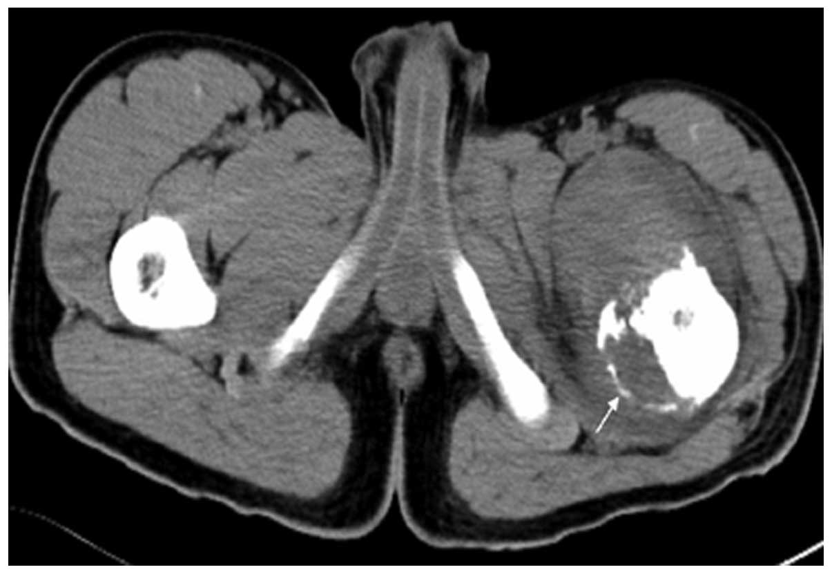
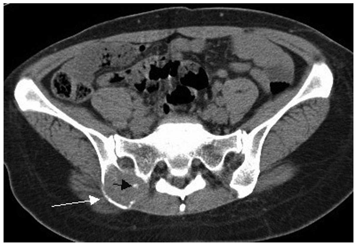
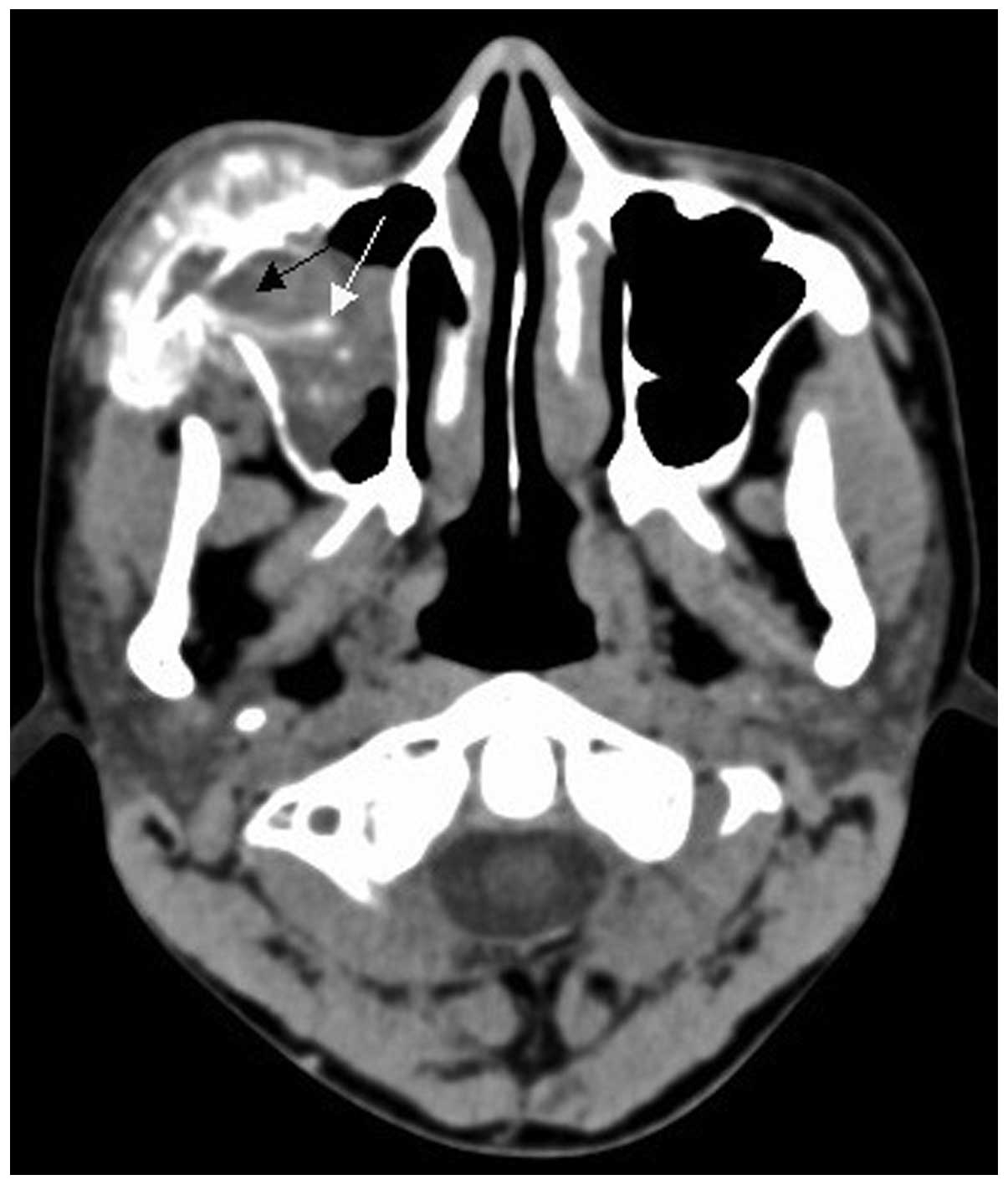
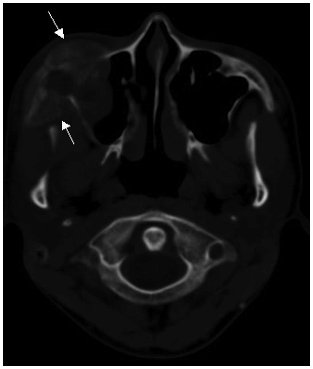
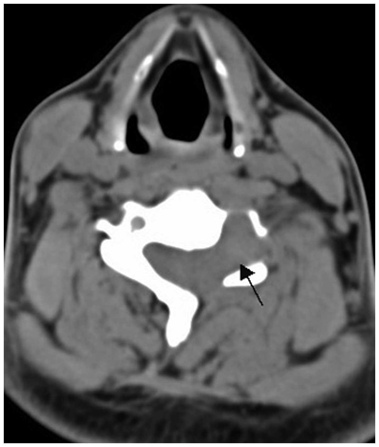
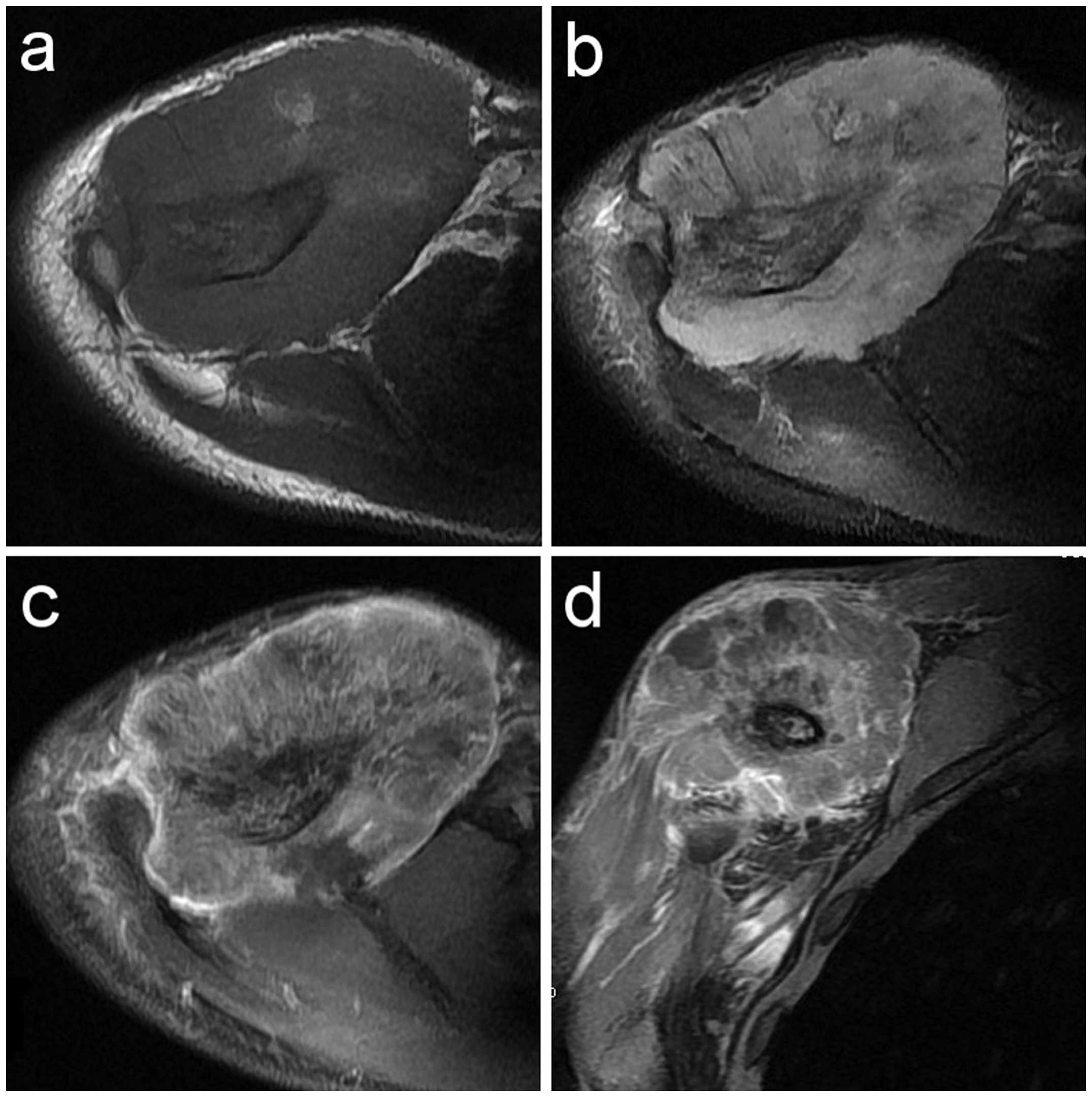
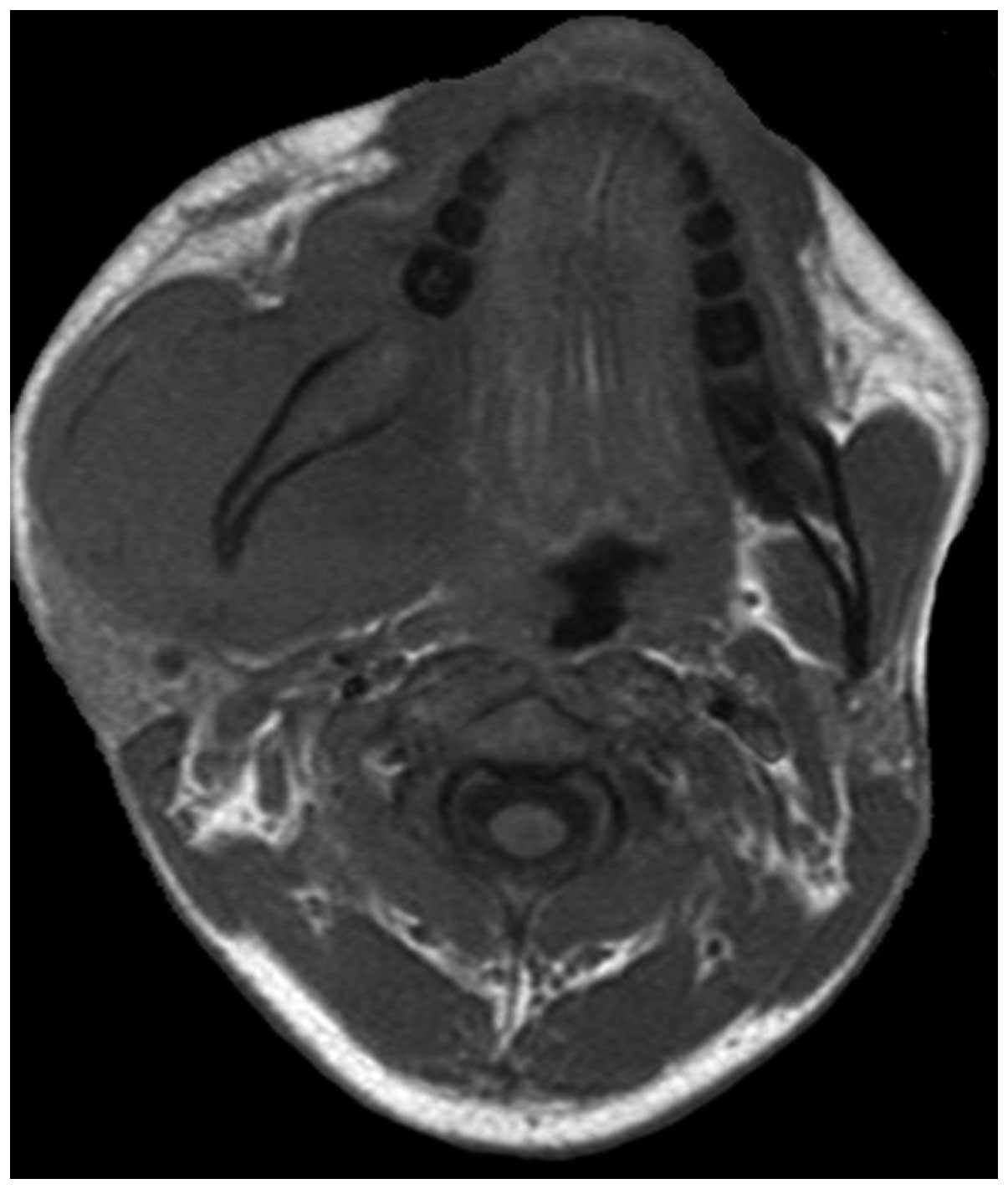
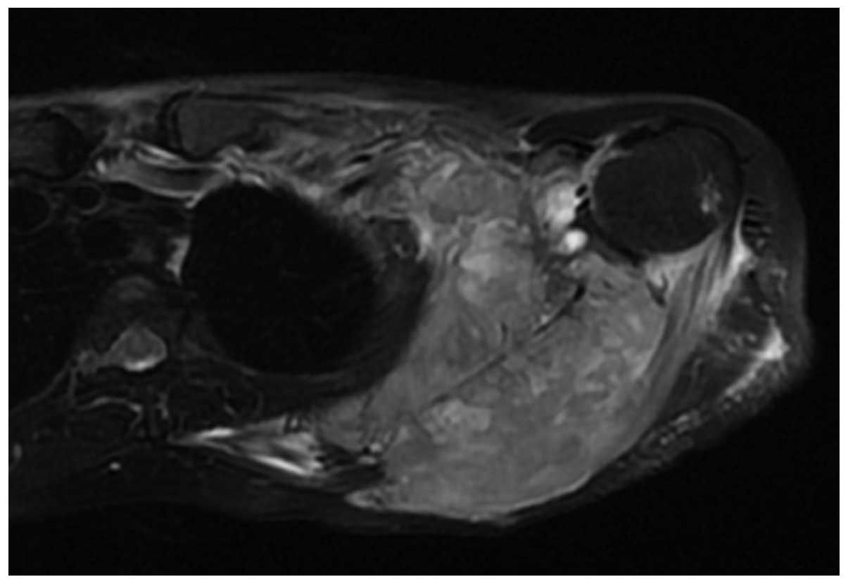
Peripheral primitive neuroectodermal tumor (pPNET) is a rare and highly malignant undifferentiated type of tumor. The aim of the present study was to analyze the computed tomography (CT), magnetic resonance imaging (MRI) and clinical findings of osseous pPNET. The present study retrospectively analyzed the clinical data and CT findings from 15 patients with osseous pPNET; the MRI findings from 11 of these 15 patients were confirmed by histopathological examination. The 15 patients included 9 men and 6 women. The mean patient age was 29 years (range, 16‑64 years) and 11 cases were aged <30 years. A CT scan was performed in 15 cases and the findings included a lytic lesion (13 cases), a lytic lesion with irregular sclerosis and dilation (2 cases), a soft tissue mass (15 cases), calcification (2 cases) and periosteal reaction (5 cases). A total of 9 cases of soft tissue mass were heterogeneous, with different sizes of lower‑density necrotic areas. An enhanced MRI scan was performed in 11 cases. On T1‑weighted images (WI), the soft tissue mass was isointense (8 cases) and marginally hyperintense (3 cases). On T2WI, aggressive soft tissue masses were heterogeneous iso‑ or hyperintense (11 cases). On contrast‑enhanced T1WI, marked heterogeneous enhancement was present in 10 cases and intermediate heterogeneous enhancement in 1 case. The results indicated that osseous pPNET mainly affects male adolescents and young adults. The CT findings of osseous pPNET were destructive lesions with a soft tissue mass and, occasionally, with periosteal reaction. The tumor was often isodense, with patchy hypodense areas. Tumor calcification was uncommon. The MRI findings were those of an aggressive soft tissue mass exhibiting isointensity on T1WI and iso‑ or hyperintensity on T2WI, with marked heterogeneous enhancement. Although the imaging characteristics of pPNETs may be non‑specific, CT and MRI may be useful in delineating the extent of the tumor, identifying distant metastases, predicting resectability and monitoring treatment.
View Figures |
Figure 1
|
|
Figure 2
|
|
Figure 3
|
|
Figure 4
|
|
Figure 5
|
|
Figure 6
|
|
Figure 7
|
|
Figure 8
|
|
Figure 9
|
|
Figure 10
|
|
Figure 11
|
View References
1
|
Schulman H, Newman-Heinman N, Kurtzbart E,
Maor E, Zirkin H and Laufer L: Thoracoabdominal peripheral
primitive neuroectodermal tumors in childhood: radiological
features. Eur Radiol. 10:1649–1652. 2000. View Article : Google Scholar : PubMed/NCBI
|
2
|
Gong J, Zhang Y, Zuo M, et al: Imaging
findings of abdominal peripheral primitive neuroectodermal tumor:
report of four cases with pathological correlation. Clin Imaging.
33:196–199. 2009. View Article : Google Scholar : PubMed/NCBI
|
3
|
Ibarburen C, Haberman JJ and Zerhouni EA:
Peripheral primitive neuroectodermal tumors. CT and MRI evaluation.
Eur J Radiol. 21:225–232. 1996. View Article : Google Scholar : PubMed/NCBI
|
4
|
Stout AP: A tumor of the ulnar nerve. Proc
NY Pathol Soc. 12:2–12. 1918.
|
5
|
Ewing J: Diffuse endothelioma of bone.
Proc NY Pathol Soc. 21:17–24. 1921.
|
6
|
Jaffe R: The neuroectodermal tumor of
bone. Am J Surg Pathol. 8:885–898. 1984. View Article : Google Scholar : PubMed/NCBI
|
7
|
Linnoila RI, Tsokos M, Triche TJ, Marangos
PJ and Chandra RS: Evidence for neural origin and PAS-positive
variants of the malignant small cell tumor of thoracopulmonary
region (‘Askin tumor’). Am J Surg Pathol. 10:124–133. 1986.
View Article : Google Scholar : PubMed/NCBI
|
8
|
Carvajal R and Meyers P: Ewing's sarcoma
and primitive neuroectodermal family of tumors. Hematol Oncol Clin
North Am. 19:501–525. 2005. View Article : Google Scholar : PubMed/NCBI
|
9
|
de Alava E and Gerald WL: Molecular
biology of the Ewing's sarcoma/primitive neuroectodermal tumor
family. J Clin Oncol. 18:204–213. 2000.PubMed/NCBI
|
10
|
Jones JE and McGill T: Peripheral
primitive neuroectodermal tumors of the head and neck. Arch
Otolaryngol Head Neck Surg. 121:1392–1395. 1995. View Article : Google Scholar : PubMed/NCBI
|
11
|
Dick EA, McHugh K, Kimber C and Michalski
A: Imaging of non-central nervous system primitive neuroectodermal
tumours: diagnostic features and correlation with outcome. Clin
Radiol. 56:206–215. 2001. View Article : Google Scholar : PubMed/NCBI
|
12
|
Khong PL, Chan GC, Shek TW, Tam PK and
Chan FL: Imaging of peripheral PNET: common and uncommon locations.
Clin Radiol. 57:272–277. 2002. View Article : Google Scholar : PubMed/NCBI
|
13
|
Yeh CH, Yeow KM, Chu SY, et al: Imaging
findings in mandibular primitive neuroectodermal tumour: a report
of a rare case and review of the literature. Dentomaxillofac
Radiol. 40:451–456. 2011. View Article : Google Scholar : PubMed/NCBI
|
14
|
Hormozi AK, Ghazisaidi MR and Hosseini SN:
Unusual presentation of peripheral primitive neuroectodermal tumor
of the maxilla. J Craniofac Surg. 21:1761–1763. 2010. View Article : Google Scholar : PubMed/NCBI
|
15
|
Zhang WD, Chen YF, Li CX, Zhang L, Xu ZB
and Zhang FJ: Computed tomography and magnetic resonance imaging
findings of peripheral primitive neuroectodermal tumors of the head
and neck. Eur J Radiol. 80:607–611. 2011. View Article : Google Scholar : PubMed/NCBI
|
16
|
Mulligan ME and Kransdorf MJ: Sequestra in
primary lymphoma of bone: prevalence and radiologic features. AJR
Am J Roentgenol. 160:1245–1248. 1993. View Article : Google Scholar : PubMed/NCBI
|