Upregulation of the MCL‑1S protein variant following dihydroartemisinin treatment induces apoptosis in cholangiocarcinoma cells
- Authors:
- Published online on: September 29, 2015 https://doi.org/10.3892/ol.2015.3760
- Pages: 3545-3550
Metrics: Total
Views: 0 (Spandidos Publications: | PMC Statistics: )
Total PDF Downloads: 0 (Spandidos Publications: | PMC Statistics: )
Abstract
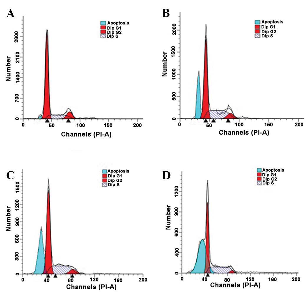
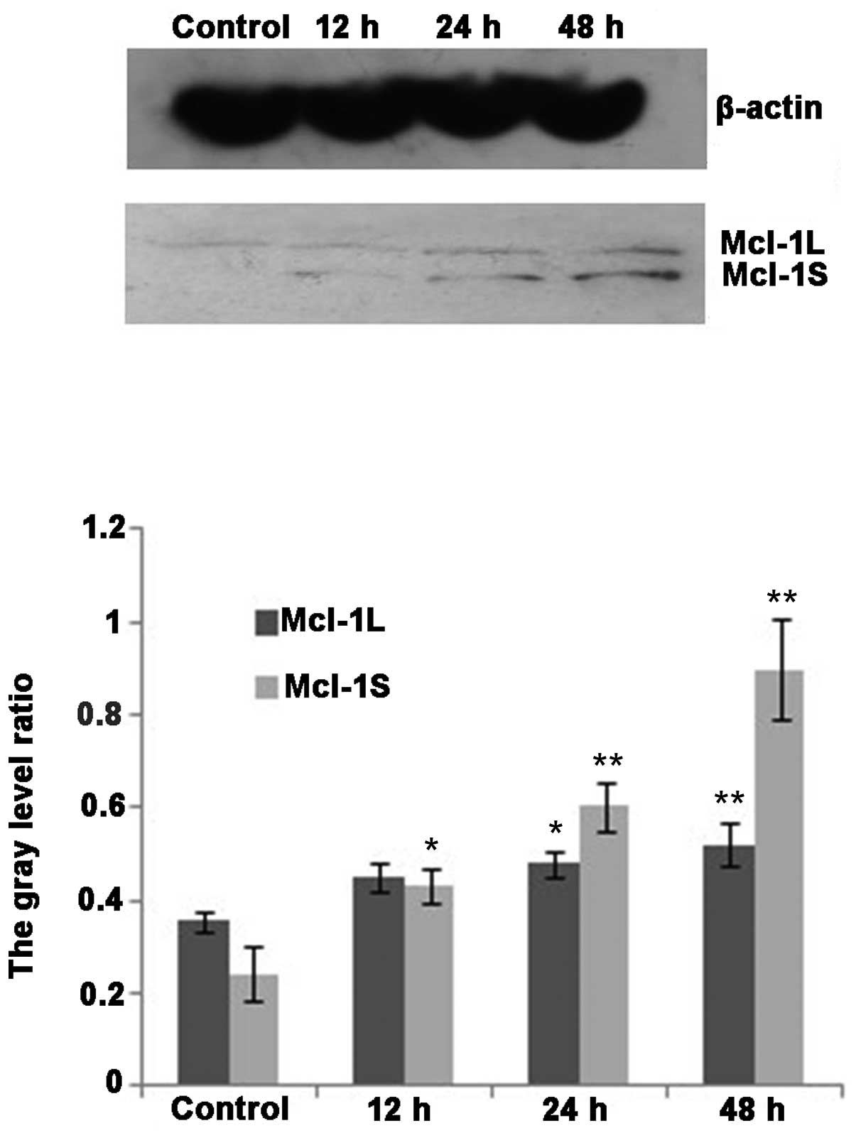
The aim of the present study was to determine whether dihydroartemisinin (DHA) induces apoptosis in the human cholangiocarcinoma QBC939 cell line through the regulation of myeloid cell leukemia‑1 (MCL‑1) expression. The inhibitory rates of DHA on QBC939 cell proliferation and the effects of DHA on the cell death rates at various DHA concentrations and following various treatment times were examined. The rate of apoptosis and cell cycle changes following DHA treatment were examined and the changes in the expression of MCL‑1 mRNAs and MCL‑1 proteins following DHA treatment were also examined. The MTT assay and trypan blue staining demonstrated that DHA significantly inhibited the proliferation (P<0.05) and promoted the death of QBC939 cells (P<0.05). The DNA ladder assay and flow cytometry (FCM) analysis demonstrated that the rate of apoptosis in the experimental group was significantly increased following DHA treatment (P<0.01). FCM analysis also demonstrated that DHA treatment led to a reduction in the percentage of QBC939 cells in the G0/G1 and G2/M phases, and the majority of the DNA‑treated cells were arrested in the S phase of the cell cycle (P<0.01). Western blot analysis demonstrated that DHA treatment significantly upregulated the expression of the pro‑apoptotic MCL‑1S protein. In contrast, no significant difference in the expression of the anti‑apoptotic MCL‑1L protein was observed following DHA treatment. DHA affected the expression of the apoptosis‑associated protein MCL‑1 through multiple mechanisms. DHA treatment increased the ratio of MCL‑1S/MCL‑1L protein, thus inducing apoptosis in cholangiocarcinoma cells.