Magnetic resonance imaging and computed tomography features of alveolar soft-part sarcoma in the right deltoid muscle: A case report
- Authors:
- Published online on: March 1, 2016 https://doi.org/10.3892/ol.2016.4290
- Pages: 2857-2860
Metrics: Total
Views: 0 (Spandidos Publications: | PMC Statistics: )
Total PDF Downloads: 0 (Spandidos Publications: | PMC Statistics: )
Abstract
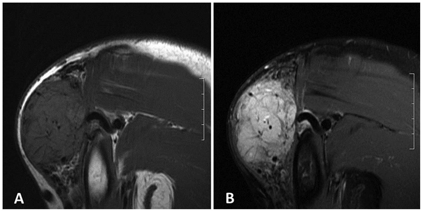
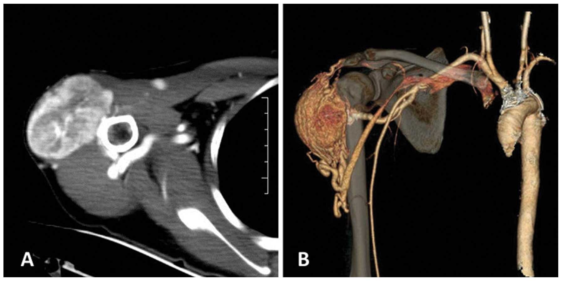
Alveolar soft-part sarcoma (ASPS) is a rare, highly vascular malignant soft-tissue tumor that predominantly affects young adults. The tumor can occur in any bodily region, but is most frequently observed in the lower deep soft tissues of the extremities, and is rarely observed in the upper extremities. The present study reported a case of ASPS of the right deltoid muscle. The patient presented with a 3‑year history of a mass on the right shoulder that exhibited rapid growth in the month prior to diagnosis. Computed tomography (CT) and magnetic resonance imaging (MRI) were performed. The lesion mainly demonstrated isointense or mildly hyperintense signals compared with the muscle on the T1‑weighted images of the MRI, and heterogeneous high signal intensity on the T2-weighted images. CT enhancement showed a homogeneous enhanced mass. The tumor was resected and submitted for histopathological examination. The diagnosis was verified as ASPS by microscopic examination and immunohistochemical analysis. No distant metastases were noted. No evidence of local tumor recurrence was seen at 6 weeks following the wide surgical excision. The CT scan revealed no metastatic nodules in either lung during the follow‑up. ASPS should be considered as a possible diagnosis when a slow‑growing, large mass is detected in young adults in the soft tissue of the extremities, with high signal intensity and numerous signal voids on T1-weighted images (T1WI) and T2WI, and strong contrast-enhancement.