Detection value of tumor cells in cerebrospinal fluid in the diagnosis of meningeal metastasis from lung cancer by immuno-FISH technology
- Authors:
- Published online on: October 25, 2016 https://doi.org/10.3892/ol.2016.5314
- Pages: 5080-5084
-
Copyright: © Lv et al. This is an open access article distributed under the terms of Creative Commons Attribution License.
Metrics: Total
Views: 0 (Spandidos Publications: | PMC Statistics: )
Total PDF Downloads: 0 (Spandidos Publications: | PMC Statistics: )
Abstract
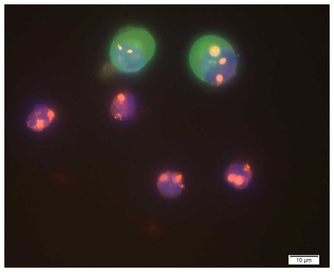
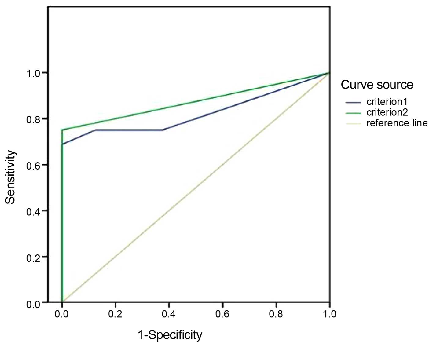
To investigate the detection value of tumor cells in cerebrospinal fluid (CSF) in the adjuvant diagnosis of meningeal metastasis from lung cancer by immunofluorescence in situ hybridization (immuno‑FISH) technology. The circulating tumor cells (CTCs) in the CSF of 16 patients with meningeal metastasis from lung cancer and 8 with non‑tumor diseases in the brain were detected using immuno‑FISH technology. The diagnosis of meningeal metastasis from lung cancer was based on neurological symptoms, enhanced magnetic resonance imaging (MRI) scans and CSF cytological examination. The number of CTCs in the patients with meningeal metastasis from lung cancer was significantly higher than those with non‑tumor diseases in the brain (P<0.01). The critical point of the maximum correct diagnostic index (Youden index) was regarded as the judging criterion for positive tumor cells in CSF according to the receiver operating characteristic curve. When there was one tumor cell in 7.5 ml CSF, the area under curve was 0.875 (95% confidence interval, 0.705~1.000). The diagnostic sensitivity, specificity, effectiveness, positive and negative predictive values were 75.0, 100.0, 83.3, 100.0 and 66.7%, respectively. There may be great clinical value in the detection of CTCs in CSF for the diagnosis of meningeal metastasis from lung cancer by immuno-FISH technology.