Milia may originate from the outermost layers of the hair bulge of the outer root sheath: A case report
- Authors:
- Ichiro Kurokawa
- Ayako Kakuno
- Airo Tsubura
View Affiliations
Affiliations: Department of Dermatology, Acne Clinical Research Center, Meiwa Hospital, Nishinomiya, Hyōgo 663‑8186, Japan, Department of Pathology, Meiwa Hospital, Nishinomiya, Hyōgo 663‑8186, Japan, Department of Pathology, Kansai Medical University, Moriguchi, Osaka 570‑8506, Japan
- Published online on: November 2, 2016 https://doi.org/10.3892/ol.2016.5335
-
Pages:
5190-5192
Metrics: Total
Views: 0 (Spandidos Publications: | PMC Statistics: )
Metrics: Total PDF Downloads: 0 (Spandidos Publications: | PMC Statistics: )
This article is mentioned in:
Abstract
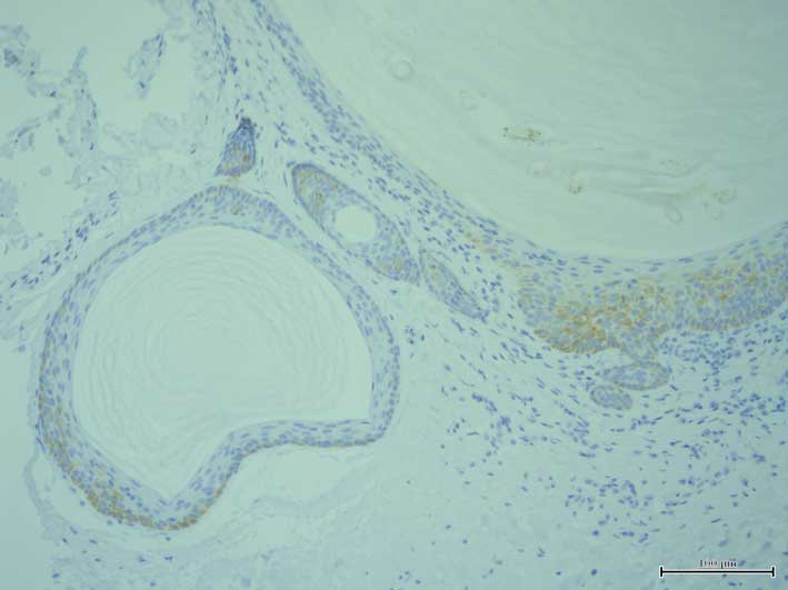
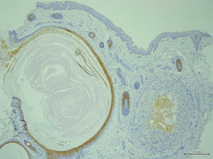
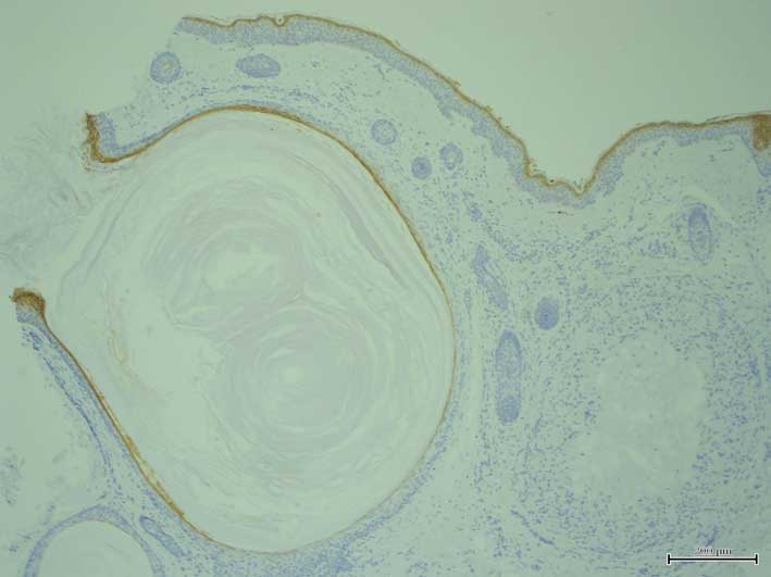
It has been hypothesized that milia originate from the hair bulge of the outer root sheath. To elucidate the histogenesis of milia, an immunohistochemical study was performed using anti‑keratin and anti‑filaggrin antibodies to determine the levels of keratin and filaggrin expression. Keratin expression was evaluated using anti‑keratin antibodies against K1, K7, K8, K10, K14, K15, K16, K17, K18, K19 and K20. K1 and K10 expression were detected in the suprabasal layers of the more superficial section of the cyst walls, but not in the deeper section of the cyst walls. However, K14 and K17 were expressed in all layers of the cyst walls. Notably, K15 was expressed in the outermost layer of the deeper section of the cyst walls and hair germ structure, whereas filaggrin was expressed in the superficial layer of the more superficial section of the cyst walls. Therefore, the pattern of keratin and filaggrin expression indicates that milia may originate from the outermost cells of the hair bulge of the outer root sheath.
View Figures |
Figure 1
|
|
Figure 2
|
|
Figure 3
|
|
Figure 4
|
|
Figure 5
|
|
Figure 6
|
View References
1
|
Berk DR and Bayliss SJ: Milia: A review
and classification. J Am Acad Dermatol. 59:1050–1063. 2008.
View Article : Google Scholar : PubMed/NCBI
|
2
|
Epstein W and Kligman AM: The pathogenesis
of milia and benign tumors of the skin. J Invest Dermatol. 26:1–11.
1956. View Article : Google Scholar : PubMed/NCBI
|
3
|
Broekaert D, Leigh IM, Lane EB, Van Muijen
GN, Ramaekers FC, De Bersaques J and Coucke P: An
immunohistochemical and histochemical study of cytokeratin,
involucrin and transglutaminase in seborrhoeic keratosis. Arch
Dermatol Res. 285:482–490. 1993. View Article : Google Scholar : PubMed/NCBI
|
4
|
van der Putte SC: The pathogenesis of
familial multiple cylindromas, trichoepitheliomas, milia, and
spiradenomas. Am J Dermatopathol. 17:271–280. 1995. View Article : Google Scholar : PubMed/NCBI
|
5
|
Kurokawa I, Takahashi K, Moll I and Moll
R: Expression of keratins in cutaneous epithelial tumors and
related disorders-distribution and clinical significance. Exp
Dermatol. 20:217–228. 2011. View Article : Google Scholar : PubMed/NCBI
|
6
|
Kanitakis J, Bourchany D, Faure M and
Claudy A: Expression of the hair stem cell-specific keratin 15 in
pilar tumors of the skin. Eur J Dermatol. 9:363–365.
1999.PubMed/NCBI
|