Differentiation between gastrointestinal schwannomas and gastrointestinal stromal tumors by computed tomography
- Authors:
- Ming‑Yan He
- Rong Zhang
- Zhenpeng Peng
- Yin Li
- Ling Xu
- Mengjie Jiang
- Zi‑Ping Li
- Shi‑Ting Feng
View Affiliations
Affiliations: Department of Radiology, The First Affiliated Hospital, Sun Yat‑Sen University, Guangzhou, Guangdong 510080, P.R. China, Department of Radiology, The Tumor Hospital, Sun Yat‑Sen University, Guangzhou, Guangdong 510080, P.R. China, Department of Gastrointestinal Surgery, The First Affiliated Hospital, Sun Yat‑Sen University, Guangzhou, Guangdong 510080, P.R. China, Faculty of Medicine and Dentistry, University of Western Australia, Perth, WA 6009, Australia
- Published online on: March 29, 2017 https://doi.org/10.3892/ol.2017.5955
-
Pages:
3746-3752
Metrics: Total
Views: 0 (Spandidos Publications: | PMC Statistics: )
Metrics: Total PDF Downloads: 0 (Spandidos Publications: | PMC Statistics: )
This article is mentioned in:
Abstract
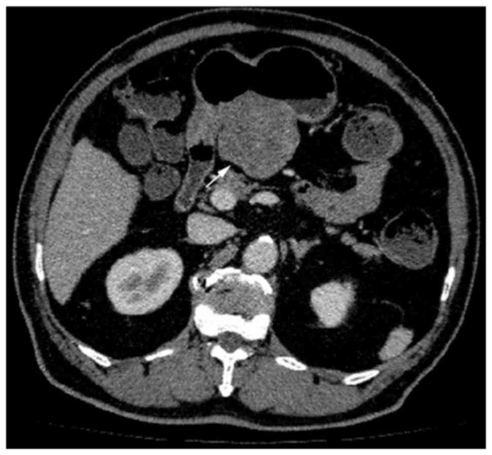
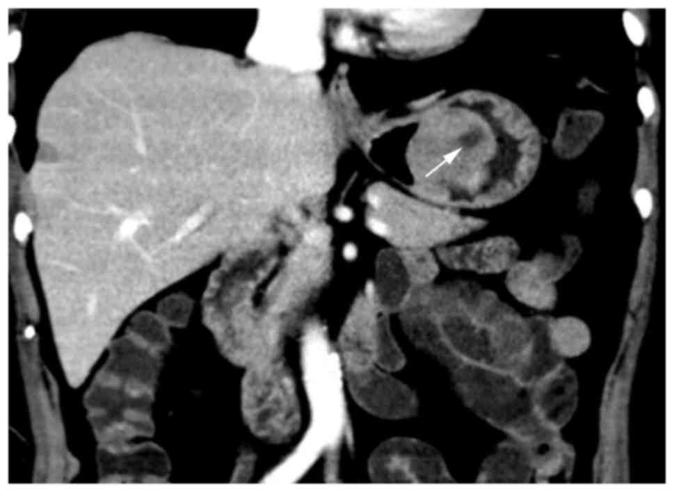
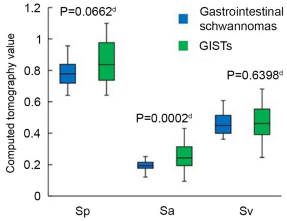
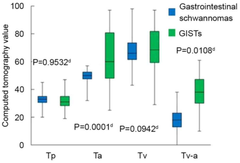
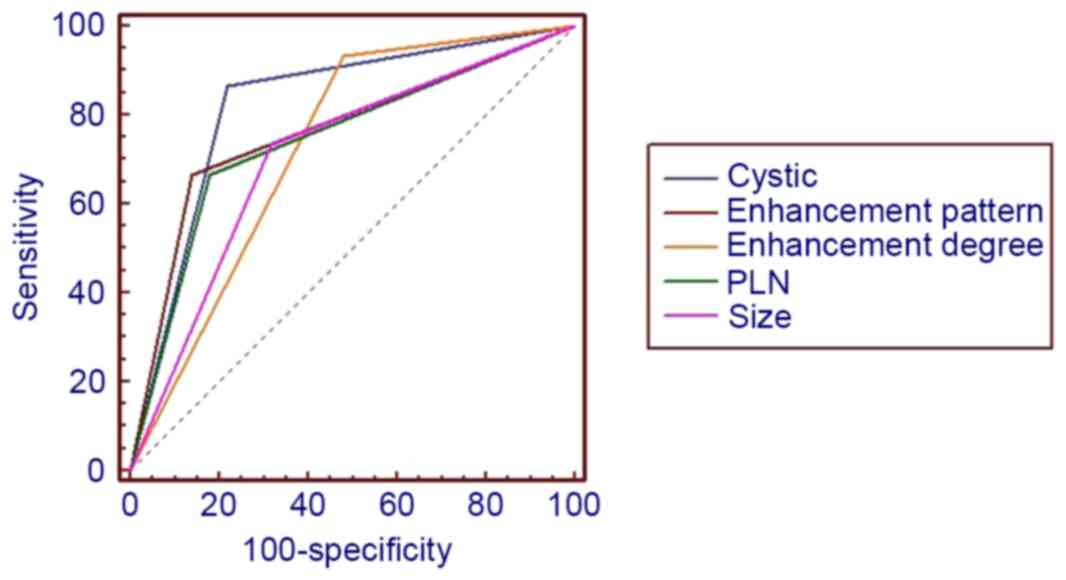
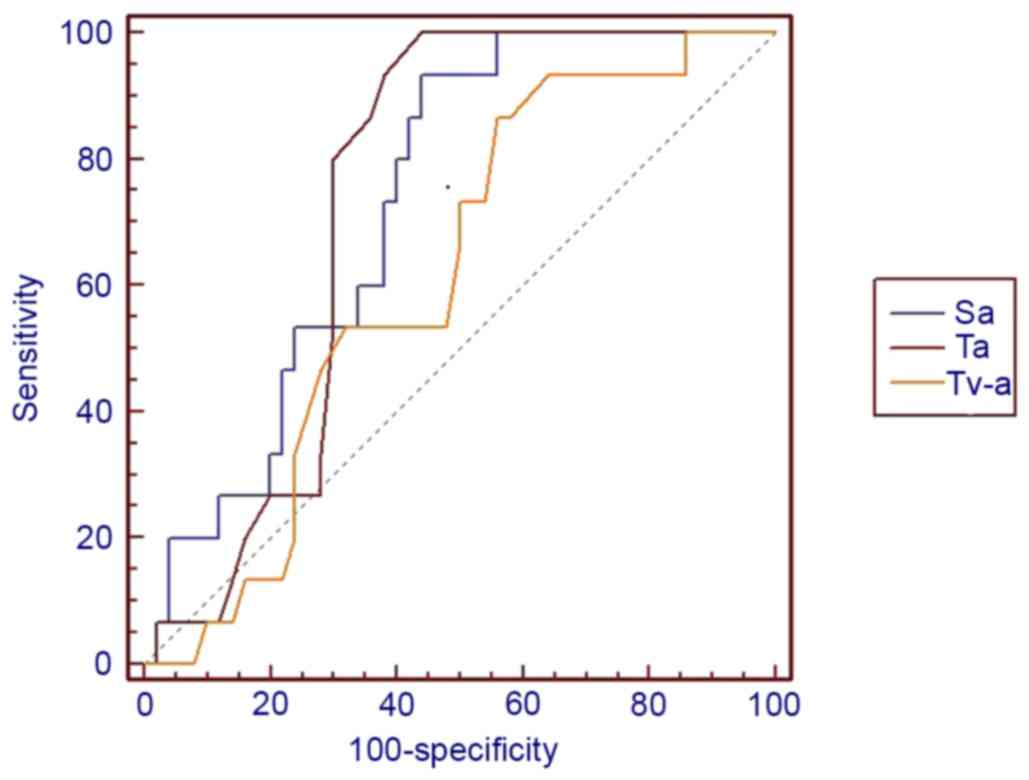
The aim of the present study was to identify computed tomography (CT) features to assist in differentiating gastrointestinal schwannomas from gastrointestinal stromal tumors (GISTs). CT images of gastrointestinal schwannomas (n=15) and GISTs (n=50) were analyzed. The absolute CT values of tumor/aorta during plain scan/arterial phase/venous phase were recorded as tumor plain scan (Tp)/aorta plain scan (Ap), tumor arterial phase (Ta)/aorta arterial phase (Aa) and tumor venous phase (Tv)/aorta venous phase (Av), respectively, and normalized CT values of the three phases were calculated as Sp=Tp/Ap, Sa=Ta/Aa and Sv=Tv/Av, respectively. The difference in tumor CT value between arterial and venous phases was calculated and recorded as Tv‑a. CT data including tumor size, contour, margin, growth pattern, presence of calcification, cystic change, hemorrhage, ulceration, perilesional lymph nodes (PLNs), local invasion to surrounding structures, metastasis, ascites, vasculatures, enhancement pattern/degree, Tp/Ta/Tv and Sp/Sa/Sv were evaluated for each patient. Receiver operating characteristic (ROC) curve analysis was used to assess the ability of the CT data to differentiate gastrointestinal schwannomas from GISTs. Compared with GISTs, gastrointestinal schwannomas more frequently demonstrated round contouring, relatively smaller tumor size, a homogeneous enhancement pattern, with the presence of PLNs and a higher level of vasculature (P<0.05), whilst the presence of cystic changes were more common in GISTs compared with gastrointestinal schwannomas (P<0.05). The Sa, Ta and Tv‑a of gastrointestinal schwannomas were less compared with those of GISTs (P<0.05). The difference in margin, growth pattern, intra‑tumoral calcifications and hemorrhage were insignificant (P>0.05). ROC analysis indicated that tumor size, cystic change, the presence of PLNs, tumor enhancement pattern and Sa demonstrated improved diagnostic potential compared with others [area under the curve (AUC) >0.7], amongst which cystic change demonstrated the best diagnostic ability (AUC=0.82). Size exhibited the highest sensitivity, 90%, and cystic change, Sa exhibited the best specificity, 87%. Quantitative analysis indicated that certain features aided the differentiation between gastrointestinal schwannomas and GISTs using CT imaging.
View Figures |
Figure 1
|
|
Figure 2
|
|
Figure 3
|
|
Figure 4
|
|
Figure 5
|
|
Figure 6
|
|
Figure 7
|
|
Figure 8
|
View References
1
|
Daimaru Y, Kido H, Hashimoto H and Enjoji
M: Benign schwannoma of the gastrointestinal tract: A
clinicopathologic and immunohistochemical study. Hum Pathol.
19:257–264. 1988. View Article : Google Scholar : PubMed/NCBI
|
2
|
Inagawa S, Hori M, Shimazaki J, Matsumoto
S, Ishii H, Itabashi M, Adachi S, Kawamoto T and Fukao K: Solitary
schwannoma of the colon: Report of two cases. Surg Today.
31:833–838. 2001. View Article : Google Scholar : PubMed/NCBI
|
3
|
Matsuki A, Kosugi S, Kanda T, Komukai S,
Ohashi M, Umezu H, Mashima Y, Suzuki T and Hatakeyama K: Schwannoma
of the esophagus: A case exhibiting high 18F-fluorodeoxyglucose
uptake in positron emission tomography imaging. Dis Esophagus.
22:E6–E10. 2009. View Article : Google Scholar : PubMed/NCBI
|
4
|
Melvin WS and Wilkinson MG: Gastric
schwannoma. Clinical and pathologic considerations. Am Surg.
59:293–296. 1993.PubMed/NCBI
|
5
|
Miettinen M, Sarlomo-Rikala M and Lassota
J: gastrointestinal stromal tumors: Recent advances in
understanding of their biology. Hum Pathol. 30:1212–1220. 1999.
View Article : Google Scholar
|
6
|
Hong HS, Ha HK, Won HJ, Byun JH, Shin YM,
Kim AY, Kim PN, Lee MG, Lee GH and Kim MJ: Gastric schwannomas:
Radiological features with endoscopic and pathological correlation.
Clin Radiol. 63:536–542. 2008. View Article : Google Scholar : PubMed/NCBI
|
7
|
Levy AD, Remotti HE, Thompson WM, Sobin LH
and Miettinen M: Gastrointestinal stromal tumors: Radiologic
features with pathologic correlation. Radiographics. 23:283–304,
456; quiz 532. 2003. View Article : Google Scholar : PubMed/NCBI
|
8
|
Loffeld RJ, Balk TG, Oomen JL and van der
Putten AB: Upper gastrointestinal bleeding due to a malignant
Schwannoma of the stomach. Eur J Gastroenterol Hepatol. 10:159–162.
1998. View Article : Google Scholar : PubMed/NCBI
|
9
|
Watanabe A, Ojima H, Suzuki S, Mochida Y,
Hirayama I, Hosouchi Y, Nishida Y, Kashiwabara K, Ohno T, Mochiki E
and Kuwano H: An individual with gastric schwannoma with
pathologically malignant potential surviving two years after
laparoscopy-assisted partial gastrectomy. Case Rep Gastroenterol.
5:502–507. 2011. View Article : Google Scholar : PubMed/NCBI
|
10
|
Zuo HD, Zhang XM and Zhai ZH: Nerve Sheath
tumor of stomach: Two Cases Report. J Med Cases. 3:68–72. 2012.
|
11
|
Ludwig DJ and Traverso LW: Gut stromal
tumors and their clinical behavior. Am J Surg. 173:390–394. 1997.
View Article : Google Scholar : PubMed/NCBI
|
12
|
Janowitz P, Meier F and Reisig J: Gastric
schwannoma as a rare differential diagnosis of pleural effusion. Z
Gastroenterol. 40:925–928. 2002. View Article : Google Scholar : PubMed/NCBI
|
13
|
Choi JW, Choi D, Kim KM, Sohn TS, Lee JH,
Kim HJ and Lee SJ: Small submucosal tumors of the stomach:
Differentiation of gastric schwannoma from gastrointestinal stromal
tumor with CT. Korean J Radiol. 13:425–433. 2012. View Article : Google Scholar : PubMed/NCBI
|
14
|
Choi YR, Kim SH, Kim SA, Shin CI, Kim HJ,
Kim SH, Han JK and Choi BI: Differentiation of large (≥5 cm)
gastrointestinal stromal tumors from benign subepithelial tumors in
the stomach: Radiologists' performance using CT. Eur J Radiol.
83:250–260. 2014. View Article : Google Scholar : PubMed/NCBI
|
15
|
Hou YY, Tan YS, Xu JF, Wang XN, Lu SH, Ji
Y, Wang J and Zhu XZ: Schwannoma of the gastrointestinal tract: A
clinicopathological, immunohistochemical and ultrastructural study
of 33 cases. Histopathology. 48:536–545. 2006. View Article : Google Scholar : PubMed/NCBI
|
16
|
Atmatzidis S, Chatzimavroudis G, Dragoumis
D, Tsiaousis P, Patsas A and Atmatzidis K: Gastric schwannoma: A
case report and literature review. Hippokratia. 16:280–282.
2012.PubMed/NCBI
|
17
|
Prévot S, Bienvenu L, Vaillant JC and de
Saint-Maur PP: Benign schwannoma of the digestive tract: A
clinicopathologic and immunohistochemical study of five cases,
including a case of esophageal tumor. Am J Surg Pathol. 23:431–436.
1999. View Article : Google Scholar : PubMed/NCBI
|