Introduction
As one of the more common malignant bone tumor,
osteosarcoma has high degree of malignancy, and relatively poor
prognosis (1). The pathogenesis and
development process has not yet been studied fully, and the current
clinical diagnosis of osteosarcoma lacks more specific indicators
(2). PTEN as a tumor suppressor gene,
is associated with the occurrence and development of a variety of
malignant tumors (3,4). Overexpression of NF-κB is associated
physiologically and pathologically with many tumors, and is a
bi-directional regulatory factor (5–7). There are
few studies on the expression and correlation of PTEN and NF-κB in
osteosarcoma to date. The purpose of this study is to use
immunohistochemical method to detect the expression of PTEN and
NF-κB in tumor tissues, explore their expression changes at genes
and protein levels, and analyze statistically the expression of
both and the prognosis of patients, to explore their expression
correlation in osteosarcoma and the potential use in clinical
diagnosis of osteosarcoma.
Materials and methods
Materials
Experimental materials
Pathologically confirmed osteosarcoma tumor tissues
(73 cases) and adjacent tissues (73 cases) were selected. All
patients signed an informed consent form. The osteosarcoma tissue
and adjacent tissues were treated with paraffin embedding, and then
paraffin sectioned with a thickness of ~4 µm. The patients with
osteosarcoma were follow-up for 5-years.
Reagents
Rabbit antihuman nuclear factor-κB (NF-κB)
monoclonal antibody (Beijing Dingguo Changsheng Biotechnology Co.,
Ltd., Beijing, China) rabbit anti-human phosphatase and tensin
homolog deleted in chromosome 10 (PTEN) polyclonal antibody, rabbit
anti-human glyceraldehyde 3-phosphate dehydrogenase (GAPDH)
polyclonal antibody (cat. no. SPC-1303, SPC-1331 and SPC-689;
StressMarq Biosciences Inc., Victoria, BC, Canada), DAB coloring
reagent (Shanghai Runwell Technology Co., Ltd., Shanghai, China),
citrate buffer powder (Shanghai X-Y Biotechnology Co., Ltd.,
Shanghai, China), reverse transcription kit (GeneCopoeia,
Rockville, MD, USA), real-time fluorescence quantitative PCR and
Western Blot test kit (Shanghai Biological BestBio Bebo, Shanghai,
China), goat anti-rabbit IgG secondary polyclonal antibody (cat.
no. ab150077; Abcam, Cambridge, UK), BCA protein quantitative kit
(Nanjing SenBeiJia Biotechnology Co., Ltd., Nanjing, China).
Experimental methods
Immunohistochemical staining
The paraffin sections were subjected to dewaxing and
then washed with phosphate-buffered saline. To reduce the
nonspecific background staining caused by endogenous peroxidase,
blocking was continued for 20 min in the blocking buffer, then
blocked in 10% serum for 10 min at room temperature, and incubated
overnight at 4°C with the addition of primary antibody dilution
buffer (rabbit anti-human NF-κB monoclonal antibody and PTEN
polyclonal antibody, 1:50 dilution). It was washed with phosphate
buffer solution, adding the goat anti-rabbit IgG secondary
polyclonal antibody dilution buffer (1:50 dilution) and incubated
at room temperature for 30 min. It was washed again with phosphate
buffer, and then incubated with Streptavidin antibiotic
protein-peroxidase solution at room temperature for 30 min, washed
with phosphate buffer solution, colored with DAB, washed with
distilled water, stained and sealed.
Evaluation of results
One hundred cells were randomly selected to observe
the visual field under a light microscope, and the average number
of cells in the field of view was obtained as the positive cells
expressing the protein in the tissue. PTEN staining was mainly
located in the cytoplasm, while the positive expression of NF-κB
was mainly located in the nucleus. Depth score: scores 0 to 2, was
assigned for no coloring, weak coloring and strong coloring,
respectively. Cells stained positive score: 1 to 25% was recorded
as 1, 26 to 50% as 2, 51 to 75% as 3, 76 to 100% as 4. The scores
of the depth score and the positive rate were multiplied, 1 to 2
were negative, and 3 to 8 were positive.
Real-time PCR method to determine the expression
of NF-κB and PTEN mRNA
Total RNA was extracted from both tissues and the
concentration and purity were determined. Table I shows the primer sequence used in the
synthesis of NF-κB and PTEN gene amplification by Nanjing CoBioer
Biotechnology Co., Ltd. (Nanjing, China). Reverse transcription
synthesis of cDNA, reaction system 20 µl.
 | Table I.NF-κB and PTEN gene amplification
primer. |
Table I.
NF-κB and PTEN gene amplification
primer.
| Gene | Primer sequence |
|---|
| NF-κB | F:
5′-TACCCTGAGGCTATAACTC-3′ |
|
| R:
5′-GACACTTGATAAGGCTTTG-3′ |
| PTEN | F:
5′-AGTTCCCTCAGCCGTTACCT-3′ |
|
| R:
5′-ATTTGACGGCTCCTCTACTG-3′ |
| GAPDH | F:
5′-TGGGTGTGAACCACGAGAA-3′ |
|
| R:
5′-GGCATGGACTGTGGTCATGA-3′ |
PCR reaction system: 25 µl, reaction conditions:
95°C 10 min, 95°C 30 sec, and 59.4°C 30 sec, 40 cycles, 95°C 15
sec, and maintained at 65°C. With GAPDH as the internal control,
RT-PCR instrument automatically calculated the NF-κB and PTEN mRNA
relative expression.
Western blot assay for detection of protein
The total protein in the two tissues was extracted
according to the instructions in the tissue total protein
extraction kit. The concentration of the extracted protein was
determined by BCA protein assay, and stored at −70°C. Sodium
dodecyl sulfate-polyacrylamide gel electrophoresis (SDS-PAGE) was
performed on 10% separation gel and 5% concentrated gel. The gel
position of the two proteins was selected according to the marker
strip. After transmembrane, the PVDF membrane was washed with 1X
TBST solution for 5 min, 5% nonfat dry milk blocking at room
temperature (1:1,000 dilution) for 1 h, 1X TBST solution for 1 h,
1X TBST solution for 1 h, 1X TBST solution 5 min, total cleaning 3
times. Anti-dark drop ECL luminescent liquid, dark environment for
2 min for development. Finally, the Multi Gauge Ver. 3.0 imaging
system was scanned, and ImageJ professional image analysis software
(National Institutes of Health, Bethesda, MD, USA) was used for
image analysis and the absorbance value was recorded.
Statistical analysis
The statistical software SPSS 17.0 (Beijing Xinmei
Jiahong Technology Co., Ltd., Beijing, China) was used in this
study for data analysis. The correlation between the expression of
NF-κB and PTEN was tested by Spearmans method, using χ2
to test the difference of positive rates between arms. The survival
analysis was analyzed by GraphPad Prism5 software (GraphPad
Software Inc., La Jolla, CA, USA). The difference was considered
statistically significant when p<0.05.
Results
The expression of PTEN and NF-κB in
osteosarcoma tissues and adjacent tissues
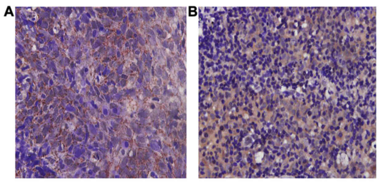
The immunohistochemical staining showed that PTEN
had more positive expression granules in osteosarcoma tissues,
while in adjacent tissues, PTEN showed diffuse granule-like
expression with expression level significantly higher than that of
osteosarcoma tissues. The notable difference of the positive
expression rate of PTEN in osteosarcoma tissues and adjacent
tissues was statistically significant (p<0.05) (Fig. 1).
Among 73 cases of osteosarcoma tissues, 49 cases
were positive in PTEN expression, with positive rate of 67.1%;
while in adjacent tissues, 66 cases were positive in PTEN
expression, with positive rate of 90.4% (Table II).
 | Table II.PTEN expression comparison in both
tissues. |
Table II.
PTEN expression comparison in both
tissues.
|
|
| PTEN |
|
|
|
|---|
|
|
|
|
|
|
|
|---|
| Arm | No. of cases | + | − | Positive rate % | χ2 | P-value |
|---|
| Osteosarcoma | 73 | 49 | 24 | 67.1 | 31.02 | <0.05 |
| Adjacent tissues | 73 | 66 | 7 | 90.4 |
|
|
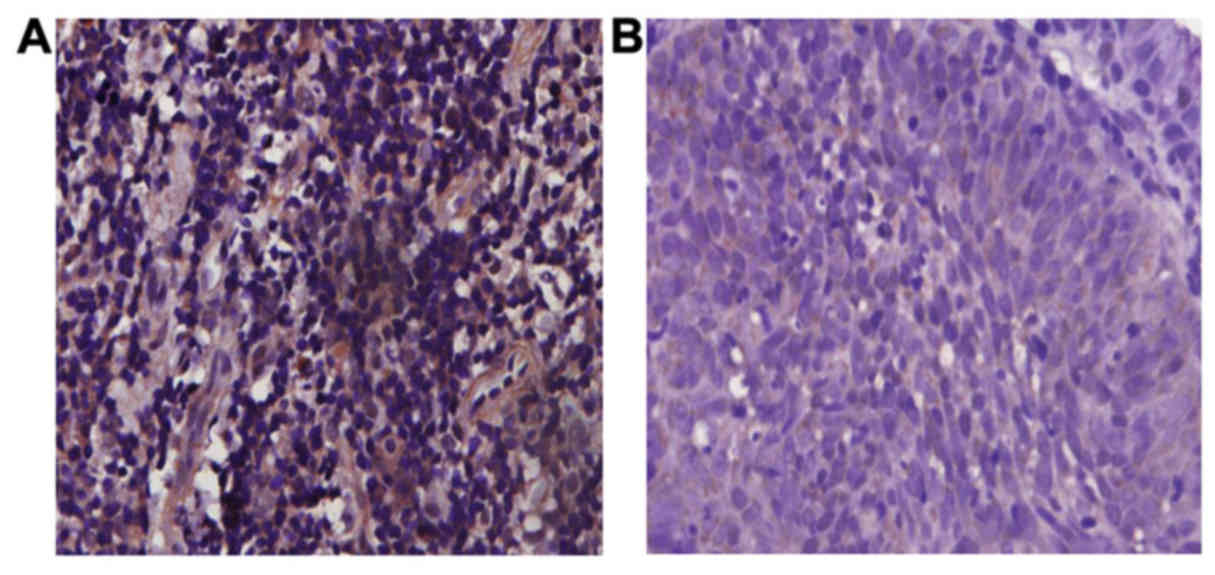
Immunohistochemical staining showed that NF-κB had a
significant positive expression of granulocyte in osteosarcoma
tissues, while NF-κB expression was not obvious in adjacent
tissues. The positive expression of NF-κB in osteosarcoma tissues
was statistically significantly higher than that in adjacent
tissues (p<0.05) (Fig. 2).
Out of 73 osteosarcoma tissues, 53 were positive in
NF-κB expression. The positive rate was 75.3%; while 24 adjacent
tissues were positive in NF-κB expression. The positive rate was
32.9% (Table III).
 | Table III.Comparison of NF-κB expression in both
tissues. |
Table III.
Comparison of NF-κB expression in both
tissues.
|
|
| NF-κB |
|
|
|
|---|
|
|
|
|
|
|
|
|---|
| Arm | No. of cases | + | − | Positive rate% | χ2 | P-value |
|---|
| Osteosarcoma | 73 | 55 | 18 | 75.3 | 40.57 | <0.01 |
| Adjacent tissues | 73 | 24 | 49 | 32.9 |
|
|
Correlative analysis of PTEN and NF-κB
expression in osteosarcoma
Out of 73 osteosarcoma tissues, 53 were positive in
NF-κB expression. Among them, 16 cases were also positive for PTEN,
at a rate of 29.1%. The statistical analysis shows a negative
correlation between NF-κB and PTEN (r=−0.502, p<0.05) (Table IV and Fig.
3).
 | Table IV.Correlation of NF-κB and PTEN
expression. |
Table IV.
Correlation of NF-κB and PTEN
expression.
|
| PTEN |
|
|
|
|---|
|
|
|
|
|
|
|---|
| NF-κB | + | − | Total | r-value | P-value |
|---|
| + | 16 | 39 | 55 | −0.502 | <0.05 |
| − | 14 | 4 | 18 |
|
|
| Total | 30 | 43 | 73 |
|
|
Detecting NF-κB and PTEN mRNA
expression by RT-PCR
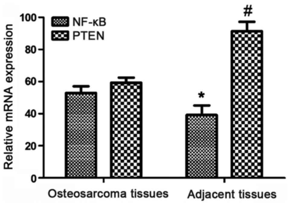
The relative expression of NF-κB mRNA in
osteosarcoma tissues (52.9±4.17) was significantly higher than that
in adjacent tissues (39.2±5.94), the difference was statistically
significant (p<0.05); and the relative expression of PTEN mRNA
(59.3±3.19) was significantly lower than that of adjacent tissues
(91.4±5.82), and the difference was statistically significant
(p<0.05) (Table V and Figs. 4 and 5).
 | Table V.The relative expression of NF-κB and
PTEN mRNA. |
Table V.
The relative expression of NF-κB and
PTEN mRNA.
| Arm | NF-κB | PTEN |
|---|
| Osteosarcoma |
52.9±4.17a |
59.3±3.19c |
| Adjacent tissues |
39.2±5.94b |
91.4±5.82d |
| P-values | <0.05 |
|
Detecting NF-κB and PTEN mRNA
expression by western blot method
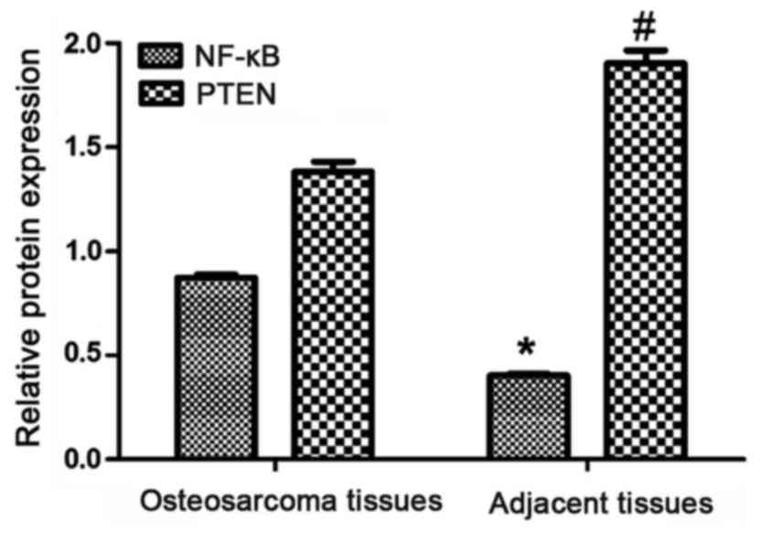
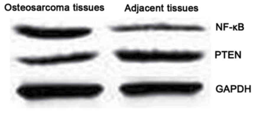
The western blot test indicates the expression of
NF-κB protein in osteosarcoma tissues was higher (0.872±0.015), and
the expression of κB in adjacent tissues was lower (0.403±0.008).
The expression of PTEN protein was lower in osteosarcoma tissues
(1.383±0.047), and higher in adjacent tissues (1.902±0.064). The
difference in expression between the two proteins was statistically
significant (p<0.05) (Table
VI).
 | Table VI.Expression of NF-κB and PTEN
protein. |
Table VI.
Expression of NF-κB and PTEN
protein.
| Arm | NF-κB | PTEN |
|---|
| Osteosarcoma |
0.872±0.015a |
1.383±0.047c |
| Adjacent tissues |
0.403±0.008b |
1.902±0.064d |
| P-values | <0.05 |
|
Prognosis of NF-κB and PTEN
expression
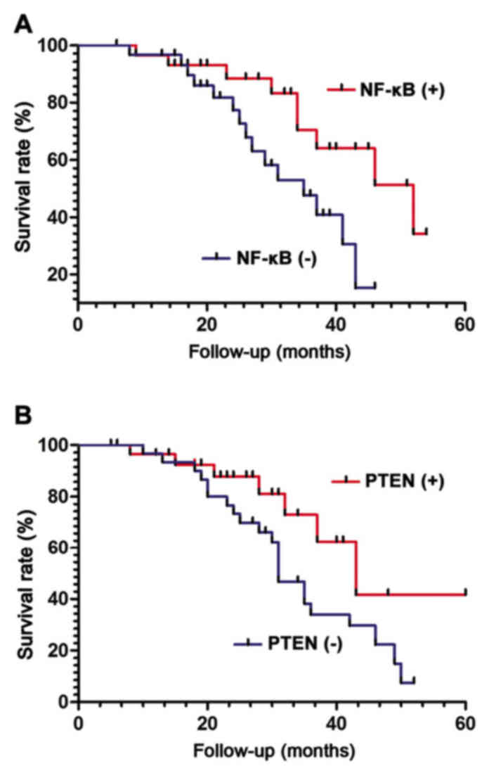
The survival rate of patients with NF-κB and PTEN
positive and negative expression was statistically analyzed.
Survival analysis using statistical software showed, the survival
rate of those with NF-κB positive expression was lower than that of
negative expression (χ2=8.014, p<0.05) (Fig. 6A). However, the 5-year survival of
patients with PTEN positive expression was significantly higher
than that of negative expression. The result was statistically
significant (χ2=7.625, p<0.05) (Fig. 6B).
Discussion
Osteosarcoma is a type of malignant tumor occurring
more frequently in adolescents. Osteosarcoma is developed from the
interstitial cell line, with strong infiltration and transfer
capacity (8–10) PTEN is a tumor suppressor gene with
phosphatase activity (11) and it has
been reported that PTEN expression is reduced in a variety of human
malignancies, such as breast cancer (12) and prostate cancer (13). PTEN has many important functions in
the body, such as the inhibition of cell growth and differentiation
(14) and promotion of cell apoptosis
(15), making it one of the most
noticeable tumor suppressor genes after the p53 gene. NF-κB is a
transcription factor with a two-way transcriptional regulation that
plays an important role in many pathophysiological processes
(16–18). Loftus et al (19) reported a significant increase in NF-κB
p65 expression in hepatocellular carcinoma. The study by Lu et
al (20) showed that the
expression of NF-κB protein in osteosarcoma negative correlates
with the apoptotic index of osteosarcoma, and significantly
inhibited the apoptosis of osteosarcoma cells. However, reports on
the correlation of NF-κB expression and PTEN in osteosarcoma are
scarce.
Immunohistochemical method was used in this study.
the study results indicated that in osteosarcoma tissues, the
positive expression rate of PTEN was significantly lower than that
in the adjacent tissues, and the positive expression rate of NF-κB
was significantly higher. The difference between the two are
statistically significant (r=−0.502, p<0.05). Therefore, we
hypothesized that the decrease of PTEN elevated the expression of
NF-κB through certain mechanism; or the increased expression of
NF-κB leads to decreased expression of PTEN through certain
mechanism. In the osteosarcoma tissues and the adjacent tissues,
the mRNA expression of NF-κB was 52.9±4.17 and 39.2±5.94,
respectively; the mRNA expression of PTEN was 59.3±3.19 and
91.4±5.82, respectively. The difference in expression of NF-κB and
PTEN mRNA was statistically significant (p<0.05). The expression
of NF-κB protein in osteosarcoma tissues was higher than that in
adjacent tissues, and the expression of PTEN protein in
osteosarcoma tissues was lower. The experimental results at both
gene and protein levels have proved the NF-κB and PTEN expression
was negatively correlated, suggesting these two molecules can act
as potential biomarkers for the clinical detection of
osteosarcoma.
The patients with osteosarcoma were follow-up for
five-years. The results showed that the 5-year survival rate was
higher in patients with NF-κB negative expression and those with
PTEN positive expression. The difference was statistically
significant. The result indicates that combining with detection of
NF-κB and PTEN expression can better assess clinically the
prognosis of patients with osteosarcoma, which provides a new
direction for the clinical treatment of osteosarcoma.
Acknowledgements
This study was supported by the Natural Science
Program of Science and Technology commission of Tianjin (no.
043609011) and the Launching fund project of Youth Doctor from
Logistics College of Chinese People's Armed Police Forces (no.
WYB201109).
References
|
1
|
Gao KT and Lian D: Long non-coding RNA
MALAT1 is an independent prognostic factor of osteosarcoma. Eur Rev
Med Pharmacol Sci. 20:3561–3565. 2016.PubMed/NCBI
|
|
2
|
Sun XZ, Liao Y and Zhou CM: NKD2 a novel
marker to study the progression of osteosarcoma development. Eur
Rev Med Pharmacol Sci. 20:2799–2804. 2016.PubMed/NCBI
|
|
3
|
Yan M, Ni J, Song D, Ding M and Huang J:
Activation of unfolded protein response protects osteosarcoma cells
from cisplatin-induced apoptosis through NF-κB pathway. Int J Clin
Exp Pathol. 8:10204–10215. 2015.PubMed/NCBI
|
|
4
|
Xia M, Tong JH, Ji NN, Duan ML, Tan YH and
Xu JG: Tramadol regulates proliferation, migration and invasion via
PTEN/PI3K/AKT signaling in lung adenocarcinoma cells. Eur Rev Med
Pharmacol Sci. 20:2573–2580. 2016.PubMed/NCBI
|
|
5
|
Ersahin A, Acet M, Acet T and Yavuz Y:
Disturbed endometrial NF-κB expression in women with recurrent
implantation failure. Eur Rev Med Pharmacol Sci. 20:5037–5040.
2016.PubMed/NCBI
|
|
6
|
Wang L, Yang L, Lu Y, Chen Y, Liu T, Peng
Y, Zhou Y, Cao Y, Bi Z, Liu T, et al: Osthole induces cell cycle
arrest and inhibits migration and invasion via PTEN/Akt pathways in
osteosarcoma. Cell Physiol Biochem. 38:2173–2182. 2016. View Article : Google Scholar : PubMed/NCBI
|
|
7
|
Ma T, Guo CJ, Zhao X, Wu L, Sun SX and Jin
QH: The effect of curcumin on NF-κB expression in rat with lumbar
intervertebral disc degeneration. Eur Rev Med Pharmacol Sci.
19:1305–1314. 2015.PubMed/NCBI
|
|
8
|
Shen L, Chen XD and Zhang YH: MicroRNA-128
promotes proliferation in osteosarcoma cells by downregulating
PTEN. Tumour Biol. 35:2069–2074. 2014. View Article : Google Scholar : PubMed/NCBI
|
|
9
|
Pang PC, Shi XY, Huang WL and Sun K:
miR-497 as a potential serum biomarker for the diagnosis and
prognosis of osteosarcoma. Eur Rev Med Pharmacol Sci. 20:3765–3769.
2016.PubMed/NCBI
|
|
10
|
Ju L, Zhou YM and Yang GS: Up-regulation
of long non-coding RNA BCAR4 predicts a poor prognosis in patients
with osteosarcoma, and promotes cell invasion and metastasis. Eur
Rev Med Pharmacol Sci. 20:4445–4451. 2016.PubMed/NCBI
|
|
11
|
Sun H, Zheng X, Wang Q, Yan J, Li D, Zhou
Y, Lin Y, Zhang L and Wang X: Concurrent blockade of NF-κB and Akt
pathways potentiates cisplatin's antitumor activity in vivo.
Anticancer Drugs. 23:1039–1046. 2012. View Article : Google Scholar : PubMed/NCBI
|
|
12
|
Mongre RK, Sodhi SS, Ghosh M, Kim JH, Kim
N, Sharma N and Jeong DK: A new paradigm to mitigate osteosarcoma
by regulation of microRNAs and suppression of the NF-κB signaling
cascade. Dev Reprod. 18:197–212. 2014. View Article : Google Scholar : PubMed/NCBI
|
|
13
|
Liu CJ, Yu KL, Liu GL and Tian DH: MiR 214
promotes osteosarcoma tumor growth and metastasis by decreasing the
expression of PTEN. Mol Med Rep. 12:6261–6266. 2015. View Article : Google Scholar : PubMed/NCBI
|
|
14
|
Kawano M, Tanaka K, Itonaga I, Ikeda S,
Iwasaki T and Tsumura H: microRNA-93 promotes cell proliferation
via targeting of PTEN in osteosarcoma cells. J Exp Clin Cancer Res.
34:762015. View Article : Google Scholar : PubMed/NCBI
|
|
15
|
Shang ZM, Tang JD, Jiang QQ, Guo A, Zhang
N, Gao ZX and Ji WS: Role of Δ133p53 in tumor necrosis
factor-induced survival of p53 functions in MKN45 gastric cancer
cell line. Eur Rev Med Pharmacol Sci. 19:2416–2422. 2015.PubMed/NCBI
|
|
16
|
Tian Z, Guo B, Yu M, Wang C, Zhang H,
Liang Q, Jiang K and Cao L: Upregulation of micro-ribonucleic
acid-128 cooperating with downregulation of PTEN confers metastatic
potential and unfavorable prognosis in patients with primary
osteosarcoma. Onco Targets Ther. 7:1601–1608. 2014.PubMed/NCBI
|
|
17
|
Hu Y, Xu S, Jin W, Yi Q and Wei W: Effect
of the PTEN gene on adhesion, invasion and metastasis of
osteosarcoma cells. Oncol Rep. 32:1741–1747. 2014. View Article : Google Scholar : PubMed/NCBI
|
|
18
|
Song D, Ni J, Xie H, Ding M and Wang J:
DNA demethylation in the PTEN gene promoter induced by
5-azacytidine activates PTEN expression in the MG-63 human
osteosarcoma cell line. Exp Ther Med. 7:1071–1076. 2014. View Article : Google Scholar : PubMed/NCBI
|
|
19
|
Loftus JP, Cavatorta D, Bushey JJ, Levine
CB, Sevier CS and Wakshlag JJ: The 5-lipoxygenase inhibitor
tepoxalin induces oxidative damage and altered PTEN status prior to
apoptosis in canine osteosarcoma cell lines. Vet Comp Oncol.
14:e17–e30. 2016. View Article : Google Scholar : PubMed/NCBI
|
|
20
|
Lu Y, Li F, Xu T and Sun J: Tetrandrine
prevents multidrug resistance in the osteosarcoma cell line, U-2OS,
by preventing Pgp overexpression through the inhibition of NF-κB
signaling. Int J Mol Med. 39:993–1000. 2017. View Article : Google Scholar : PubMed/NCBI
|


