|
1
|
Coussens LM and Werb Z: Inflammation and
cancer. Nature. 420:860–867. 2002. View Article : Google Scholar : PubMed/NCBI
|
|
2
|
Ohshima H, Tazawa H, Sylla BS and Sawa T:
Prevention of human cancer by modulation of chronic inflammatory
processes. Mutat Res. 591:110–122. 2005. View Article : Google Scholar : PubMed/NCBI
|
|
3
|
Okayasu I: Development of ulcerative
colitis and its associated colorectal neoplasia as a model of the
organ-specific chronic inflammation-carcinoma sequence. Pathol Int.
62:368–380. 2012. View Article : Google Scholar : PubMed/NCBI
|
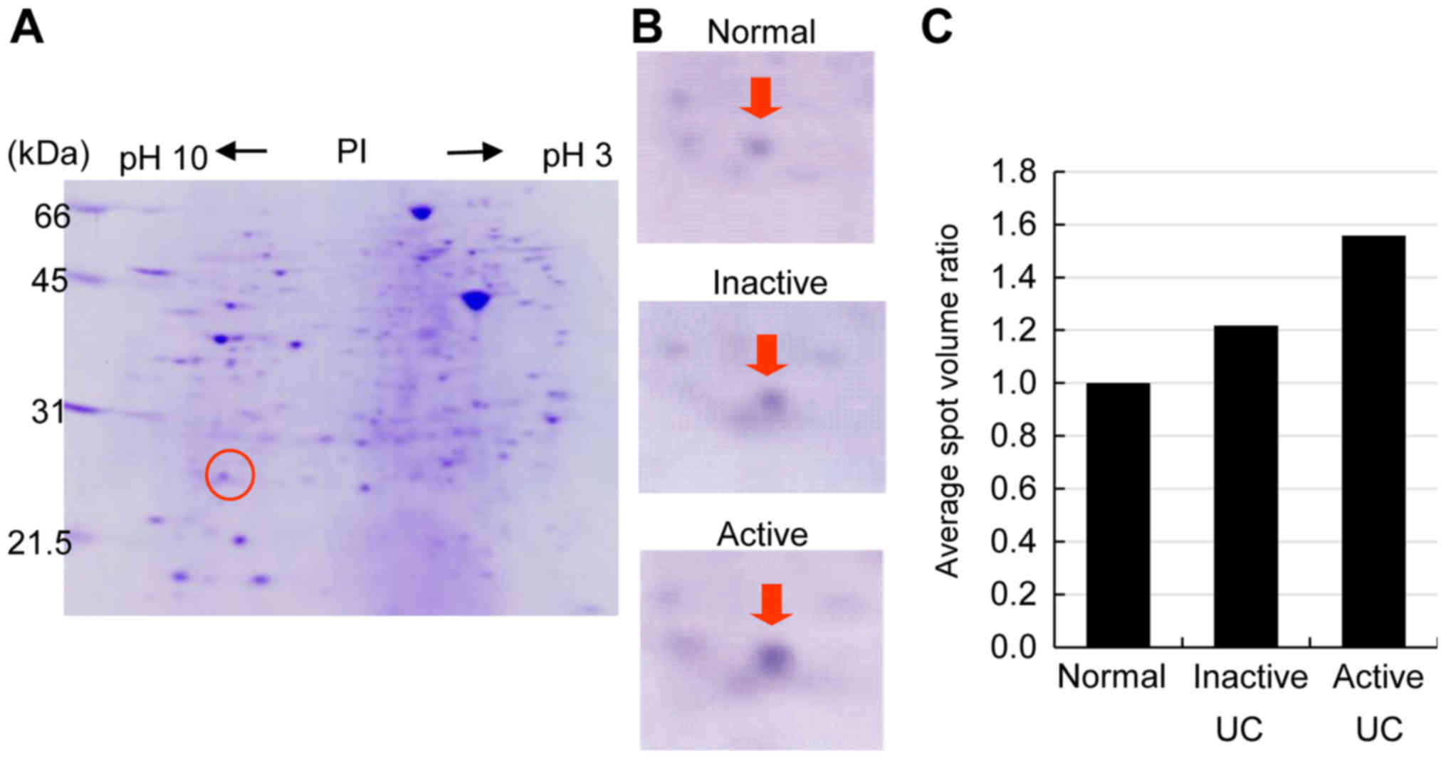
|
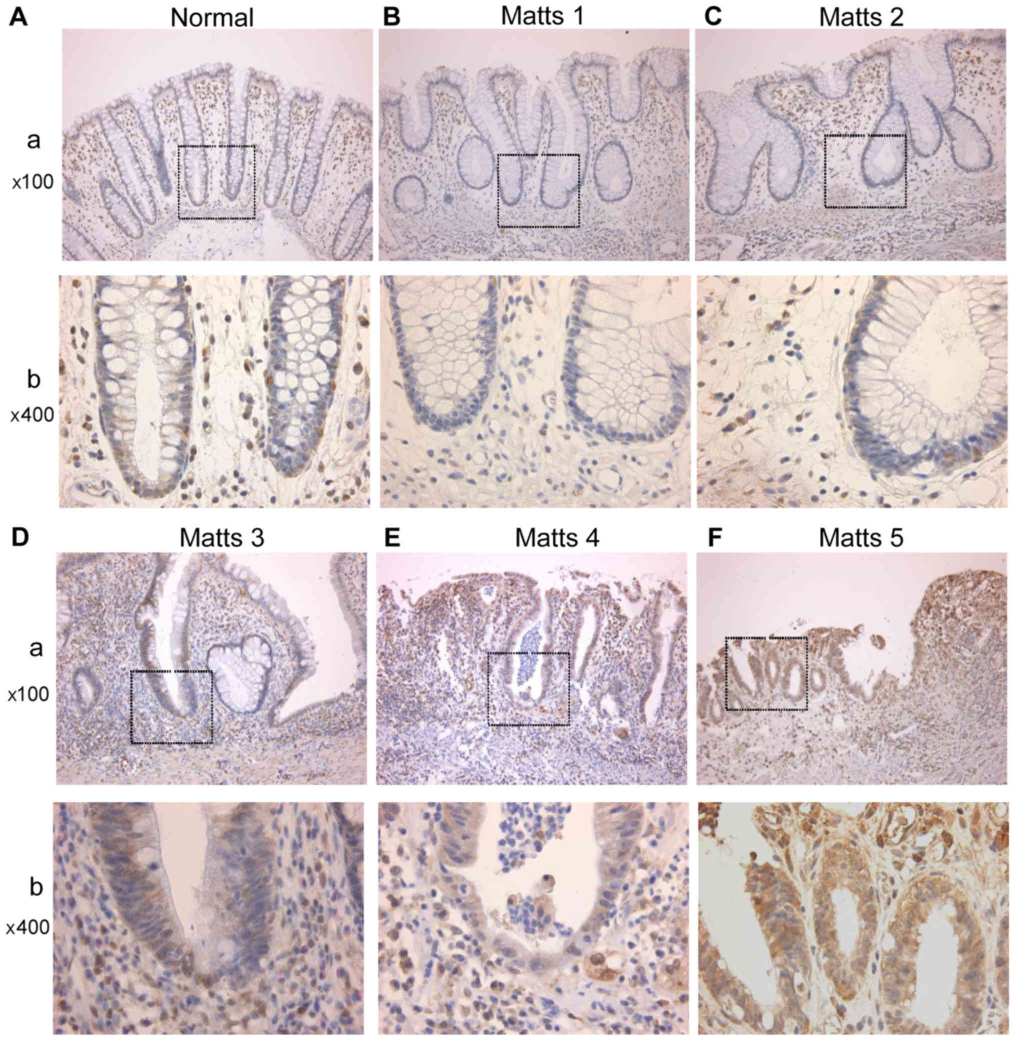
4
|
Wong NA and Harrison DJ: Colorectal
neoplasia in ulcerative colitis-recent advances. Histopathology.
39:221–234. 2001. View Article : Google Scholar : PubMed/NCBI
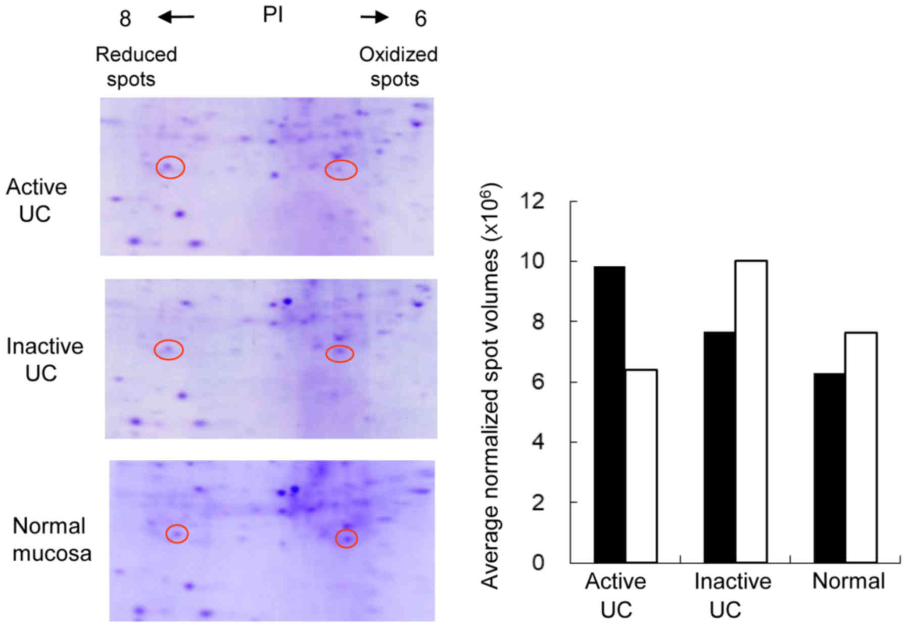
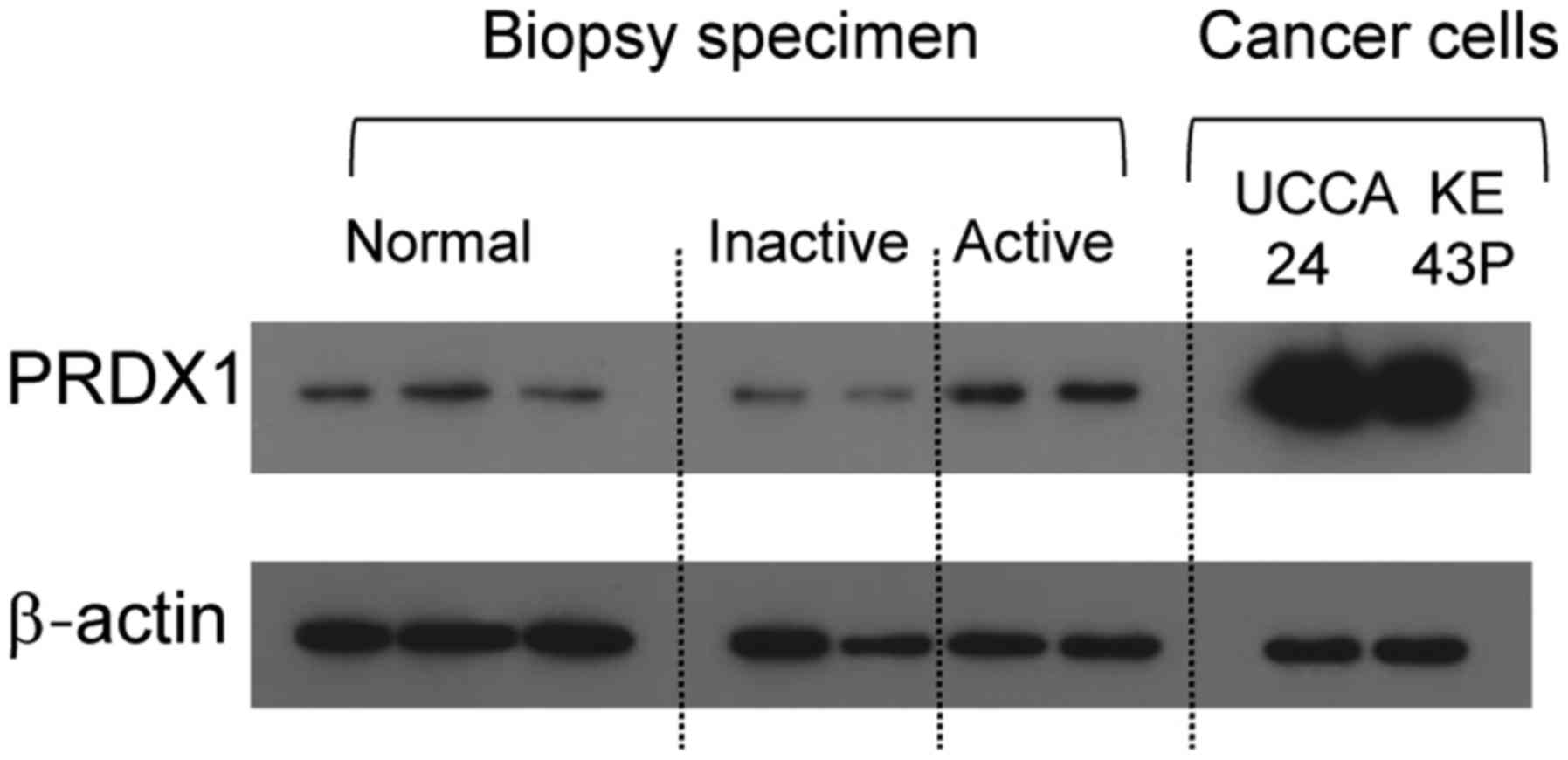
|
|
5
|
Okayasu I, Hana K, Yoshida T, Mikami T,
Kanno J and Fujiwara M: Significant increase of colonic mutated
crypts in ulcerative colitis correlatively with duration of
illness. Cancer Res. 62:2236–2238. 2002.PubMed/NCBI
|
|
6
|
Rhodes JM and Campbell BJ: Inflammation
and colorectal cancer: Ibd-associated and sporadic cancer compared.
Trends Mol Med. 8:10–16. 2002. View Article : Google Scholar : PubMed/NCBI
|
|
7
|
Sada M, Igarashi M, Yoshizawa S, Kobayashi
K, Katsumata T, Saigenji K, Otani Y, Okayasu I and Mitomi H: Dye
spraying and magnifying endoscopy for dysplasia and cancer
surveillance in ulcerative colitis. Dis Colon Rectum. 47:1816–1823.
2004. View Article : Google Scholar : PubMed/NCBI
|
|
8
|
Fujii S, Katsumata D and Fujimori T:
Limits of diagnosis and molecular markers for early detection of
ulcerative colitis-associated colorectal neoplasia. Digestion. 77
Suppl 1:S2–S12. 2008. View Article : Google Scholar
|
|
9
|
Vermeire S, Van Assche G and Rutgeerts P:
C-reactive protein as a marker for inflammatory bowel disease.
Inflamm Bowel Dis. 10:661–665. 2004. View Article : Google Scholar : PubMed/NCBI
|
|
10
|
Sachar DB, Smith H, Chan S, Cohen LB,
Lichtiger S and Messer J: Erythrocytic sedimentation rate as a
measure of clinical activity in inflammatory bowel disease. J Clin
Gastroenterol. 8:647–650. 1986. View Article : Google Scholar : PubMed/NCBI
|
|
11
|
Gabay C: Interleukin-6 and chronic
inflammation. Arthritis Res Ther. 8 Suppl 2:S32006. View Article : Google Scholar : PubMed/NCBI
|
|
12
|
Braegger CP, Nicholls S, Murch SH,
Stephens S and MacDonald TT: Tumour necrosis factor alpha in stool
as a marker of intestinal inflammation. Lancet. 339:89–91. 1992.
View Article : Google Scholar : PubMed/NCBI
|
|
13
|
Song M, Wu K, Ogino S, Fuchs CS,
Giovannucci EL and Chan AT: A prospective study of plasma
inflammatory markers and risk of colorectal cancer in men. Br J
Cancer. 108:1891–1898. 2013. View Article : Google Scholar : PubMed/NCBI
|
|
14
|
Schoepfer AM, Beglinger C, Straumann A,
Trummler M, Vavricka SR, Bruegger LE and Seibold F: Fecal
calprotectin correlates more closely with the simple endoscopic
score for crohn's disease (SES-CD) than CRP, blood leukocytes and
the CDAI. Am J Gastroenterol. 105:162–169. 2010. View Article : Google Scholar : PubMed/NCBI
|
|
15
|
Gisbert JP and McNicholl AG: Questions and
answers on the role of faecal calprotectin as a biological marker
in inflammatory bowel disease. Dig Liver Dis. 41:56–66. 2009.
View Article : Google Scholar : PubMed/NCBI
|
|
16
|
Lehmann FS, Burri E and Beglinger C: The
role and utility of faecal markers in inflammatory bowel disease.
Therap Adv Gastroenterol. 8:23–36. 2015. View Article : Google Scholar : PubMed/NCBI
|
|
17
|
Judd TA, Day AS, Lemberg DA, Turner D and
Leach ST: Update of fecal markers of inflammation in inflammatory
bowel disease. J Gastroenterol Hepatol. 26:1493–1499. 2011.
View Article : Google Scholar : PubMed/NCBI
|
|
18
|
D'Haens G, Ferrante M, Vermeire S, Baert
F, Noman M, Moortgat L, Geens P, Iwens D, Aerden I, Van Assche G,
et al: Fecal calprotectin is a surrogate marker for endoscopic
lesions in inflammatory bowel disease. Inflamm Bowel Dis.
18:2218–2224. 2012. View Article : Google Scholar : PubMed/NCBI
|
|
19
|
van Rheenen PF, Van de Vijver E and Fidler
V: Faecal calprotectin for screening of patients with suspected
inflammatory bowel disease: Diagnostic meta-analysis. BMJ.
341:c33692010. View Article : Google Scholar : PubMed/NCBI
|
|
20
|
Okayasu I, Yoshida T, Mikami T, Hana K,
Yokozawa M, Araki K, Mitsuhashi J, Kikuchi M, Adachi E and Sada M:
Mucosal remodeling in long-standing ulcerative colitis with
colorectal neoplasia: Significant alterations of NCAM+ or
alpha-SMA+ subepithelial myofibroblasts and interstitial cells.
Pathol Int. 59:701–711. 2009. View Article : Google Scholar : PubMed/NCBI
|
|
21
|
Araki K, Mikami T, Yoshida T, Kikuchi M,
Sato Y, Oh-Ishi M, Kodera Y, Maeda T and Okayasu I: High expression
of hsp47 in ulcerative colitis-associated carcinomas: Proteomic
approach. Br J Cancer. 101:492–497. 2009. View Article : Google Scholar : PubMed/NCBI
|
|
22
|
Matts SG: The value of rectal biopsy in
the diagnosis of ulcerative colitis. Q J Med. 30:393–407.
1961.PubMed/NCBI
|
|
23
|
Nagashio R, Sato Y, Jiang SX, Ryuge S,
Kodera Y, Maeda T and Nakajima T: Detection of tumor-specific
autoantibodies in sera of patients with lung cancer. Lung Cancer.
62:364–373. 2008. View Article : Google Scholar : PubMed/NCBI
|
|
24
|
Nagashio R, Sato Y, Matsumoto T, Kageyama
T, Satoh Y, Ryuge S, Masuda N, Jiang SX and Okayasu I: Significant
high expression of cytokeratins 7, 8, 18, 19 in pulmonary large
cell neuroendocrine carcinomas, compared to small cell lung
carcinomas. Pathol Int. 60:71–77. 2010. View Article : Google Scholar : PubMed/NCBI
|
|
25
|
Yamashita K, Yasuda S, Kuba T, Otani Y,
Fujiwara M and Okayasu I: Unique characteristics of rectal
carcinoma cell lines derived from invasive carcinomas in ulcerative
colitis patients. Cancer Sci. 95:211–217. 2004. View Article : Google Scholar : PubMed/NCBI
|
|
26
|
Tokuyama W, Mikami T, Fujiwara M, Matsui T
and Okayasu I: Midkine expression in colorectal tumors: Correlation
with ki-67 labeling in sporadic, but not ulcerative
colitis-associated ones. Pathol Int. 57:260–267. 2007. View Article : Google Scholar : PubMed/NCBI
|
|
27
|
Rhee SG and Woo HA: Multiple functions of
peroxiredoxins: Peroxidases, sensors and regulators of the
intracellular messenger H2O2 and protein chaperones. Antioxid Redox
Signal. 15:781–794. 2011. View Article : Google Scholar : PubMed/NCBI
|
|
28
|
Karihtala P, Mantyniemi A, Kang SW,
Kinnula VL and Soini Y: Peroxiredoxins in breast carcinoma. Clin
Cancer Res. 9:3418–3424. 2003.PubMed/NCBI
|
|
29
|
Poynton RA and Hampton MB: Peroxiredoxins
as biomarkers of oxidative stress. Biochim Biophys Acta.
1840:906–912. 2014. View Article : Google Scholar : PubMed/NCBI
|
|
30
|
Zhang B, Wang Y and Su Y: Peroxiredoxins,
a novel target in cancer radiotherapy. Cancer Lett. 286:154–160.
2009. View Article : Google Scholar : PubMed/NCBI
|
|
31
|
Cha MK, Suh KH and Kim IH: Overexpression
of peroxiredoxin I and thioredoxin1 in human breast carcinoma. J
Exp Clin Cancer Res. 28:932009. View Article : Google Scholar : PubMed/NCBI
|
|
32
|
Zhou J, Shen W, He X, Qian J, Liu S and Yu
G: Overexpression of prdx1 in hilar cholangiocarcinoma: A predictor
for recurrence and prognosis. Int J Clin Exp Pathol. 8:9863–9874.
2015.PubMed/NCBI
|
|
33
|
Hoshino I, Matsubara H, Akutsu Y,
Nishimori T, Yoneyama Y, Murakami K, Sakata H, Matsushita K and
Ochiai T: Tumor suppressor prdx1 is a prognostic factor in
esophageal squamous cell carcinoma patients. Oncol Rep. 18:867–871.
2007.PubMed/NCBI
|
|
34
|
Ambrosone CB: Oxidants and antioxidants in
breast cancer. Antioxid Redox Signal. 2:903–917. 2000. View Article : Google Scholar : PubMed/NCBI
|
|
35
|
Ding C, Fan X and Wu G: Peroxiredoxin 1-an
antioxidant enzyme in cancer. J Cell Mol Med. 21:193–202. 2017.
View Article : Google Scholar : PubMed/NCBI
|
|
36
|
Chu G, Li J, Zhao Y, Liu N, Zhu X, Liu Q,
Wei D and Gao C: Identification and verification of prdx1 as an
inflammation marker for colorectal cancer progression. Am J Transl
Res. 8:842–859. 2016.PubMed/NCBI
|
|
37
|
Liu CH, Kuo SW, Hsu LM, Huang SC, Wang CH,
Tsai PR, Chen YS, Jou TS and Ko WJ: Peroxiredoxin 1 induces
inflammatory cytokine response and predicts outcome of cardiogenic
shock patients necessitating extracorporeal membrane oxygenation:
An observational cohort study and translational approach. J Transl
Med. 14:1142016. View Article : Google Scholar : PubMed/NCBI
|
|
38
|
Serada S, Fujimoto M, Terabe F, Iijima H,
Shinzaki S, Matsuzaki S, Ohkawara T, Nezu R, Nakajima S, Kobayashi
T, et al: Serum leucine-rich alpha-2 glycoprotein is a disease
activity biomarker in ulcerative colitis. Inflamm Bowel Dis.
18:2169–2179. 2012. View Article : Google Scholar : PubMed/NCBI
|
|
39
|
Arai Y, Arihiro S, Matsuura T, Kato T,
Matsuoka M, Saruta M, Mitsunaga M, Matsuura M, Fujiwara M, Okayasu
I, et al: Prostaglandin e-major urinary metabolite as a reliable
surrogate marker for mucosal inflammation in ulcerative colitis.
Inflamm Bowel Dis. 20:1208–1216. 2014. View Article : Google Scholar : PubMed/NCBI
|
|
40
|
Arai Y, Matsuura T, Matsuura M, Fujiwara
M, Okayasu I, Ito S and Arihiro S: Prostaglandin e-major urinary
metabolite as a biomarker for inflammation in ulcerative colitis:
Prostaglandins revisited. Digestion. 93:32–39. 2016. View Article : Google Scholar : PubMed/NCBI
|
|
41
|
Hagiwara SI, Okayasu I, Fujiwara M,
Matsuura M, Ohnishi H, Ito S, Kishimoto H, Nambu R and Kagimoto S:
Prostaglandin e-major urinary metabolite as a biomarker for
pediatric ulcerative colitis activity. J Pediatr Gastroenterol
Nutr. 64:955–961. 2017. View Article : Google Scholar : PubMed/NCBI
|
|
42
|
Marks PA: Thioredoxin in cancer-role of
histone deacetylase inhibitors. Semin Cancer Biol. 16:436–443.
2006. View Article : Google Scholar : PubMed/NCBI
|
|
43
|
Mitsumoto A, Takanezawa Y, Okawa K,
Iwamatsu A and Nakagawa Y: Variants of peroxiredoxins expression in
response to hydroperoxide stress. Free Radic Biol Med. 30:625–635.
2001. View Article : Google Scholar : PubMed/NCBI
|