Introduction
Diffuse large B-cell lymphoma (DLBCL) is an
aggressive malignant lymphoma and despite significant therapeutic
advances in recent years, relapsed/refractory DLBCL occurs in
30–40% of patients due to DLBCL morphological and molecular
heterogeneity (1–4). According to the World Health
Organization, DLBCL subgroups are classified based on gene
expression profiling or cell of origin (5) and primarily stratified into germinal
center B-cell (GCB) and activated B-cell (ABC) subtypes. In
comparison with the GCB subtype of DLBCL, the ABC subtype is
associated with a poor prognosis, with a high expression of BCL2,
cMYC and BCL6, and a typical progression of clinical features
(6,7). Despite the effectiveness of
lenalidomide, BTK and PI3K inhibitors (8–13),
ABC-type DLBCL still displays an inadequate response to standard
immunotherapy (6). Furthermore,
although the pathogenesis of these two DLBCL subtypes involves
different mutated genes and activated signaling pathways, with
corresponding drugs already available, the mechanisms remain
unclear and require investigation.
Oxidative stress and related regulatory genes are
considered hallmarks of cancer progression and therefore can be
used to assess disease course and prognosis. The Kelch-like
ECH-associated protein 1 (Keap1)-nuclear factor erythroid 2-related
factor 2 (Nrf2) signaling pathway is a typical antioxidant stress
pathway that exhibits abnormalities in several human malignant
tumors, such as breast cancer, lung cancer, liver cancer, thyroid
cancer, ovarian cancer and gastric cancer (14–17).
Nrf2 is a transcriptional regulatory factor for antioxidant stress
and is induced by oxidative stress to enter the nucleus to activate
downstream antioxidant genes to protect cells from oxidative and
electrophilic stress. Conversely, upregulation of Nrf2 activity
within cancer cells suppresses drug-induced reactive oxygen species
(ROS) production, leading to the development of drug resistance,
thereby facilitating cancer cell survival and proliferation
(18). Recent studies have reported
that Nrf2 is overexpressed in cancer cells, indicating that it may
serve an oncogenic role in carcinogenesis (15). Moreover, due to its dual role, Nrf2
and its antagonist Keap1 have become the subject of debate
regarding their specific roles in preventing or promoting tumor
progression (18). Exploring the
related gene expression of this pathway is of great significance
for cancer prevention and treatment. In addition, previous studies
have also reported the association between Nrf2 and autophagy. The
autophagy receptor p62 is also a target of Nrf2, whereby induction
of autophagy leads to increased p62 levels. Consequently, p62
interacts with Keap1, activating Nrf2, further promoting cancer
cell survival (19–22).
Currently, there is limited data regarding the
impact of Nrf2 expression in different DLBCL subtypes on subsequent
treatment or clinical outcomes. Therefore, the present study
assessed the gene expression of NFE2L2 (Nrf2), KEAP1
(Keap1) and MAP1LC3B (LC3B), as well as ROS, in the cells of
different subtypes (ABC and GCB) of newly diagnosed patients with
DLBCL. Subsequently, the present study analyzed the correlation
between these expression profiles and clinical pathological data,
as well as the correlations among diverse genes to assess the
impact of varying gene expression on DLBCL subtypes to identify
potential prognostic biomarkers.
Materials and methods
Public databases for gene expression
analysis
DLBCL samples from the Cancer Genome Atlas (TCGA)
(https://www.cancer.gov/ccg/research/genome-sequencing/tcga)
and the Genotype-Tissue Expression (GTEx) (https://gtexportal.org/home/) databases were analyzed
using Gene Expression Profiling Interactive Analysis 2 (http://gepia2.cancer-pku.cn/) and the University of
Alabama at Birmingham Cancer data analysis Portal (https://ualcan.path.uab.edu/) software (23,24).
The gene expression data included NFE2L2, KEAP1 and
MAP1LC3B and the value |log2FoldChange|>1 and q values
<0.01 were considered to indicate differential expression. These
databases provide tumor/normal differential expression analysis to
aid in the analysis of RNA-sequencing data.
Study design
Following approval by the Institutional Review Board
of Kaohsiung Medical University Chung-Ho Memorial Hospital
[Kaohsiung, Taiwan; approval nos. KMUHIRB-E(I)-20210119 and
KMUHIRB-E(I)-20220298], pathological specimens of patients
diagnosed with DLBCL at Kaohsiung Medical University Chung-Ho
Memorial Hospital between July 2015 and December 2021 were
collected, along with clinical and biochemical laboratory data for
subsequent analysis. A total of 43 specimens were obtained and
DLBCL subtypes were classified as ABC or GCB subtypes using
immunohistochemistry (IHC) analysis (25). The inclusion criteria included the
following: i) Histopathologically-confirmed DLBCL; ii) age of ≥18
years; and iii) pathological tissue sections measuring ≥1×1×5 mm.
The exclusion criteria included the following: i) Concurrent
malignancies; ii) aged of <18 years; and iii) adequate
pathological specimens were unavailable or too small for
analysis.
IHC
The staining process was automated using a BOND-MAX
Automated IHC Staining System (Leica Biosystems) following a
standardized protocol (26).
Briefly, pre-existing formalin-fixed paraffin-embedded blocks were
sectioned to 4-µm thick, deparaffinized with xylene at 72°C and
pre-treated for permeabilization using Epitope Retrieval Solution 1
(citrate, pH 6.0; Leica Biosystems) at 100°C for 20 min.
Hydroperoxide blocking was performed for 5 min using the Bond
Polymer Refine Detection Kit (Leica Biosystems Newcastle Ltd,
United Kingdom), incubated with the following primary antibodies at
room temperature for 30 min: Primary antibodies targeting Nrf2
(EP1808Y; monoclonal; 1:50; cat. no. ab62352; Abcam), Keap1 (lB4;
monoclonal; 1:150; cat. no. ab119403; Abcam), LC3B (polyclonal;
1:100; cat. no. ab63817; Abcam) and nitrotyrosine (39B6;
monoclonal; 1:100; cat. no. sc-32757; Santa Cruz Biotechnology,
Inc.). Afterward, the tissue was incubated with the secondary
antibody from the BOND Polymer Refine Detection Kit (Leica
Biosystems) at 25°C for 15 min. Polymer incubation lasted for 15
min before development with 3,3′-diaminobenzidine
tetrahydrochloride hydrate (DAB) chromogen for 10 min. The
specimens were counterstained with hematoxylin at 25°C for 5 min.
Positive and negative controls consisted of squamous cell carcinoma
tissues. The staining of cytoplasmic Nrf2, Keap1, LC3B and
nitrotyrosine was visualized using the TissueFAXS PLUS system
(version 4.2; TissueGnostics GmbH) and the Zeiss Observer
microscope with a 20X objective lens (Ziess GmbH). HistoQuest
software (version 4.0; TissueGnostics GmbH) was used to quantify
cytoplasmic Nrf2, Keap1, LC3B, nitrotyrosine and hematoxylin
staining (Fig. S1). Regions of
interest (ROIs) within each slide were selected for analysis, with
≥3 representative areas measured for consistency. Positive cell
expression within the ROIs was quantified as %. Staining intensity
was assessed by assigning arbitrary numbers based on grayscale
pixel conversion, with the numbers of DAB-positive and
hematoxylin-positive events used for optimization. This approach
ensured robust analysis of immunohistochemically stained samples
adhering to established protocols for accurate interpretation. Due
to the limitations of imaging software in accurately distinguishing
between nuclear and cytoplasmic Nrf2, pathologists qualitatively
assessed the nuclear expression of Nrf2 in DLBCL using optical
microscopy. A positivity threshold of ≥10% was used to define a
sample as positive.
Cell culture
Human DLBCL cancer cell lines, U2932 (Guangzhou
Ubigene Biosciences Co., Ltd.) and HT (cat. no. 60486; Bioresource
Collection and Research Center, Taiwan) were cultured in RPMI-1640
supplemented with 10% FBS (Merck KGaA), 2 mM L-glutamine (Gibco;
Thermo Fisher Scientific, Inc.), 10 mM HEPES (BioConcept AG), 1 mM
sodium pyruvate (Gibco; Thermo Fisher Scientific, Inc.) and 1%
penicillin/streptomycin (Gibco; Thermo Fisher Scientific, Inc.) in
5% CO2 at 37°C. A total of 2×10⁶ cells were
initially seeded into a 75T flask with 10 ml medium. Every 3–4
days, the cells and medium were transferred to a 15 ml centrifuge
tube and centrifuged at 150 × g for 5 min at 25°C. After
centrifugation, the supernatant was carefully discarded and half of
the cells were resuspended in fresh medium.
Reverse transcription-quantitative
(q)PCR
Total RNA was isolated from the U2932 and HT cell
lines using the TOOLSmart RNA Extractor reagent (cat. no. DPT-BD24;
BIOTOOLS Co., Ltd.) and 1 µg RNA reverse transcribed using the
High-Capacity cDNA Reverse Transcription Kit (cat. no. 4368814;
Thermo Fisher Scientific, Inc.) at 25°C for 10 min, followed by
37°C for 120 min, 85°C for 5 min and maintenance at 4°C. Each 20 µl
reaction contained 2× SYBR Green Master Mix (cat. no. A46012;
Applied Biosystems; Thermo Fisher Scientific, Inc.), 1 µl forward
and reverse primers (KEAP1 forward: 5′-CGTAGCCCCCATGAAGCA-3′
and reverse: 5′-ACTCCACACTGTCCAGGAACGT-3′; GAPDH forward:
5′-GCACCACCAACTGCTTAGCA-3′ and reverse: 5′-TCTTCTGGGTGGCAGTGATG-3′)
and 2 µl cDNA. qPCR was performed using the QuantStudio real-time
PCR system (Applied Biosystems; Thermo Fisher Scientific, Inc.) and
the thermocycling conditions were as follows: 95°C for 1 min,
followed by 40 cycles at 95°C for 10 sec and 60°C for 60 sec; 65°C
for 10 sec for melting curves; and 40°C for 30 sec for cooling.
Target gene expression was quantified using the 2−ΔΔCq
method with GAPDH used as the internal control (27,28).
The PCR reactions were performed in triplicate.
Statistical analysis
Statistical analyses were performed using SPSS
software (version 19; IBM Corp.). Pearson's correlation coefficient
was used to assess the correlation between Nrf2, Keap1, LC3B and
nitrotyrosine. Continuous variables were compared between groups
using an unpaired t-test. Fisher's exact test or the χ2
test were used to evaluate the association of categorical
variables. Multiple logistic regression models were also used to
assess the association between nuclear Nrf2 and clinical
characteristics. Independent variables with P<0.05 in
univariable analysis were selected in the multivariable analysis. A
forward selection approach determined the final multivariable
model. Continuous data are presented as mean ± standard deviation.
P<0.05 was considered to indicate a statistically significant
difference.
Results
Differential distribution of Nrf2 in
subtypes and stages of DLBCL
Nrf2 is a crucial gene involved in mitigating ROS
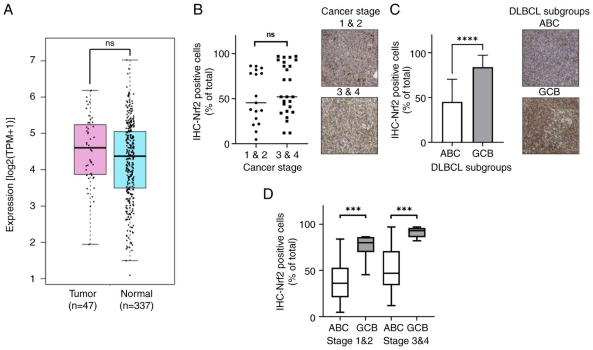
and is present in several cancers (Fig. S2). In DLBCL, the NFE2L2
(Nrf2) mRNA level also demonstrated a significantly higher
expression, with transcripts per million (TPM) of 23.29, compared
with 19.69 TPM in normal tissues, as revealed by analysis of data
from the TCGA and GTEx databases (Fig.
1A). Furthermore, a higher proportion of Nrf2-positive cancer
cells was demonstrated in the advanced cancer stages (Ann Arbor
stages 3 and 4) (29), where 60.88%
cells were Nrf2-positive, compared with 51.26% in the early cancer
stages (stages 1 and 2) of DLBCL, as determined by IHC analysis
(Fig. 1B). Furthermore, when
comparing the subtypes of DLBCL, it was demonstrated that the GCB
subtype (84.20%) had a significantly higher frequency of
Nrf2-positive cancer cells than the ABC subtype (45.32%)
(P<0.0001; Fig. 1C). This trend
persisted across disease stages, where the GCB subtype had a
significantly greater proportion of Nrf2-positive cells compared
with the ABC subtype in both the early stages (GCB, 75.98%; and
ABC, 37.78%) and the late stages (GCB, 91.25%; and ABC, 49.69%)
(Fig. 1D). The results revealed
that Nrf2 was prominently distributed in the advanced stages of
DLBCL and showed a higher frequency in the GCB subtype compared
with that in the ABC subtype.
Active Nrf2 is predominantly located
in the nucleus of the DLBCL ABC subtype
Nrf2 functions as a transcription factor activated
within the cell nucleus (30).
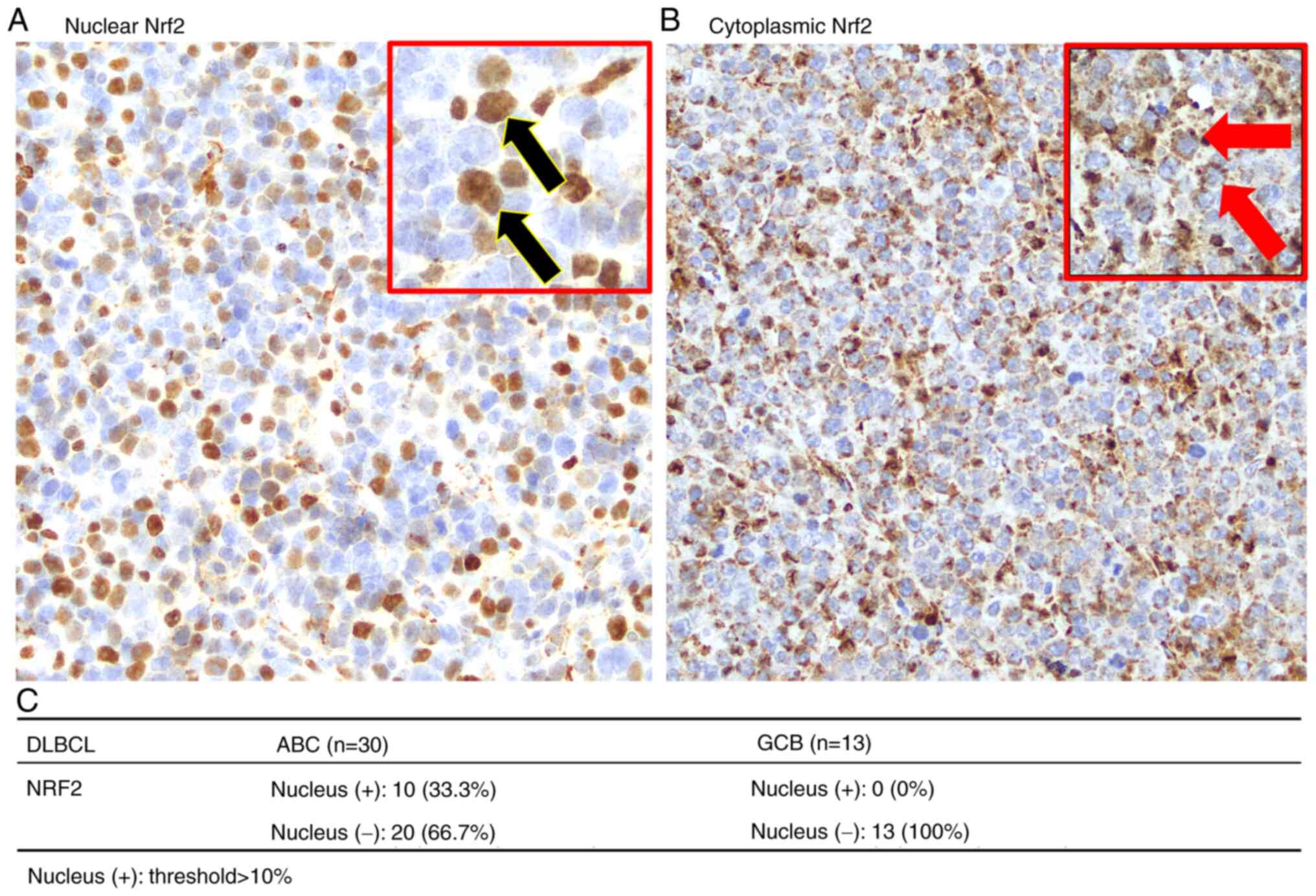
Therefore, the present study further assessed the nuclear
expression of Nrf2 in DLBCL. Fig. 2A
and B present images captured at ×40 magnification, of Nrf2
localization in the nucleus and cytoplasm of DLBCL cells,
respectively. Notably, the analysis revealed that nuclear Nrf2 was
exclusively present in the ABC subtype, accounting for 33.3% of
cases (n=30), whilst no nuclear Nrf2 was observed in the GCB
subtype (n=13; Fig. 2C).
KEAP1 mRNA and protein expression in
advanced cancer stages and the DLBCL ABC subtype
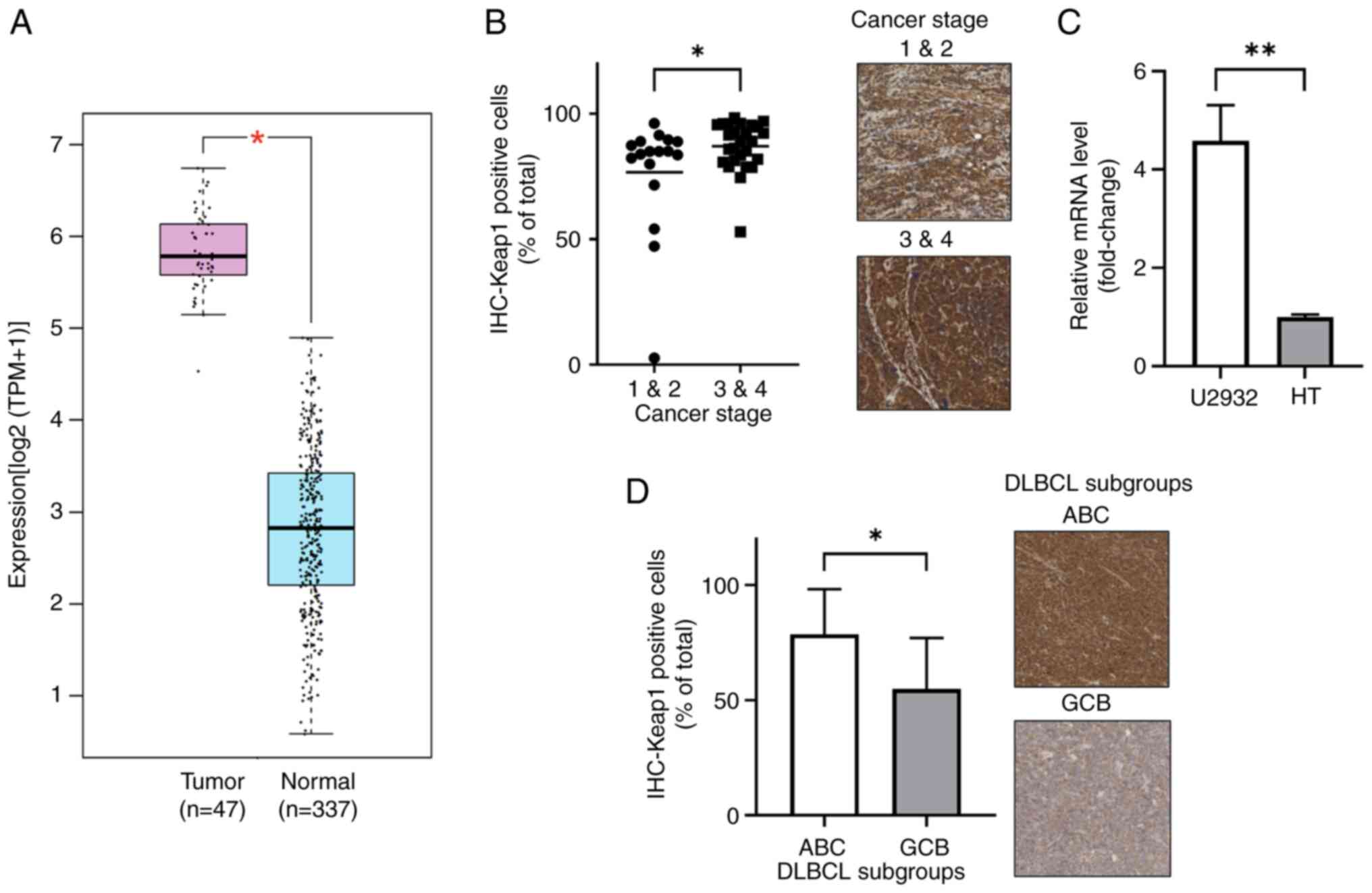
A comparative assessment of mRNA levels from the
public databases of TCGA and GTEx revealed higher KEAP1
expression in tumor samples compared with that in controls.
Specifically, KEAP1 exhibited an average expression of 54.07
TPM in tumor samples, significantly elevated compared with 6.1 TPM
in normal controls (P<0.05; Fig.
3A). Immunohistochemical staining for Keap1 also demonstrated
higher expression in advanced stages (stages 3 and 4), with 87.10%
Keap1-positive cells, compared with 76.66% in the early stages
(stages 1 and 2; P=0.046; Fig. 3B).
Additionally, the KEAP1 expression ratios in DLBCL cell
lines revealed a 4.3-fold higher expression in U2932 cells (ABC)
compared with that in HT cells (GCB) when normalized to GAPDH
(P<0.01; Fig. 3C). Moreover,
assessment of Keap1 expression across DLBCL subtypes indicated a
higher proportion of Keap1-expressing cells in tumor samples from
patients with ABC (78.69%) compared with those with the GCB subtype
(54.86%; P=0.014; Fig. 3D).
LC3B expression and association with
Nrf2 in DLBCL
Nrf2 has been implicated in autophagy (31,32),
therefore the present study evaluated the expression of the
autophagosome marker LC3B in DLBCL and its relationship with Nrf2.
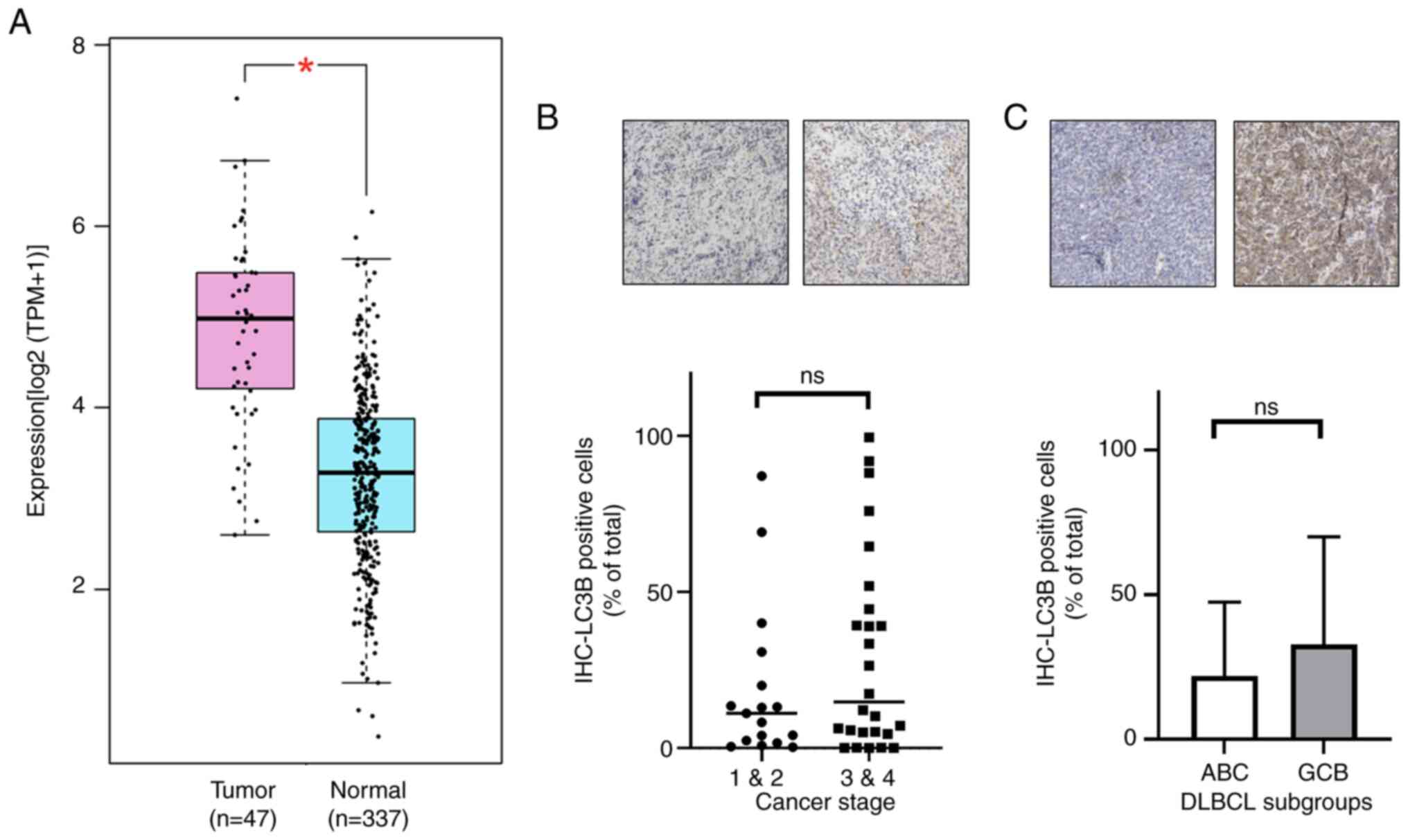
Analysis of TCGA and GTEx data revealed that MAP1LC3B (LC3B) mRNA
expression was significantly elevated in DLBCL compared with normal
tissues (30.58 TPM vs. 8.77 TPM; P<0.05; Fig. 4A). In IHC-stained samples, although
statistical significance was not achieved, LC3B-positive cells were
notably more prevalent in advanced stages (stages 3 and 4) and in
the GCB subtype of DLBCL compared with that in early stages (stages
1 and 2) and the ABC subtype, respectively (Fig. 4B and C). Furthermore, a significant
positive correlation was observed between Nrf2 and LC3B expression
in DLBCL samples (Pearson correlation=0.345; P=0.023; data not
shown). These results indicate that LC3B expression in advanced
cancer stages and the GCB subtype parallels that of Nrf2.
Nitrotyrosine expression slightly
increases in advanced stages and ABC subtype of DLBCL
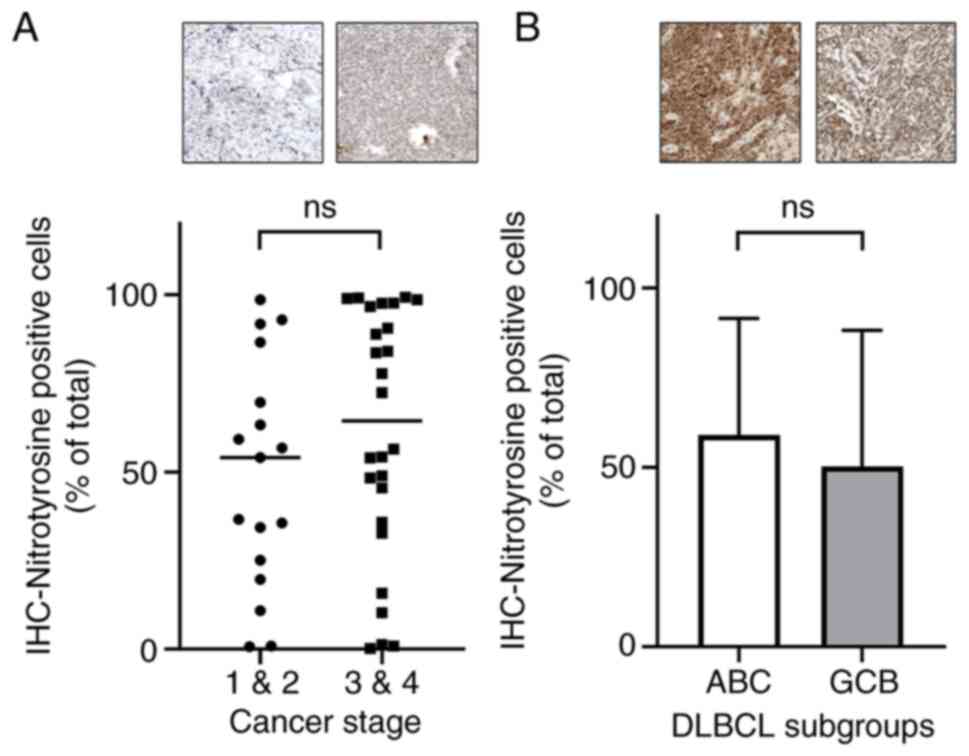
To evaluate whether Nrf2 suppresses ROS,
nitrotyrosine was used as a marker to assess ROS levels in DLBCL.
IHC analysis revealed a trend of markedly increased nitrotyrosine
levels in advanced stages of DLBCL compared with that in early
stages (Fig. 5A). Additionally, the
frequency of nitrotyrosine-positive cells was notably higher in the
ABC subtype compared with that in the GCB subtype, although this
difference was not statistically significant (Fig. 5B).
Clinical differences between ABC and
GCB subtypes of DLBCL
Given the classification of DLBCL into ABC and GCB
subtypes, a comparative analysis of the clinical data was performed
to elucidate the differences between these subgroups. Table I presents the clinical
characteristics of the ABC and GCB subtypes. The findings revealed
that the ABC subtype was significantly associated with a lower
white blood cell (WBC) count compared with those with the GCB
subtype (P=0.0096). Moreover, patients in the ABC subtype underwent
a significantly greater number of chemotherapy cycles compared with
those with the GCB subtype (P=0.0173). Additionally, several
clinical parameters, including Eastern Cooperative Oncology Group
performance status (33), overall
survival (OS), progression-free survival (PFS), platelet count,
lactate dehydrogenase, albumin, glutamic oxaloacetic transaminase
(GOT), glutamic-pyruvic transaminase (GPT), blood urea nitrogen,
creatinine and ionized calcium levels, were markedly lower in the
ABC subtype compared with that of the GCB subtype. These data
indicate that the ABC subgroup presented with worse clinical
values.
 | Table I.Basic clinical characteristics of the
diffuse large B-cell lymphoma activated B-cell and germinal center
B-cell subtypes. |
Table I.
Basic clinical characteristics of the
diffuse large B-cell lymphoma activated B-cell and germinal center
B-cell subtypes.
| Clinical
characteristic | ABC (n=30) | GCB (n=13) | P-value |
|---|
| Sex |
|
| 0.1854 |
|
Male | 13 | 9 |
|
|
Female | 17 | 4 |
|
| Age, years | 58.5 | 62.5 | 0.4313 |
| Ann Arbor
stage |
|
| 0.4970 |
|
1-2 | 10 | 4 |
|
|
3-4 | 20 | 9 |
|
| ECOG performance
status |
|
| 0.3607 |
|
<2 | 25 | 10 |
|
| ≥2 | 3 | 3 |
|
|
N.D. | 2 | 0 |
|
| OS, months | 33.0±30.8 | 34.1±43.0 | 0.9240 |
| PFS, months | 28.2±32.0 | 34.1±43.0 | 0.6215 |
| HBsAg |
|
| 0.6972 |
|
Positive | 6 | 2 |
|
|
Negative | 19 | 10 |
|
|
N.D. | 5 | 1 |
|
| HBcAb |
|
| 0.4340 |
|
Positive | 15 | 8 |
|
|
Negative | 10 | 2 |
|
|
N.D. | 5 | 3 |
|
| HCV |
|
| 0.6594 |
|
Positive | 4 | 3 |
|
|
Negative | 21 | 9 |
|
|
N.D. | 5 | 1 |
|
| B
symptomsb |
|
| 0.2953 |
|
Positive | 14 | 4 |
|
|
Negative | 11 | 8 |
|
|
N.D. | 5 | 1 |
|
| WBC, µl |
6,189.6±1,912.2 |
9,545.0±5,543.8 | 0.0096a |
| Platelet,
×103/µl | 230.5±118.9 | 257.7±129.1 | 0.5311 |
| LDH, U/l | 378.6±437.6 | 673.5±1091.9 | 0.2459 |
| β-2 microglobulin,
µg/l | 435.9±483.1 | 306.9±215.9 | 0.4055 |
| Albumin, g/dl | 3.9±0.6 | 3.9±0.4 | 0.8598 |
| GOT, U/l | 31.6±18.9 | 45.9±46.2 | 0.1860 |
| GPT, U/l | 26.9±24.7 | 30.1±19.4 | 0.6960 |
| Blood urea
nitrogen, mg/dl | 16.7±9.6 | 19.4±14.4 | 0.4981 |
| Creatinine,
mg/dl | 1.38±2.1 | 1.52±2.2 | 0.8558 |
| Ionized calcium,
mg/dl | 4.68±0.4 | 4.94±0.4 | 0.0945 |
| Chemotherapy
courses | 5.93±2.25 | 3.75±3.05 | 0.0173a |
Patients with nuclear expression of
Nrf2 have worse clinical biomarkers
Previous findings indicated the presence of nuclear
Nrf2 in the ABC subtype of DLBCL (Fig.
2C). To further assess the impact of nuclear Nrf2 expression on
patient outcomes, clinical data were collected and analyzed by
dividing patients into two groups based on the presence or absence
of nuclear Nrf2. The comparison revealed significant associations
with several clinical characteristics (Table II). Specifically, hepatitis B
surface antigen (HBsAg), body weight loss (BWL) and ionized calcium
were significantly higher in the nuclear Nrf2-positive group than
in the Nrf2-negative group (P<0.05). Additionally, there was a
marked trend towards lower WBC and platelet counts in the nuclear
Nrf2-positive group compared with that of the nuclear Nrf2-negative
group, whilst higher levels of GOT and GPT were observed in the
nuclear Nrf2-positive group compared with that of the nuclear
Nrf2-negative group. These findings demonstrate that nuclear Nrf2
expression had a notable impact on the clinical biomarkers of
patients with DLBCL.
 | Table II.Clinical analysis of nuclear factor
erythroid 2-related factor 2 expression in diffuse large B-cell
lymphoma. |
Table II.
Clinical analysis of nuclear factor
erythroid 2-related factor 2 expression in diffuse large B-cell
lymphoma.
| Clinical
characteristic | Nuclear
Nrf2-positive | Nuclear
Nrf2-negative | P-value |
|---|
| Subtype |
|
| 0.0196a |
|
ABC | 10 | 20 |
|
|
GCB | 0 | 13 |
|
| HBsAg |
|
| 0.0487a |
|
Positive | 4 | 4 |
|
|
Negative | 4 | 25 |
|
|
N.D. | 2 | 4 |
|
| BWL |
|
| 0.0421a |
|
Yes | 6 | 9 |
|
| No | 2 | 20 |
|
|
N.D. | 2 | 4 |
|
| WBC, µl | 6471.3±1290.6 | 7500.0±4220.8 | 0.5043 |
| Platelet,
×103/µl | 201.1±83.8 | 239.8±136.6 | 0.3207 |
| GOT, U/l | 37.1±29.5 | 36.0±31.5 | 0.9621 |
| GPT, U/l | 35.4±38.0 | 25.9±17.1 | 0.3044 |
| Ionized calcium,
mg/dl | 5.1±0.4 | 4.7±0.4 | 0.0134a |
Discussion
DLBCL, the most common subtype of non-Hodgkin's
lymphoma in Asia, is regarded as a severe form of non-Hodgkin's
lymphoma and is increasing in incidence (34). DLBCL can be further categorized into
ABC and GCB subtypes based on cell of origin and gene expression
analysis (35). The frequency of
the ABC subtype is 60–70% in Asian countries, which is markedly
higher than the 37–40% observed in Western countries, and it is
associated with a worse prognosis (36,37).
The distinction between ABC and GCB subtypes notably impacts the
prognosis of patients with DLBCL (38), as the expression of antioxidant
genes in cancer cells can attenuate the efficacy of drug therapy,
leading to drug resistance and relapse (39). Despite extensive research on DLBCL,
there are limited studies focusing on the association between DLBCL
subtypes, antioxidant genes like Nrf2 and Keap1, and clinical data.
Therefore, the present study assessed the relationship between the
different DLBCL subtypes (ABC and GCB), clinical data and the
expression of Nrf2, Keap1, nitrotyrosine and LC3B.
Nrf2, a transcription factor with antioxidant
capabilities, activates in the nucleus and is associated with worse
treatment outcomes and prognosis in cancer therapy (30). In the dataset in the present study,
~23.26% (10/43) of DLBCL tissue specimens exhibited nuclear Nrf2
expression in tumor cells, particularly in the ABC subtype with
worse prognosis, where it accounted for 33.33% (10/33), potentially
contributing to chemotherapy resistance and worsening treatment
outcomes. Previous studies have reported an association between
Nrf2 expression and drug resistance in cancers, as well as its
association with clinical characteristics (40). Generally, higher Nrf2 expression is
associated with a worse patient prognosis, suggesting its potential
as a cancer biomarker (30,39,41).
The clinical data analysis in the present study revealed a higher
prevalence of HBsAg in patients with nuclear Nrf2 expression,
indicating a potential link between Nrf2 activation and HBsAg
presence in DLBCL. Previous studies have also reported that
Hepatitis B virus (HBV) regulatory proteins hepatitis B virus X
protein (HBx) and large hepatitis B virus surface protein activate
Nrf2 via the c-Raf and MEK pathways, thereby protecting
HBV-positive cells from oxidative damage (42–44).
Moreover, previous studies reported that a marked reduction in
HBsAg release was associated with decreased Nrf2 activity (45,46).
Conversely, other research suggested that during HBV infection, ROS
production was induced, with HBsAg contributing to ROS formation.
HBx further activated Nrf2 by increasing its protein levels and
enhancing nuclear localization. However, excessive Nrf2 expression
markedly suppressed HBV core promoter activity, leading to reduced
viral replication (47,48). These findings suggest that nuclear
Nrf2 activation, associated with HBsAg presence in DLBCL, may
protect cancer cells from oxidative stress, indicating its
potential as a biomarker for identifying patients with
HBsAg-positive DLBCL. Furthermore, in the present study, patients
with DLBCL with nuclear Nrf2 expression exhibited significant
weight loss, suggesting that Nrf2 activation may contribute to this
outcome. Previous studies using a diabetic mouse (db/db) model
reported that Nrf2 activation by the inducer CDDO-Im markedly
suppressed high-fat diet-induced obesity and alleviated diabetes,
leading to weight loss. This is a process reversed by Nrf2 gene
disruption (49,50). This mechanism involves insulin
secretion by β-cells, where ROS generated from glucose metabolism
activate Nrf2. In turn, Nrf2 stimulates the expression of
downstream genes such as glucose-6-phosphate dehydrogenase and
phosphogluconate dehydrogenase in the pentose phosphate pathway,
producing NADPH, which enhances insulin secretion, glucose
metabolism and energy expenditure, ultimately affecting body weight
(51,52). The association between nuclear Nrf2
expression and weight loss in patients with DLBCL suggests that
Nrf2 activation may influence metabolism, reinforcing its potential
as a biomarker for assessing clinical outcomes.
Under normal conditions, the transcription factor
Nrf2 is bound by the Keap1-dependent E3 ubiquitin ligase complex,
which includes Keap1, Cullin 3 and RING box protein 1, along with
an E2 ubiquitin-conjugating enzyme. This interaction facilitates
the ubiquitination and subsequent proteasomal degradation of Nrf2.
However, under stress conditions, sensor cysteine residues within
Keap1 are modified, allowing Nrf2 to escape degradation,
translocate to the nucleus and initiate its antioxidant
transcriptional program (53). The
findings of the present study, consistent with those reported by Yi
et al (41), demonstrated
significant Keap1 expression in DLBCL, particularly in advanced
stages of the disease. In the ABC subtype, which is characterized
by elevated oxidative stress (54),
Nrf2 predominantly localized to the nucleus and was positively
associated with HBsAg expression and BWL, suggesting increased
oxidative stress in this subtype. Furthermore, despite elevated
Keap1 expression in ABC subtype, its regulatory function appeared
to be impaired, resulting in enhanced nuclear translocation of
Nrf2. This dysregulation likely contributed to the activation of
aberrant metabolic and stress response pathways in ABC, negatively
influencing clinical outcomes. In contrast, Nrf2 was primarily
confined to the cytoplasm in the GCB subtype, indicating that the
inhibitory function of Keap1 remained intact. This suggests that
the Keap1-Nrf2 axis was more effectively regulated in GCB, allowing
for a more controlled cellular response to oxidative stress. These
findings highlight substantial differences in the regulation of the
Nrf2-Keap1 pathway between the ABC and GCB subtypes of DLBCL. In
ABC, the compromised inhibitory function of Keap1 may serve a
pivotal role in disease progression by promoting oxidative
stress-induced cellular damage, whereas in GCB, Keap1 retained its
suppressive role, potentially contributing to the less aggressive
disease phenotype observed in this subtype. Previous studies have
reported abnormal expression of Keap1 and Nrf2 in solid tumors,
possibly due to mutations in the NFE2L2 or KEAP1
genes, preventing Keap1 from binding to Nrf2, thereby allowing Nrf2
to enter the nucleus and be activated (55,56).
Additionally, abnormalities in NFE2L2 and KEAP1 in
DLBCL may not solely result from genetic mutations but may involve
other factors. Both GCB and ABC DLBCL cell lines exhibit
autophagy-dependent characteristics when treated with autophagy
inhibitors, with no difference in LC3B expression between subtypes
(57). The results of the present
study also indicated a positive correlation between Nrf2 and LC3B.
As a major autophagy-related gene, LC3B expression is regulated by
p62, which promotes autophagy generation and regulates LC3B
expression, whilst also activating Nrf2 by bypassing Keap1 through
non-canonical pathways (31,32,58).
In summary, abnormalities in the NFE2L2 or KEAP1
genes may lead to Nrf2 activation without Keap1 inhibition and may
also be regulated by non-canonical pathways involving sequestosome
1 (SQSTM1/p62). These possibilities suggest the activation
of nuclear Nrf2 and cytoplasmic Keap1 expression in ABC subtype
DLBCL. Nrf2 induction is associated with ROS generation. In the
present study, immunohistochemical analysis revealed no differences
in ROS levels across different cancer stages or DLBCL subtypes.
This may be due to ROS being detected by nitrotyrosine, which
specifically represents tyrosine nitration and may not
comprehensively reflect total ROS, potentially introducing
measurement bias (59).
Additionally, the similar ROS expression indicates that the amount
of ROS generated under oxidative stress is consistent across
different DLBCL subtypes. Alternatively, whilst the ROS levels
appear comparable, the activated Nrf2 in the ABC subtype may have
already suppressed a portion of the ROS. Consequently, the original
ROS levels in the ABC subtype would be higher; however, the
retrospective data of the present study cannot demonstrate
this.
Patients with DLBCL of the ABC subtype have a worse
prognosis, as confirmed by the analysis of patient medical records
in the present study. Despite being older on average, patients with
the DLBCL GCB subtype demonstrated better OS and PFS compared with
those with the ABC subtype (Table
I). Furthermore, patients with GCB exhibited higher WBC counts
and required fewer chemotherapy sessions, indicating better
treatment outcomes in patients with DLBCL GCB than in patients with
DLBCL ABC. The worse prognosis of patients with the DLBCL ABC
subtype compared with those with the GCB subtype may be attributed
to differences in nuclear Nrf2 expression. Whilst the GCB subtype
exhibited cytoplasmic accumulation of Nrf2, this inactive form did
not influence the disease progression in GCB patients. This
hypothesis is supported by the clinical data in Table II, which demonstrates that patients
with nuclear Nrf2 expression have worse clinical outcomes.
The present study has certain limitations. The small
sample size of patients with the GCB subtype limits the
generalizability of the conclusions regarding Nrf2 expression. The
difficulty in obtaining pathological specimens and the lack of
complete clinical records had also contributed to the inability of
the present study to collect a larger sample size. Additionally, in
Asian countries, the ratio of the DLBCL ABC subtype to GCB subtype
is ~2:1 (37), which further
explains the limited number of GCB subtype samples in the present
study. Despite the smaller sample size, the present study
demonstrated certain trends in Nrf2 expression in patients with ABC
subtype DLBCL were associated with clinical data, warranting
further investigation. However, there was no associated
demonstrated between distinct genes, such as Nrf2 and Keap1, and
there were no statistically significant differences in OS between
the DLBCL ABC and GCB subtypes in the cohort due to the limited
sample size and salvage therapy. Typically, patients with the ABC
subtype receive salvage therapy after disease progression, and the
diverse treatment options, including novel pharmacological agents,
hematopoietic stem cell transplantation and cellular therapy, may
also contribute to the absence of statistically significant
differences in OS between the subtypes (9,10,13).
Furthermore, whilst IHC staining was performed using automated
staining equipment with control groups for comparison, the staining
intensity remains subject to interpretation. Moreover, gene
expression levels were analyzed by calculating the % stained cells,
and human subjective decisions were still required to define the
regions of interest for calculating changes in cell staining even
with automated quantification using the HistoQuest software.
Efforts were made to reduce bias by randomly selecting ≥3 regions
of interest for averaging. Future directions should focus on
efficient computer-based identification to reduce subjectivity and
potential bias. In future studies, multi-center and prospective
research should be performed to collect a larger sample size and
more diverse patient populations. This will enable a more detailed
analysis of the differences in Nrf2 expression in DLBCL subtypes
and clinical characteristics, and will help mitigate the impact of
sample size on the interpretation of results, thereby validating
and expanding upon the findings of the present study.
In conclusion, the proportion of Nrf2-positive cells
was predominantly distributed in advanced stages of DLBCL and the
GCB subtype, correlating with LC3B expression, whereas activated
nuclear Nrf2 was exclusively detected in the nuclei of cells within
the ABC subtype. Patients with nuclear Nrf2-positive DLBCL were
more frequently associated with clinical symptoms such as HBsAg
positivity and significant BWL. Additionally, Keap1 expression
increased with disease progression and was significantly elevated
in the GCB subtype. These findings suggest that the oxidative
stress marker Nrf2 may serve as a potential biomarker for the ABC
subtype in DLBCL, aiding in the identification of therapeutic
targets.
Supplementary Material
Supporting Data
Acknowledgements
The authors would like to thank Dr Ming-Yen Lin from
the Division of Medical Statistics and Bioinformatics, Department
of Medical Research, Kaohsiung Medical University Hospital,
Kaohsiung Medical University for assisting with correctly applying
statistical methods. The authors would also like to thank Ms. Li Li
Lin from the Biobank, Kaohsiung Medical University Hospital,
Department of Medical Research, Kaohsiung Medical University
Hospital, Kaohsiung Medical University, for her assistance with the
IHC stain.
Funding
The present study was supported by the Kaohsiung Medical
University Hospital [grant nos. KMUH109-M907, KMUH111-1R16,
KMUH111-1M17, KMUH112-2M12, KMUH112-2M13 and KMUH-DK(B)111002-3]
and the Ministry of Health and Welfare (grant no.
MOHW112-TDU-B-212-144006).
Availability of data and materials
The data generated in the present study may be
requested from the corresponding author.
Authors' contributions
CMH, KCC and HHH designed the present study and
contributed to the conceptualization. HHH contributed to the
project administration. CMH and CHY cultured the DLBCL cell lines.
CHY, YT and LCH performed qPCR. CHY, YT, SYH and LCH scanned the
IHC-stained samples using TissueFAXS PLUS. SYH, LCH, SYK, CEH, YT
and KCC quantified the IHC staining through HistoQuest analysis.
SFC, HCW, TJY, JSD, MHW, TYH and YCL collected the clinical data.
SYK, HCW, KCC and HHH organized the patient data. SFC, HCW, TJY,
JSD, MHW, TYH and YCL analyzed the patient data and performed
statistical analyses. SYK, CEH, SYH, LCH, KCC and HHH revised and
edited the manuscript. CMH, YT and HHH validated the data. CMH
wrote the original draft. HHH and CMH confirm the authenticity of
all the raw data. All authors have read and approved the final
version of the manuscript.
Ethics approval and consent to
participate
The present study was performed in accordance with
the Declaration of Helsinki, and approved by the Institutional
Review Board of Kaohsiung Medical University Chung-Ho Memorial
Hospital [approval nos. KMUHIRB-E(I)-20210119 and
KMUHIRB-E(I)-20220298]. Due to the retrospective nature of the
study, the requirement for informed patient consent was waived.
Patient consent for publication
Not applicable.
Competing interests
The authors declare that they have no competing
interests.
References
|
1
|
Park C, Lee HS, Kang KW, Lee WS, Do YR,
Kwak JY, Shin HJ, Kim SY, Yi JH, Lim SN, et al: Combination of
acalabrutinib with lenalidomide and rituximab in
relapsed/refractory aggressive B-cell non-Hodgkin lymphoma: A
single-arm phase II trial. Nat Commun. 15:27762024. View Article : Google Scholar : PubMed/NCBI
|
|
2
|
Liu Y and Barta SK: Diffuse large B-cell
lymphoma: 2019 update on diagnosis, risk stratification, and
treatment. Am J Hematol. 94:604–616. 2019. View Article : Google Scholar : PubMed/NCBI
|
|
3
|
Silkenstedt E, Salles G, Campo E and
Dreyling M: B-cell non-Hodgkin lymphomas. Lancet; 2024, View Article : Google Scholar
|
|
4
|
Sehn LH and Salles G: Diffuse Large B-Cell
Lymphoma. N Engl J Med. 384:842–858. 2021. View Article : Google Scholar : PubMed/NCBI
|
|
5
|
Alaggio R, Amador C, Anagnostopoulos I,
Attygalle AD, Araujo IBO, Berti E, Bhagat G, Borges AM, Boyer D,
Calaminici M, et al: The 5th edition of the World Health
Organization Classification of Haematolymphoid Tumours: Lymphoid
Neoplasms. Leukemia. 36:1720–1748. 2022. View Article : Google Scholar : PubMed/NCBI
|
|
6
|
Eriksen PRG, de Groot F, Clasen-Linde E,
de Nully Brown P, de Groen R, Melchior LC, Maier AD, Minderman M,
Vermaat JSP, von Buchwald C, et al: Sinonasal DLBCL: Molecular
profiling identifies subtypes with distinctive prognosis and
targetable genetic features. Blood Adv. 8:1946–1957. 2024.
View Article : Google Scholar : PubMed/NCBI
|
|
7
|
Xia M, David L, Teater M, Gutierrez J,
Wang X, Meydan C, Lytle A, Slack GW, Scott DW, Morin RD, et al:
BCL10 mutations define distinct dependencies guiding precision
therapy for DLBCL. Cancer Discov. 12:1922–1941. 2022.PubMed/NCBI
|
|
8
|
Nowakowski GS, LaPlant B, Macon WR, Reeder
CB, Foran JM, Nelson GD, Thompson CA, Rivera CE, Inwards DJ,
Micallef IN, et al: Lenalidomide combined with R-CHOP overcomes
negative prognostic impact of Non-germinal center B-cell phenotype
in newly diagnosed diffuse Large B-Cell lymphoma: A phase II study.
J Clin Oncol. 33:251–257. 2015. View Article : Google Scholar : PubMed/NCBI
|
|
9
|
Lenz G, Hawkes E, Verhoef G, Haioun C,
Thye Lim S, Seog Heo D, Ardeshna K, Chong G, Haaber J, Shi W, et
al: Single-agent activity of phosphatidylinositol 3-kinase
inhibition with copanlisib in patients with molecularly defined
relapsed or refractory diffuse large B-cell lymphoma. Leukemia.
34:2184–2197. 2020. View Article : Google Scholar : PubMed/NCBI
|
|
10
|
Wilson WH, Young RM, Schmitz R, Yang Y,
Pittaluga S, Wright G, Lih CJ, Williams PM, Shaffer AL, Gerecitano
J, et al: Targeting B cell receptor signaling with ibrutinib in
diffuse large B cell lymphoma. Nat Med. 21:922–926. 2015.
View Article : Google Scholar : PubMed/NCBI
|
|
11
|
Wilson WH, Gerecitano JF, Goy A, de Vos S,
Kenkre VP, Barr PM, Blum KA, Shustov AR, Advani RH and Lih J: The
Bruton's tyrosine kinase (BTK) inhibitor, ibrutinib (PCI-32765),
has preferential activity in the ABC subtype of relapsed/refractory
de novo diffuse large B-cell lymphoma (DLBCL): Interim results of a
multicenter, open-label, phase 2 study. Blood. 120:6862012.
View Article : Google Scholar
|
|
12
|
Goy A, Ramchandren R, Ghosh N, Munoz J,
Morgan DS, Dang NH, Knapp M, Delioukina M, Kingsley E, Ping J, et
al: Ibrutinib plus lenalidomide and rituximab has promising
activity in relapsed/refractory non-germinal center B-cell-like
DLBCL. Blood. 134:1024–1036. 2019. View Article : Google Scholar : PubMed/NCBI
|
|
13
|
Czuczman MS, Trneny M, Davies A, Rule S,
Linton KM, Wagner-Johnston N, Gascoyne RD, Slack GW, Brousset P,
Eberhard DA, et al: A Phase 2/3 multicenter, randomized, open-label
study to compare the efficacy and safety of lenalidomide versus
Investigator's choice in patients with relapsed or refractory
diffuse large B-Cell lymphoma. Clin Cancer Res. 23:4127–4137. 2017.
View Article : Google Scholar : PubMed/NCBI
|
|
14
|
Kitamura H and Motohashi H: NRF2 addiction
in cancer cells. Cancer Sci. 109:900–911. 2018. View Article : Google Scholar : PubMed/NCBI
|
|
15
|
Gong Z, Xue L, Li H, Fan S, van Hasselt
CA, Li D, Zeng X, Tong MCF and Chen GG: Targeting Nrf2 to treat
thyroid cancer. Biomed Pharmacother. 173:1163242024. View Article : Google Scholar : PubMed/NCBI
|
|
16
|
Barrera-Rodriguez R: Importance of the
Keap1-Nrf2 pathway in NSCLC: Is It a possible biomarker? Biomed
Rep. 9:375–382. 2018.PubMed/NCBI
|
|
17
|
Liao H, Zhou Q, Zhang Z, Wang Q, Sun Y, Yi
X and Feng Y: NRF2 is overexpressed in ovarian epithelial carcinoma
and is regulated by gonadotrophin and sex-steroid hormones. Oncol
Rep. 27:1918–1924. 2012.PubMed/NCBI
|
|
18
|
Glorieux C, Enriquez C, Gonzalez C,
Aguirre-Martinez G and Buc Calderon P: The Multifaceted Roles of
NRF2 in Cancer: Friend or Foe? Antioxidants (Basel). 13:702024.
View Article : Google Scholar : PubMed/NCBI
|
|
19
|
Walker A, Singh A, Tully E, Woo J, Le A,
Nguyen T, Biswal S, Sharma D and Gabrielson E: Nrf2 signaling and
autophagy are complementary in protecting breast cancer cells
during glucose deprivation. Free Radic Biol Med. 120:407–413. 2018.
View Article : Google Scholar : PubMed/NCBI
|
|
20
|
Wang J, Liu Z, Hu T, Han L, Yu S, Yao Y,
Ruan Z, Tian T, Huang T, Wang M, et al: Nrf2 promotes progression
of non-small cell lung cancer through activating autophagy. Cell
Cycle. 16:1053–1062. 2017. View Article : Google Scholar : PubMed/NCBI
|
|
21
|
Jiang T, Harder B, Rojo de la Vega M, Wong
PK, Chapman E and Zhang DD: p62 links autophagy and Nrf2 signaling.
Free Radic Biol Med. 88:199–204. 2015. View Article : Google Scholar : PubMed/NCBI
|
|
22
|
Bartolini D, Dallaglio K, Torquato P,
Piroddi M and Galli F: Nrf2-p62 autophagy pathway and its response
to oxidative stress in hepatocellular carcinoma. Transl Res.
193:54–71. 2018. View Article : Google Scholar : PubMed/NCBI
|
|
23
|
Tang Z, Kang B, Li C, Chen T and Zhang Z:
GEPIA2: An enhanced web server for Large-scale expression profiling
and interactive analysis. Nucleic Acids Res. 47:W556–W560. 2019.
View Article : Google Scholar : PubMed/NCBI
|
|
24
|
Chandrashekar DS, Karthikeyan SK, Korla
PK, Patel H, Shovon AR, Athar M, Netto GJ, Qin ZS, Kumar S, Manne
U, et al: UALCAN: An update to the integrated cancer data analysis
platform. Neoplasia. 25:18–27. 2022. View Article : Google Scholar : PubMed/NCBI
|
|
25
|
Swerdlow SH, Campo E, Pileri SA, Harris
NL, Stein H, Siebert R, Advani R, Ghielmini M, Salles GA, Zelenetz
AD, et al: The 2016 revision of the World Health Organization
classification of lymphoid neoplasms. Blood. 127:2375–2390. 2016.
View Article : Google Scholar : PubMed/NCBI
|
|
26
|
Hsu CM, Chang KC, Chuang TM, Chu ML, Lin
PW, Liu HS, Kao SY, Liu YC, Huang CT, Wang MH, et al: High G9a
expression in DLBCL and its inhibition by niclosamide to induce
autophagy as a therapeutic approach. Cancers (Basel). 15:41502023.
View Article : Google Scholar : PubMed/NCBI
|
|
27
|
Hsu CM, Tsai Y, Wan L and Tsai FJ: Bufalin
induces G2/M phase arrest and triggers autophagy via the TNF, JNK,
BECN-1 and ATG8 pathway in human hepatoma cells. Int J Oncol.
43:338–348. 2013. View Article : Google Scholar : PubMed/NCBI
|
|
28
|
Livak KJ and Schmittgen TD: Analysis of
relative gene expression data using real-time quantitative PCR and
the 2(−Delta Delta C(T)) method. Methods. 25:402–408. 2001.
View Article : Google Scholar : PubMed/NCBI
|
|
29
|
Armitage JO: Staging non-Hodgkin lymphoma.
CA Cancer J Clin. 55:368–376. 2005. View Article : Google Scholar : PubMed/NCBI
|
|
30
|
Rojo de la Vega M, Chapman E and Zhang DD:
NRF2 and the hallmarks of cancer. Cancer Cell. 34:21–43. 2018.
View Article : Google Scholar : PubMed/NCBI
|
|
31
|
Komatsu M, Kurokawa H, Waguri S, Taguchi
K, Kobayashi A, Ichimura Y, Sou YS, Ueno I, Sakamoto A, Tong KI, et
al: The selective autophagy substrate p62 activates the stress
responsive transcription factor Nrf2 through inactivation of Keap1.
Nat Cell Biol. 12:213–223. 2010. View Article : Google Scholar : PubMed/NCBI
|
|
32
|
Zhang C, Ma S, Zhao X, Wen B, Sun P and Fu
Z: Upregulation of antioxidant and autophagy pathways via NRF2
activation protects spinal cord neurons from ozone damage. Mol Med
Rep. 23:4282021. View Article : Google Scholar : PubMed/NCBI
|
|
33
|
Azam F, Latif MF, Farooq A, Tirmazy SH,
AlShahrani S, Bashir S and Bukhari N: Performance status assessment
by using ECOG (Eastern Cooperative Oncology Group) score for cancer
patients by oncology healthcare professionals. Case Rep Oncol.
12:728–736. 2019. View Article : Google Scholar : PubMed/NCBI
|
|
34
|
Wang SS: Epidemiology and etiology of
diffuse large B-cell lymphoma. Semin Hematol. 60:255–266. 2023.
View Article : Google Scholar : PubMed/NCBI
|
|
35
|
Alizadeh AA, Eisen MB, Davis RE, Ma C,
Lossos IS, Rosenwald A, Boldrick JC, Sabet H, Tran T, Yu X, et al:
Distinct types of diffuse large B-cell lymphoma identified by gene
expression profiling. Nature. 403:503–511. 2000. View Article : Google Scholar : PubMed/NCBI
|
|
36
|
Nowakowski GS, Chiappella A, Witzig TE,
Scott DW, Spina M, Gascoyne RD, Zhang L, Russo J, Kang J, Zhang J,
et al: Variable global distribution of cell-of-origin from the
ROBUST phase III study in diffuse large B-cell lymphoma.
Haematologica. 105:e72–e75. 2020. View Article : Google Scholar : PubMed/NCBI
|
|
37
|
Shiozawa E, Yamochi-Onizuka T, Takimoto M
and Ota H: The GCB subtype of diffuse large B-cell lymphoma is less
frequent in Asian countries. Leuk Res. 31:1579–1583. 2007.
View Article : Google Scholar : PubMed/NCBI
|
|
38
|
Pileri SA, Tripodo C, Melle F, Motta G,
Tabanelli V, Fiori S, Vegliante MC, Mazzara S, Ciavarella S and
Derenzini E: Predictive and prognostic molecular factors in diffuse
large B-Cell Lymphomas. Cells. 10:2021. View Article : Google Scholar
|
|
39
|
Wang XJ, Sun Z, Villeneuve NF, Zhang S,
Zhao F, Li Y, Chen W, Yi X, Zheng W, Wondrak GT, et al: Nrf2
enhances resistance of cancer cells to chemotherapeutic drugs, the
dark side of Nrf2. Carcinogenesis. 29:1235–1243. 2008. View Article : Google Scholar : PubMed/NCBI
|
|
40
|
Yen CH and Hsiao HH: NRF2 is one of the
players involved in bone marrow mediated drug resistance in
multiple myeloma. Int J Mol Sci. 19:35032018. View Article : Google Scholar : PubMed/NCBI
|
|
41
|
Yi X, Zhao Y, Xue L, Zhang J, Qiao Y, Jin
Q and Li H: Expression of Keap1 and Nrf2 in diffuse large B-cell
lymphoma and its clinical significance. Exp Ther Med. 16:573–578.
2018.PubMed/NCBI
|
|
42
|
Schaedler S, Krause J, Himmelsbach K,
Carvajal-Yepes M, Lieder F, Klingel K, Nassal M, Weiss TS, Werner S
and Hildt E: Hepatitis B virus induces expression of antioxidant
response element-regulated genes by activation of Nrf2. J Biol
Chem. 285:41074–41086. 2010. View Article : Google Scholar : PubMed/NCBI
|
|
43
|
Severi T, Vander Borght S, Libbrecht L,
VanAelst L, Nevens F, Roskams T, Cassiman D, Fevery J, Verslype C
and van Pelt JF: HBx or HCV core gene expression in HepG2 human
liver cells results in a survival benefit against oxidative stress
with possible implications for HCC development. Chem Biol Interact.
168:128–134. 2007. View Article : Google Scholar : PubMed/NCBI
|
|
44
|
Basic M, Thiyagarajah K, Glitscher M,
Schollmeier A, Wu Q, Gorgulu E, Lembeck P, Sonnenberg J, Dietz J,
Finkelmeier F, et al: Impaired HBsAg release and
antiproliferative/antioxidant cell regulation by HBeAg-negative
patient isolates reflects an evolutionary process. Liver Int.
44:2773–2792. 2024. View Article : Google Scholar : PubMed/NCBI
|
|
45
|
Peiffer KH, Akhras S, Himmelsbach K,
Hassemer M, Finkernagel M, Carra G, Nuebling M, Chudy M, Niekamp H,
Glebe D, et al: Intracellular accumulation of subviral HBsAg
particles and diminished Nrf2 activation in HBV genotype G
expressing cells lead to an increased ROI level. J Hepatol.
62:791–798. 2015. View Article : Google Scholar : PubMed/NCBI
|
|
46
|
Kalantari L, Ghotbabadi ZR, Gholipour A,
Ehymayed HM, Najafiyan B, Amirlou P, Yasamineh S, Gholizadeh O and
Emtiazi N: A state-of-the-art review on the NRF2 in Hepatitis
virus-associated liver cancer. Cell Commun Signal. 21:3182023.
View Article : Google Scholar : PubMed/NCBI
|
|
47
|
Ariffianto A, Deng L, Abe T, Matsui C, Ito
M, Ryo A, Aly HH, Watashi K, Suzuki T, Mizokami M, et al: Oxidative
stress sensor Keap1 recognizes HBx protein to activate the Nrf2/ARE
signaling pathway, thereby inhibiting hepatitis B virus
replication. J Virol. 97:e01287232023. View Article : Google Scholar : PubMed/NCBI
|
|
48
|
Bender D and Hildt E: Effect of hepatitis
viruses on the Nrf2/Keap1-signaling pathway and its impact on viral
replication and pathogenesis. Int J Mol Sci. 20:46592019.
View Article : Google Scholar : PubMed/NCBI
|
|
49
|
Uruno A, Furusawa Y, Yagishita Y, Fukutomi
T, Muramatsu H, Negishi T, Sugawara A, Kensler TW and Yamamoto M:
The Keap1-Nrf2 system prevents onset of diabetes mellitus. Mol Cell
Biol. 33:2996–3010. 2013. View Article : Google Scholar : PubMed/NCBI
|
|
50
|
Shin S, Wakabayashi J, Yates MS,
Wakabayashi N, Dolan PM, Aja S, Liby KT, Sporn MB, Yamamoto M and
Kensler TW: Role of Nrf2 in prevention of high-fat diet-induced
obesity by synthetic triterpenoid CDDO-imidazolide. Eur J
Pharmacol. 620:138–144. 2009. View Article : Google Scholar : PubMed/NCBI
|
|
51
|
Baumel-Alterzon S, Katz LS, Brill G,
Garcia-Ocana A and Scott DK: Nrf2: The master and captain of beta
cell fate. Trends Endocrinol Metab. 32:7–19. 2021. View Article : Google Scholar : PubMed/NCBI
|
|
52
|
David JA, Rifkin WJ, Rabbani PS and
Ceradini DJ: The Nrf2/Keap1/ARE pathway and oxidative stress as a
therapeutic target in type II diabetes mellitus. J Diabetes Res.
2017:48267242017. View Article : Google Scholar : PubMed/NCBI
|
|
53
|
Baird L and Yamamoto M: The molecular
mechanisms regulating the KEAP1-NRF2 Pathway. Mol Cell Biol.
40:e00099–20. 2020. View Article : Google Scholar : PubMed/NCBI
|
|
54
|
Mai Y, Yu JJ, Bartholdy B, Xu-Monette ZY,
Knapp EE, Yuan F, Chen H, Ding BB, Yao Z, Das B, et al: An
oxidative stress-based mechanism of doxorubicin cytotoxicity
suggests new therapeutic strategies in ABC-DLBCL. Blood.
128:2797–2807. 2016. View Article : Google Scholar : PubMed/NCBI
|
|
55
|
Zavitsanou AM, Pillai R, Hao Y, Wu WL,
Bartnicki E, Karakousi T, Rajalingam S, Herrera A, Karatza A,
Rashidfarrokhi A, et al: KEAP1 mutation in lung adenocarcinoma
promotes immune evasion and immunotherapy resistance. Cell Rep.
42:1132952023. View Article : Google Scholar : PubMed/NCBI
|
|
56
|
Shibata T, Ohta T, Tong KI, Kokubu A,
Odogawa R, Tsuta K, Asamura H, Yamamoto M and Hirohashi S: Cancer
related mutations in NRF2 impair its recognition by Keap1-Cul3 E3
ligase and promote malignancy. Proc Natl Acad Sci USA.
105:13568–13573. 2008. View Article : Google Scholar : PubMed/NCBI
|
|
57
|
Mandhair HK, Radpour R, Westerhuis M, Banz
Y, Humbert M, Arambasic M, Dengjel J, Davies A, Tschan MP and Novak
U: Analysis of autophagy in DLBCL reveals subtype-specific
differences and the preferential targeting of ULK1 inhibition in
GCB-DLBCL provides a rationale as a new therapeutic approach.
Leukemia. 38:424–429. 2024. View Article : Google Scholar : PubMed/NCBI
|
|
58
|
Galluzzi L, Pietrocola F, Bravo-San Pedro
JM, Amaravadi RK, Baehrecke EH, Cecconi F, Codogno P, Debnath J,
Gewirtz DA, Karantza V, et al: Autophagy in malignant
transformation and cancer progression. EMBO J. 34:856–880. 2015.
View Article : Google Scholar : PubMed/NCBI
|
|
59
|
Cadenas-Garrido P, Schonvandt-Alarcos A,
Herrera-Quintana L, Vazquez-Lorente H, Santamaria-Quiles A, Ruiz de
Francisco J, Moya-Escudero M, Martin-Oliva D, Martin-Guerrero SM,
Rodriguez-Santana C, et al: Using redox proteomics to gain new
insights into neurodegenerative disease and protein modification.
Antioxidants (Basel). 13:1272024. View Article : Google Scholar : PubMed/NCBI
|


