Surgical considerations in regard to Denonvilliers' fascia
- Authors:
- Published online on: March 1, 2010 https://doi.org/10.3892/ol_00000069
- Pages: 389-392
Metrics: Total
Views: 0 (Spandidos Publications: | PMC Statistics: )
Total PDF Downloads: 0 (Spandidos Publications: | PMC Statistics: )
Abstract
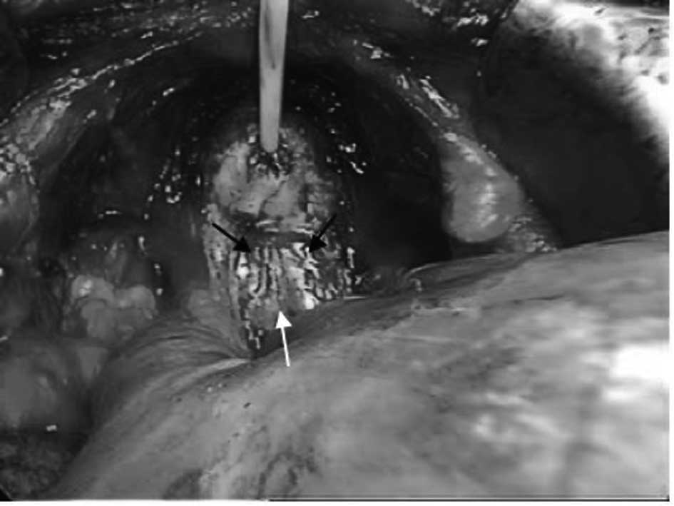
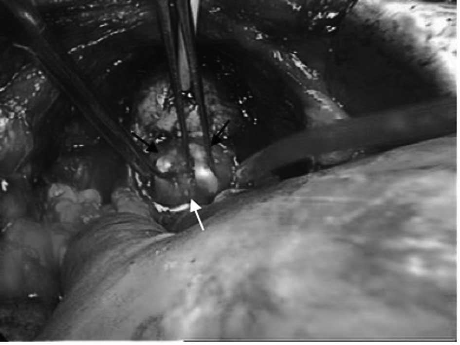
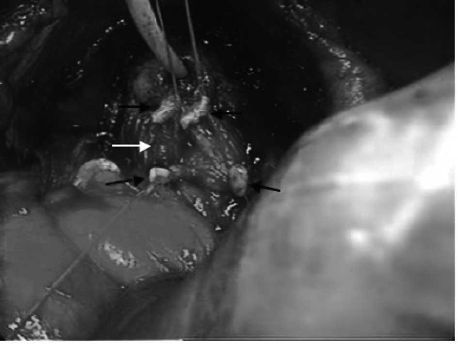
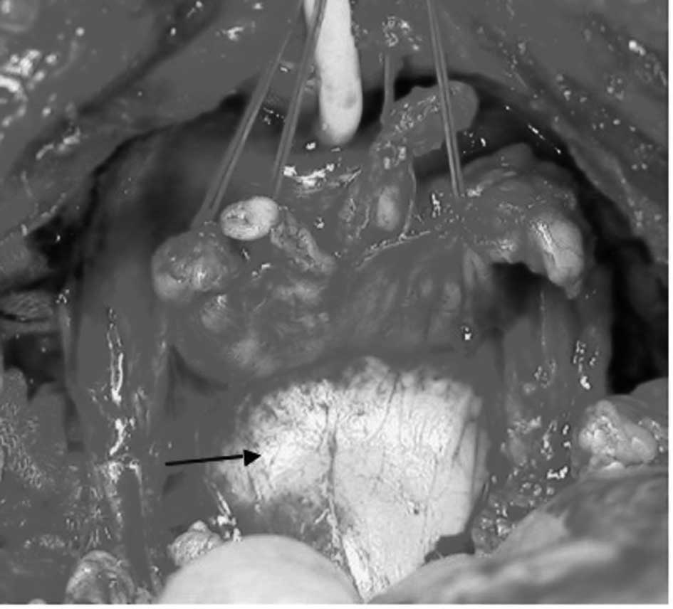
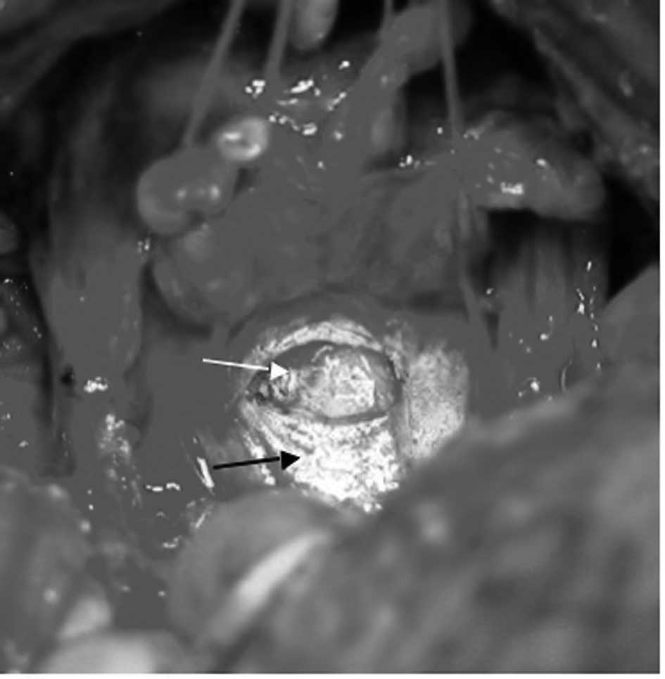
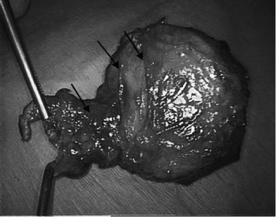
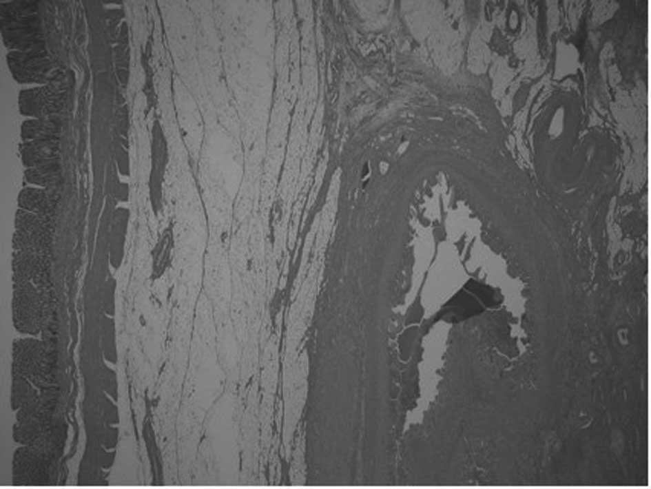
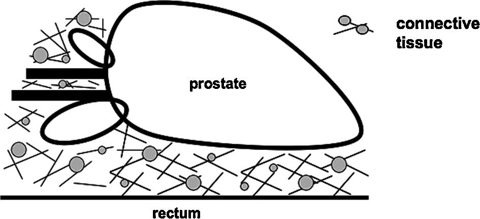
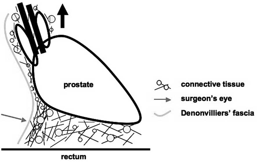
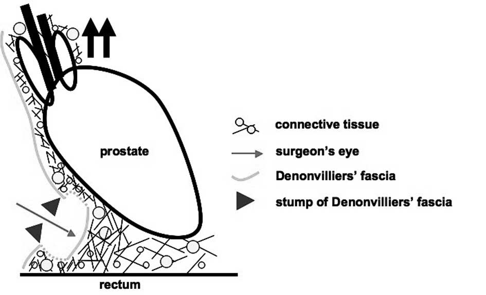
In this study, the region between the prostate and the rectum was examined. We reviewed, macroscopically, the clinical anatomy of the perioperative regions and excised specimens. Moreover, using fixed specimens from three cases of total pelvic exenteration, we histologically reviewed the regions between the prostate and rectum. Following bladder neck transection, the deferent duct was observed in connective tissues that continued to Denonvilliers' fascia. When the prostate was pulled ventrally, this connective tissue was identified as a sheet of membrane that traversed from the anterior prostate to the dorsal rectum. The dorsal surface of the specimen appeared to be covered with a thin membrane, and the regions that continued from this thin membrane were judged macroscopically as a stump. Between the prostate and the rectum, intricate connective tissue was present in the fixed specimen that was removed via a total pelvic exenteration. This connective tissue continued to the connective tissue surrounding the seminal vesicles and prostate. The complex connective tissue was present anatomically between the prostate and rectum. As a result of the surgical procedures, this connective tissue was identified surgically as Denonvilliers' fascia.