Role of vascular endothelial growth factor receptor-3/Flt-4 in early-stage cervical cancer
- Authors:
- Shiqian Zhang
- Hao Yu
- Linlin Zhang
View Affiliations
Affiliations: Department of Obstetrics and Gynecology, Qilu Hospital of Shandong University, Shandong 250012, P.R. China. zhangshiqian370112@126.com
- Published online on: May 1, 2010 https://doi.org/10.3892/ol_00000080
-
Pages:
453-456
Metrics: Total
Views: 0 (Spandidos Publications: | PMC Statistics: )
Metrics: Total PDF Downloads: 0 (Spandidos Publications: | PMC Statistics: )
This article is mentioned in:
Abstract
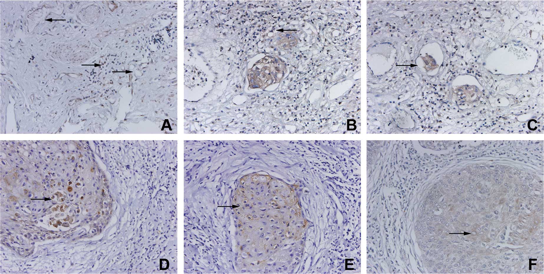
The present study was designed to investigate the role of vascular endothelial growth factor receptor (VEGFR)-3/Flt-4 in the early stages of cervical cancer. VEGFR-3/Flt-4 expression, vascular endothelial growth factor (VEGF)-C and VEGF-D in the early stages (Ia-IIa) of cervical cancer in 41 patients was examined by immunohistochemical analysis. Additionally, the VEGFR-3/Flt-4-marked vascular density (MVD) was examined and the relationship of these factors with clinicopathological factors was analyzed. VEGFR-3/Flt-4 was found to be expressed in lymphatic endothelial cells and, to a certain extent, in vascular endothelial cells. The VEGFR-3/Flt-4-positive vessels were largely distributed in the stroma surrounding the tumor tissues, and these vessels were morphologically divided into blood and lymphatic vessels, respectively. Cancer cells were found to express VEGF-C, VEGF-D and VEGFR-3/Flt-4, and their positive expression rate was 48.7% (20/41), 58.5% (24/41) and 63.4% (26/41), respectively. VEGFR-3/Flt-4 expression in the cancer cells of the cervical cancer patients in our study was found to be correlated to the clinical stage, lymph node metastasis, lymphatic invasion and expression of VEGF-C and VEGF-D. However, this expression was unrelated to menstrual status, histological grade and histological classification. MVD was correlated to the clinical stage and expression of VEGF-C and VEGF-D, but was unrelated to menstrual status, histological grade, histological classification, lymph node metastasis and lymphatic invasion. In conclusion, VEGFR-3/Flt-4 plays an important role in the early stages of cervical cancer.
View References
1
|
Su JL, Yen CJ, Chen PS, et al: The role of
the VEGF-C/VEGFR-3 axis in cancer progression. Br J Cancer.
96:541–545. 2007. View Article : Google Scholar : PubMed/NCBI
|
2
|
Jüttner S, Wissmann C, Jons T, et al:
Vascular endothelial growth factor-D and its receptor VEGFR-3: two
novel independent prognostic markers in gastric adenocarcinoma. J
Clin Oncol. 24:228–240. 2006.PubMed/NCBI
|
3
|
Weidner N, Semple JP, Welch WR and Folkman
J: Tumor angiogenesis and metastasis – correlation in invasive
breast carcinoma. N Engl J Med. 324:1–8. 1991.
|
4
|
Rouzaut A, Irigoyen M and Montuenga LM:
Lymphangiogenesis and lung cancer. J Thorac Oncol. 2:384–386. 2007.
View Article : Google Scholar : PubMed/NCBI
|
5
|
Yasuoka H, Nakamura Y, Zuo H, et al:
VEGF-D expression and lymph vessels play an important role for
lymph node metastasis in papillary thyroid carcinoma. Mod Pathol.
18:1127–1133. 2005. View Article : Google Scholar : PubMed/NCBI
|
6
|
Kawai Y, Minami T, Fujimori M, et al:
Characterization and microarray analysis of genes in human
lymphatic endothelial cells from patients with breast cancer.
Lymphat Res Biol. 5:115–126. 2007. View Article : Google Scholar : PubMed/NCBI
|
7
|
Yang J, Wu HF, Qian LX, et al: Increased
expression of vascular endothelial growth factor (VEGF), VEGF-C and
VEGF receptor-3 in prostate cancer tissue are associated with tumor
progression. Asian J Androl. 8:169–175. 2006. View Article : Google Scholar : PubMed/NCBI
|
8
|
Filho AL, Baltazar F, Bedrossian C,
Michael C and Schmitt FC: Immunohistochemical expression and
distribution of VEGFR-3 in malignant mesothelioma. Diagn
Cytopathol. 35:786–791. 2007. View
Article : Google Scholar : PubMed/NCBI
|
9
|
Saintigny P, Kambouchner M, Ly M, et al:
Vascular endothelial growth factor-C and its receptor VEGFR-3 in
non-small cell lung cancer: concurrent expression in cancer cells
from primary tumour and metastatic lymph node. Lung Cancer.
58:205–213. 2007. View Article : Google Scholar
|
10
|
Van Trappen PO, Steele D, Lowe DG, et al:
Expression of vascular endothelial growth factor (VEGF)-C and
VEGF-D, and their receptor VEGFR-3, during different stages of
cervical carcinogenesis. J Pathol. 201:544–554. 2003.PubMed/NCBI
|
11
|
Su JL, Yang PC, Shih JY, et al: The
VEGF-C/Flt-4 axis promotes invasion and metastasis of cancer cells.
Cancer Cell. 9:209–223. 2006. View Article : Google Scholar : PubMed/NCBI
|
12
|
Timoshenko AV, Rastogi S and Lala PK:
Migration-promoting role of VEGF-C and VEGF-C binding receptors in
human breast cancer cells. Br J Cancer. 97:1090–1098. 2007.
View Article : Google Scholar : PubMed/NCBI
|
13
|
Masood R, Kundra A, Zhu S, Xia G, Scalia
P, Smith DL and Gill PS: Malignant mesothelioma growth inhibition
by agents that target the VEGF and VEGF-C autocrine loops. Int J
Cancer. 104:603–610. 2003. View Article : Google Scholar : PubMed/NCBI
|
14
|
Dias S, Choy M, Alitalo K and Rafii S:
Vascular endothelial growth factor (VEGF)-C signaling through FLT-4
(VEGFR-3) mediates leukemic cell proliferation, survival and
resistance to chemotherapy. Blood. 99:2179–2184. 2002. View Article : Google Scholar : PubMed/NCBI
|
15
|
Wong SY and Hynes RO: Tumor-lymphatic
interactions in an activated stromal microenvironment. J Cell
Biochem. 101:840–850. 2007. View Article : Google Scholar : PubMed/NCBI
|