Prediction of invasion depth for submucosal differentiated gastric cancer by magnifying endoscopy with narrow-band imaging
- Authors:
- Published online on: June 26, 2012 https://doi.org/10.3892/or.2012.1889
- Pages: 841-847
Metrics: Total
Views: 0 (Spandidos Publications: | PMC Statistics: )
Total PDF Downloads: 0 (Spandidos Publications: | PMC Statistics: )
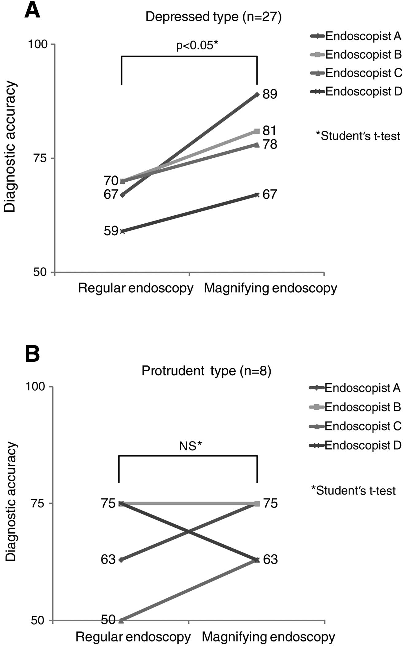
Abstract
The usefulness of determining gastric cancer invasion depth by magnifying endoscopy with narrow-band imaging (NBI-ME) has not been established. The objective of our study was to retrospectively compare NBI-ME images of differentiated submucosal (SM) 1 cancer with those of SM2 to identify the indicators of invasion depth for SM2 gastric cancer. Fifteen patients with SM1 differentiated gastric cancer and 20 with SM2 removed by endoscopic submucosal resection (ESD) were included. NBI-ME images matching the invasion depth of pathological specimens were examined to define the following three findings as diagnostic indicators of SM2: non-structure, scattery vessels and multi-caliber vessels. The relationship between indicators and invasion depth and between indicator score and invasion depth was examined in 27 patients (SM1/SM2: 11/16) with depressed-type gastric cancer (D-GC) and in 8 (SM1/SM2: 4/4) with protruding-type gastric cancer (P-GC). Diagnostic accuracy for invasion depth determined by four endoscopists using regular endoscopic images was compared with that determined by the same endoscopists using NBI-ME. In D-GC, all three indicators were significantly more frequent in SM2 than in SM1 (p<0.05). All D-GC with ≥2 points were SM2, demonstrating a significant difference in score distribution between SM1 and SM2 (p<0.05). In D-GC, diagnostic accuracy by NBI-ME was higher than that by regular endoscopy by all 4 endoscopists (p<0.05). NBI-ME findings of non-structure, scattery vessels and multi-caliber vessels can possibly serve as indicators of SM2 invasion in differentiated D-GC. Scoring of the three indicators was significant.