Introduction
Hepatic fibrosis is an outcome of numerous chronic
liver diseases, including hepatitis B and hepatitis C viral
infections, alcoholic liver disease and non-alcoholic
steatohepatitis (1). Hepatic
fibrosis is a considerable medical problem associated with
significant levels of morbidity and mortality (2). Regardless of the underlying etiology,
hepatic fibrosis is characterized by the accumulation of excess
extracellular matrix (ECM), including types I and III collagen. The
extent of matrix deposition depends on the balance between the
synthesis and degradation of ECM; when the levels of ECM synthesis
exceed those of degradation, the pathological accumulation of ECM
leads to liver fibrosis. The excessive ECM in the liver is mainly
synthesized by activated hepatic stellate cells (HSCs) (3). Therefore, suppressing the activation
of HSCs is essential for alleviation of liver fibrosis.
The Wnt/β-catenin signaling pathway is critical in
development and in adult tissue homeostasis. Wnt/β-catenin
signaling initiates a signaling cascade critical in the normal
development of a number of organs, including the brain, intestines,
skin, liver and lung (4). The
hallmark of this pathway is that it activates the transcriptional
role of the multifunctional protein β-catenin. Canonical Wnt
signaling inactivates glycogen synthase kinase (GSK)-3β, preventing
β-catenin phosphorylation. This action leads to accumulation of
hypophosphorylated β-catenin in the cytoplasm, which subsequently
translocates to the nucleus where it regulates target gene
expression through interactions with members of the T-cell factor
(TCF)/lymphoid enhancer factor (LEF) family of transcription
factors (5). Previous studies have
implicated Wnt/β-catenin signaling in abnormal wound repair and
fibrogenesis. In addition, the Wnt canonical signaling pathway,
mediated by β-catenin, has been indicated to be important in liver
development and remodelling, and HSC activation (6–7).
However, certain pathological processes, including fibrosis and
liver malignancy, may occur partly due to the activation of the Wnt
canonical pathway.
RNA interference is a powerful tool for
post-transcriptional gene silencing (9) and has opened novel avenues in gene
therapy. In the present study, the expression of β-catenin in
hepatic fibrosis was detected in cases of hepatic fibrosis of
different grades and normal hepatic tissues to confirm and explore
the association between β-catenin and the progression of hepatic
fibrosis. Furthermore, a synthetic small interfering RNA (siRNA)
was transfected into HSC-T6 cells to suppress β-catenin expression
and investigate whether inhibition of the Wnt/β-catenin signaling
pathway attenuates hepatic fibrosis.
Materials and methods
Patients
All samples, from patients who underwent liver
biopsies directed by ultrasonography within one week after
inclusion in this study, were obtained from Shanghai Changzheng
Hospital, Second Military Medical University (Shanghai, China) and
Shanghai Xinhua Hospital, Shanghai Jiaotong University School of
Medicine (Shanghai, China), between January 2006 and January 2007.
The sample donation and application procedures were approved by the
Health Human Research Ethics Committee of Shanghai Changzheng
Hospital and the Health Human Research Ethics Committee of Xinhua
Hospital. The study complied with the Declaration of Helsinki,
1995. The patients provided full written informed consent at the
time of sample acquisition and patient anonymity has been
preserved. All samples were treated by 10% formalin fixation and
paraffin imbedding. Diagnosis and classification of the hepatic
fibrosis were determined using Scheuer’s classification (10,11):
F0, no fibrosis; F1, enlarged fibrotic portal tracts; F2,
periportal or portal-portal septa, but intact architecture; F3,
fibrosis with architectural distortion, but no obvious cirrhosis;
F4, cirrhosis. All sections were blindly and independently assessed
by three pathologists and the observed results were processed using
the Kappa concordance test. The inter- and intraobserver results
agreed excellently. A total of 89 samples were included: 15 normal
cases of F0, 18 cases of F1, 20 cases of F2, 20 cases of F3 and 16
cases of F4.
Histological and immunohistochemical
examination
Part of all the paraffin-embedded liver tissues was
stained with hematoxylin and eosin and another part underwent
silver staining for reticular fibers. Immunohistochemical
examination was conducted to detect the expression of β-catenin.
Briefly, the paraffin sections of the left median hepatic lobes
were incubated with 3% H2O2 in methanol at
37°C for 10 min to suppress endogenous peroxidase activity.
Subsequent to blocking at room temperature for 20 min with 10%
bovine serum (Wako, Osaka, Japan), the sections were incubated
overnight at 4°C with antibodies against β-catenin (Santa Cruz
Biotechnology, Inc., Santa Cruz, CA, USA), followed by incubation
with horseradish peroxidase (HRP)-conjugated secondary antibody
(Daco, Kyoto, Japan) at 37°C for 20 min. The signal was amplified
by avidin-biotin complex formation and developed using
diaminobenzidine followed by counterstaining with hematoxylin. The
samples were dehydrated in alcohol and xylene and mounted onto
glass slides. Positive cells were counted in at least 10 fields at
×400 magnification. The incidence of immunoreactivity for β-catenin
was evaluated based on the mean percentage of cells positive, as
follows: High, >80% of cells positive; intermediate, 25–80% of
cells positive; low, <25% of cells positive and negative, <5%
cells positive.
Cell culture
HSC-T6, an immortalized rat HSC line exhibiting a
stable phenotype and biochemical characteristics of activated HSCs,
was donated by Dr SL Friedman (Liver Center Laboratory, San
Francisco General Hospital, CA, USA) (12). All cells were maintained in
RPMI-1640 medium with 10% fetal bovine serum in a humidified
atmosphere at 37°C and 5% CO2. The cells were divided
into four groups: A normal group (NG), in which cells were
maintained in RPMI-1640 medium without transfection; a control
group (CG), in which cells were transfected with non-specific gene
silencing effects; and two treatment groups: in which cells were
transfected with β-catenin siRNA and analyzed after either 24 or 48
h, respectively.
β-catenin siRNA preparation and
transfection
β-catenin-specific siRNAs were designed as described
by Elbashir et al (13).
The sense and antisense sequences of β-catenin siRNA were as
follows: 5′-AAACTACTGTGG ACCACAAGCCCTGTCTC-3′ and 5′-AAGCTTGTGGTC
CACAGTAGTCCTGTCTC-3′, respectively. The siRNA fragments were
synthesized using the Silencer® siRNA Construction kit
(Ambion, Austin, Texas, USA) according to the manufacturer’s
instructions. The cells were transfected with a mixture of plasmid
DNA and Lipofectamine 2000 (Invitrogen Life Technologies, Carlsbad,
CA, USA) in Opti-MEM I medium without serum (Invitrogen Life
Technologies), as recommended by the manufacturer.
Quantitative polymerase chain reaction
(qPCR)
Total RNA was extracted at different time points
following siRNA transfection using a TRIzol kit (Gibco Life
Technologies) according to the manufacturer’s instructions. The
mixture of RNA and primers was loaded into the PCR amplifier
(PE5700; Perkin-Elmer, Norwalk, CT, USA). The following sense and
antisense primers were used: Collagen I,
5′-GGTGGTTATGACTTCAGCTTCC-3′ and 5′-CATGTA GGCTACGCTGTTCTTG-3′;
collagen III, 5′-GTCTTATCA GCCCTGATGGTTC-3′ and 5′-GCTCCATTCACCAGT
GTGTTTA-3′; and β-actin, 5′-TGAAGGTCGGAGTCAACG GATTTGG-3′ and
5′-CATGTGGGCCATGAGGTCCAC CAC-3′. The PCR procedure was as follows:
Predenaturate setting at 95°C for 5 min, denature at 94°C for 45
sec, annealing at 50 °C for 1 min and extension at 72°C for 1 min.
The PCR was performed for 40 cycles followed by a final extension
at 72°C for 10 min. The PCR product was then visualized by running
it on a 1.5% agarose gel and was quantitatively analyzed with
LabWorks 4.5 analysis software (UVP Products, Upland, CA, USA).
Western blot analysis
Following transfection, the cells were harvested and
immediately prepared for protein extraction. The protein content in
the supernatant was detected using the bicinchoninic acid method
(Pierce, Rockford, IL, USA). Equal quantities of protein were run
on 10% SDS-PAGE gel and transferred to polyvinylidene fluoride
membranes. Following incubation with 10% non-fat milk for 1 h, the
membranes were probed with polyclonal rabbit anti-rat β-catenin
antibody (1:400; Sigma, St. Louis, MO, USA) overnight at 4°C and
then incubated with HRP-labeled goat anti-rabbit secondary
antibodies (diluted 1:3,000; Santa Cruz Biotechnology, Inc.). The
protein levels were normalized using β-actin as a loading control.
The relative optical density of the protein bands was measured
using a Zeineh Laser Densitometer (Biomed Instruments Inc.,
Fullerton, CA, USA) after subtracting the film background.
Immunofluorescent staining
Expression of collagen types I and III in HSC-T6
cells infected with β-catenin siRNAs was examined by
immunocytofluorescent staining using polyclonal antibodies against
collagen types I and III (Boster Biological Tech Ltd., Fremont, CA,
USA). The fixed cells were treated with the primary antibodies
(against collagen types I and III) overnight at 4°C, followed by
incubation with secondary antibodies (TRITC AffiniPure Goat
Anti-Rabbit IgG; EarthOx, LLC, San Francisco, CA, USA) at 4°C for 2
h. The cells were then stained for 30 min at room temperature with
4,6-diamidino-2-phenylindole. Following rinsing, the slides were
viewed with a Zeiss LSM-510 Laser Scanning Confocal microscope
(Carl Zeiss AG, Oberkochen, Germany). The fluorescence was
quantified with semi-quantitative analysis by image scanning.
Cell proliferation and cell cycle
analysis
The effect of siRNA-mediated downregulation of
β-catenin on HSC-T6 cell proliferation was determined by
3-(4,5-dimethylthiazol-2-yl)-2,5-diphenyl tetrazolium bromide (MTT)
assay. The cell suspension was placed into 96-well plates at 1,000
cells per well with eight repeat wells and incubated for 1, 2, 3, 4
or 5 days after transfection. The cells were then incubated with 20
μl methyl thiazolyl tetrazolium for 4 h. Following centrifugation,
150 μl dimethylsulfoxide was added to the precipitate and the
absorbance of the enzyme was measured at 490 nm with a microplate
reader (MK3; Multiskan Co., Vantaa, Finland). For the cell cycle
analysis, HSC-T6 cells infected with β-catenin siRNA were harvested
and fixed with 70% ethanol at 20°C for 24 h. Subsequently,
1×10⁵ cells were prepared to analyze the cell cycle
phases by flow cytometry (ZM Coulter Counter; Coulter Electronics
Inc., Hialeah, FL, USA).
Apoptosis assessment by Annexin V
staining
To detect the cells in the early stages of
apoptosis, the Annexin V-fluorescein isothiocyanate (FITC)
Apoptosis Detection kit (BD Biosciences, San Diego, CA, USA) was
used according to the manufacturer’s instructions. Briefly,
following transfection for 48 h, HSC-T6 cells were harvested and
stained with propidium iodide and Annexin V-FITC in 100 μl staining
solution at room temperature for 15 min in the dark. Samples were
then diluted with binding buffer and were analyzed by the
FACScalibur flow cytometer (BD Immunocytometry Systems, San Jose,
CA, USA) within 1 h.
Statistical analysis
Continuous data were expressed as the mean ±
standard deviation. For statistical analysis, the group
distributions were compared parametrically using Student’s t-test
or one-way analysis of variance, and the group distributions were
compared non-parametrically using the Mann-Whitney U test. The
results were analyzed statistically using the Kruskal-Wallis test
for the association between the incidence of immunoreactivity for
β-catenin and the histological grade. P<0.05 was considered to
indicate a statistically significant difference.
Results
Histological and immunohistochemical
assessment of the hepatic fibrosis tissues
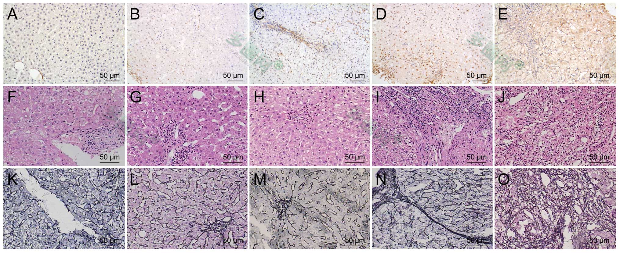
Histologically, the hepatic fibrosis tissues
exhibited connective tissue fibers extending from the central vein,
and early septal formation was established in the livers of the F2
group. The expression levels of β-catenin were examined in the
tissues by immunohistochemistry. The majority of β-catenin staining
was observed in the high grade hepatic fibrosis tissues. Little
specific β-catenin staining was observed in normal liver tissues
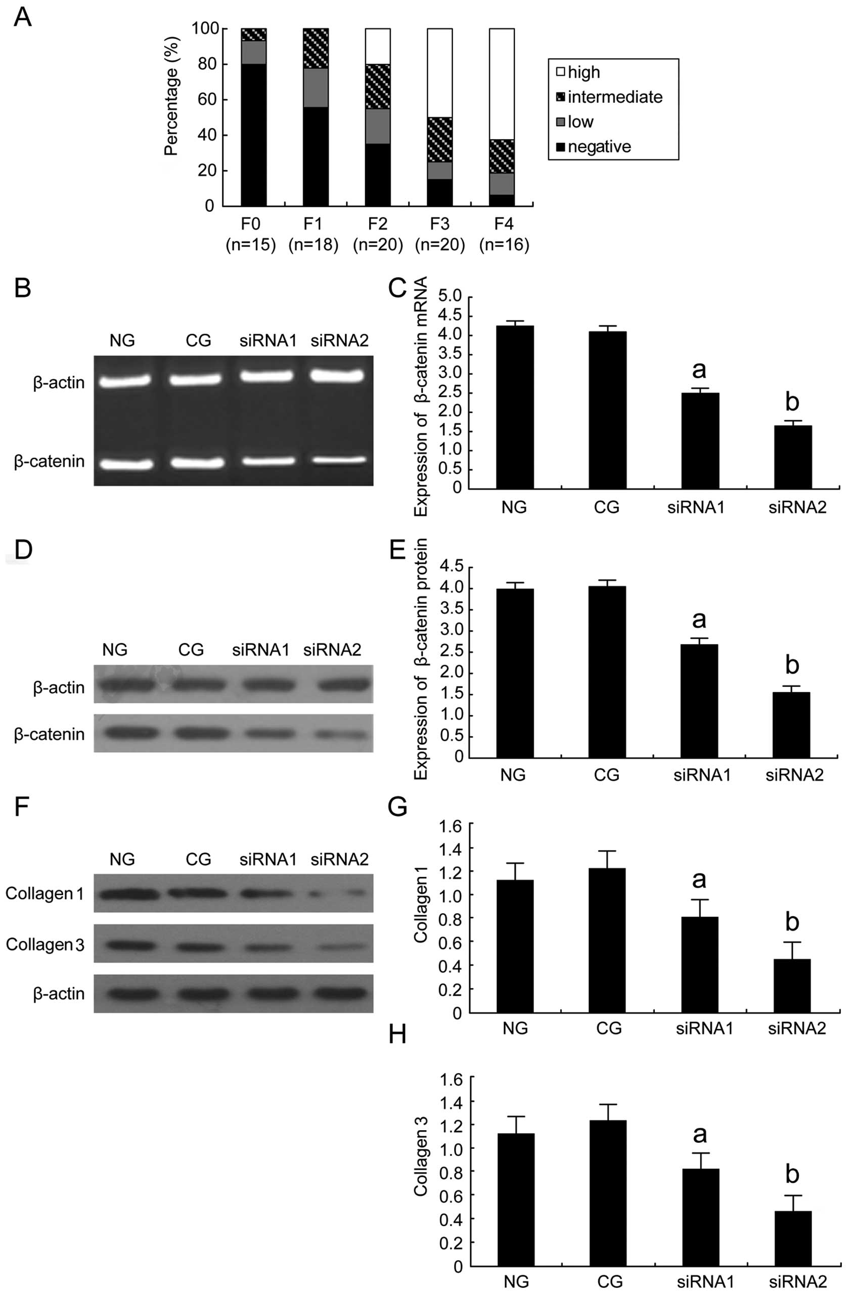
(Fig. 1). Compared with that of
the normal controls, β-catenin was overexpressed in hepatic
fibrosis and its expression level increased with the histological
grade of the fibrosis (P<0.05; Fig.
2A). The data suggested that overexpression of β-catenin may be
associated with the degree of liver fibrosis and may be important
in the initiation and progression of hepatic fibrosis.
 | Figure 1β-catenin was upregulated in liver
fibrogenesis. (A–E) Immunohistochemical analysis of β-catenin
distribution and expression in liver fibrosis specimens (original
magnification, ×400). Brown color signifies positive expression.
(A) No immunoreactivity was detected in the normal liver tissue;
(B) weak staining in liver fibrosis tissue at F1; (C and D)
moderate staining in liver fibrosis tissue at F2 and F3; (E) strong
staining in liver fibrosis tissue at F4. (F–J) Hematoxylin and
eosin was used to examine pathological alterations and collagen
deposition. Images F–J indicate fibrosis stages F0, F1, F2, F3 and
F4, respectively. (K–O) Silver staining of reticular fibers was
used to examine pathological alterations and collagen deposition.
Images K–O indicate fibrosis stages F0, F1, F2, F3 and F4,
respectively. F0, no fibrosis; F1, enlarged fibrotic portal tracts;
F2, periportal or portal-portal septa, but intact architecture; F3,
fibrosis with architectural distortion, but no obvious cirrhosis;
F4, cirrhosis. |
 | Figure 2(A) Expression levels of β-catenin in
different grades of hepatic fibrosis. F0, no fibrosis; F1, enlarged
fibrotic portal tracts; F2, periportal or portal-portal septa, but
intact architecture; F3, fibrosis with architectural distortion,
but no obvious cirrhosis; F4, cirrhosis. Incidence of
immunoreactivity for β-catenin: High, >80% of cells positive;
intermediate, 25–80% of cells positive; low, <25% of cells
positive and negative, <5% cells positive. (B–E) β-catenin siRNA
inhibited β-catenin mRNA and protein expression in HSC-T6 cells
compared with NG and CG cells. β-actin served as the internal
loading control. (B) qPCR analysis for β-catenin mRNA expression in
HSC-T6 cells following β-catenin siRNA transfection. (C)
Semi-quantitative analysis of the qPCR result. (D) Western blot
analysis of β-catenin protein expression following transfection.
(E) Semi-quantitative analysis of the western blotting results.
(F–H) β-catenin siRNA inhibited collagen types I and III mRNA
expression in HSC-T6 cells. (F) qPCR analysis for collagen types I
and III mRNA expression in HSC-T6 cells following siRNA β-catenin
transfection. β-actin served as the internal loading control. (G
and H) Semi-quantitative analysis of the qPCR result.
aP<0.05, bP<0.01 compared with NG and
CG. n, number of samples; siRNA, small interfering RNA; NG, normal
group; CG, control group; qPCR, quantitative polymerase chain
reaction. siRNA1, 24 h after transfection; siRNA2, 48 h after
transfection. |
β-catenin siRNA effectively inhibits
β-catenin expression in HSC-T6 cells
Forty-eight hours after transfection with β-catenin
siRNA, the β-catenin transcript and protein levels were reduced in
the transfected cells compared with those in the control group.
This β-catenin gene-silencing effect was reproducible and specific
as it failed to knock down the expression of an unrelated protein,
β-actin. The β-catenin siRNA used in the present study was able to
reduce β-catenin mRNA expression levels in HSC-T6 cells compared
with the levels in the NG and CG (Fig.
2B). Western blotting further confirmed the β-catenin siRNA
silencing of the β-catenin protein in the HSC-T6 cells (Fig. 2D). Semi-quantitative analysis of
the qPCR and western blotting results (Fig. 2C and E) also revealed that
β-catenin siRNA reduced the expression levels of β-catenin in
HSC-T6 cells comapared with those in the NG or CG.
β-catenin siRNA downregulates collagen
types I and III in HSC-T6 cells
To investigate the effect of β-catenin siRNA on
collagen degradation, the quantity of collagen types I and III in
HSC-T6 cells transfected with β-catenin siRNA was examined using
qPCR and immunofluorescent staining. The expression levels of
collagen types I and III mRNAs were reduced in the HSC-T6 cells
transfected with β-catenin siRNA, compared with those in the cells
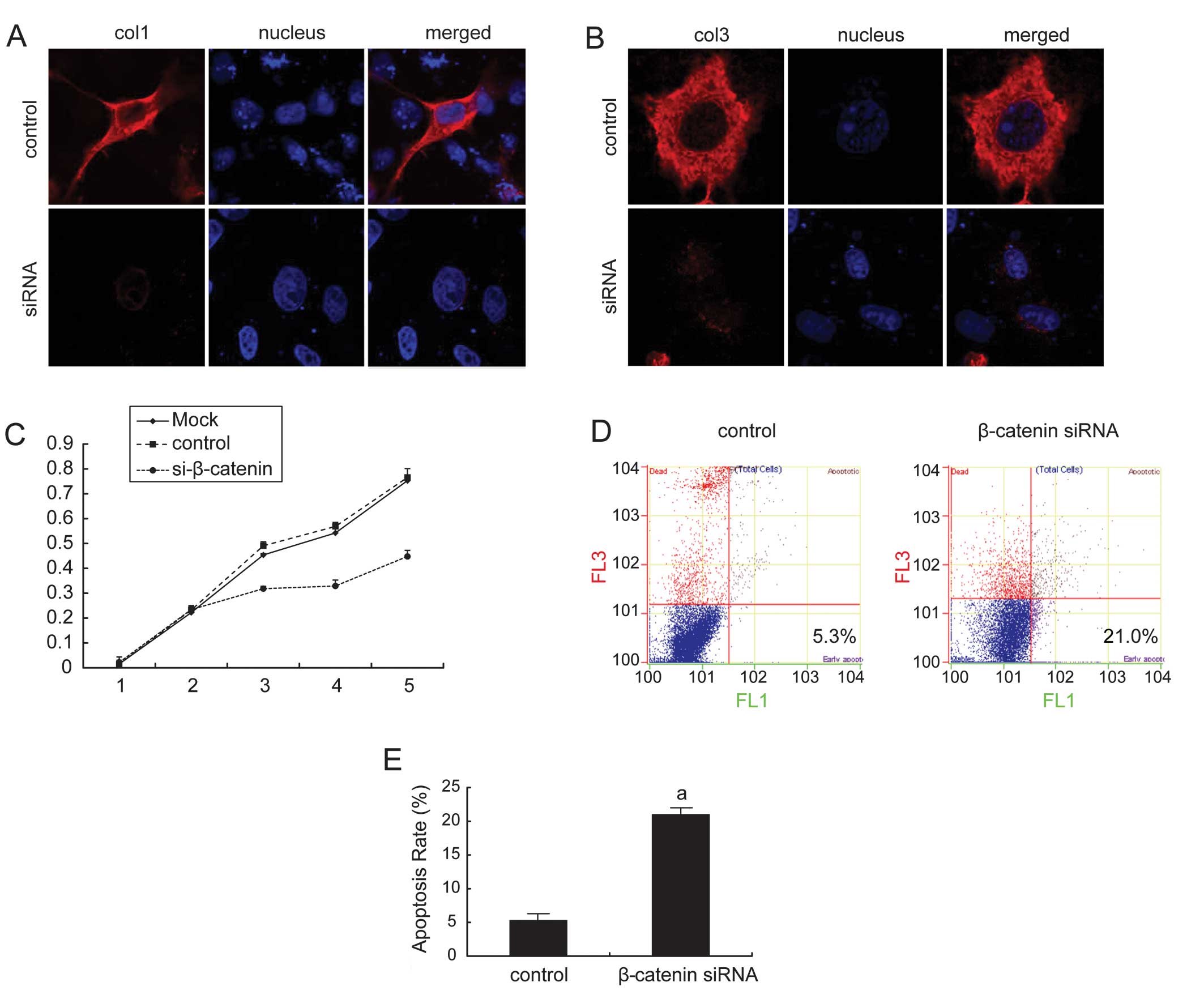
of the NG or CG (Fig. 2F–H). The
fluorescent light for collagen types I and III observed in the cell
cytoplasm was significantly reduced in the β-catenin siRNA group
compared with that in the control group. The expression of collagen
type I was reduced by 82% and the expression of collagen type III
was reduced by 78% by β-catenin siRNA treatment using
quantification of the fluorescent light intensity compared with
that in the controls (Fig.
3A–B).
β-catenin siRNA inhibits the
proliferation of HSC-T6 cells
To investigate whether the reduction in β-catenin
levels exhibits an effect on cell growth and proliferation, cell
cycle distribution and MTT assays were performed. Knockdown of
β-catenin by siRNA at 3, 4 and 5 days after transfection led to
reduced cell proliferation in the treatment group than that in the
control and mock groups (Fig. 3C).
Investigation of the cell cycle also indicated that cells were
arrested in the G0/G1 phase and the proportion of cells in the S
phase was significantly reduced compared with the control group
following downregulation of β-catenin in the HSCs (Table I).
 | Table IEffect of β-catenin siRNA on the cell
cycle (%). |
Table I
Effect of β-catenin siRNA on the cell
cycle (%).
| Cell cycle phase | β-catenin siRNA
group | Control group |
|---|
| G0/G1 | 59.42±0.46a | 43.52±0.65 |
| S | 28.68±1.28a | 42.56±1.62 |
| G2/M | 13.28±1.06 | 14.38±1.26 |
β-catenin siRNA induces apoptosis in
HSC-T6 cells
Annexin V staining revealed that the proportion of
apoptotic HSC-T6 cells transfected with β-catenin siRNA was
significantly elevated when compared with the proportion of
apoptotic cells in the control group (Fig. 3D–E). The rate of apoptosis
increased from 5.3% in the control group to 21.0% in the β-catenin
siRNA group (P<0.01). This result suggested that siRNA targeting
of β-catenin was able to induce apoptosis in HSC-T6 cells.
Discussion
Hepatic fibrosis is a chronic pathological process
that involves an inflammatory response characterized by an
excessive release of inflammatory cytokines and infiltration of
inflammatory cells (14,15). All have been demonstrated to
promote the activation of HSCs and deposition of ECM in the liver,
leading to liver fibrosis (16).
HSCs are important in ECM remodeling and have been used as an in
vitro cell model of hepatic fibrosis for over a decade
(12). HSCs are the most important
source of ECM proteins during this fibrotic process. Therefore, the
majority of antifibrotic therapies are designed to inhibit the
activation, proliferation or synthetic products of HSCs.
The Wnt signaling pathway is an evolutionarily
conserved, complex signaling pathway that is critical for
development, differentiation and cellular homeostasis (17). Three signal transduction pathways
are activated by Wnt: The canonical Wnt pathway, the planar cell
polarity pathway and the Wnt/Ca2+ pathway (18). The canonical Wnt/β-catenin pathway
is centered on regulating the levels of its predominant effector,
β-catenin (19). In the absence of
Wnt ligands, β-catenin is sequestered in the cytoplasm by a protein
complex composed of the scaffolding protein Axin, adenomatous
polyposis coli, casein kinase 1 (CK1) and GSK3β. Specific serine
and threonine residues in the amino terminal region of β-catenin
are sequentially phosphorylated by CK1 and GSK3β. Phosphorylated
β-catenin is then ubiquinated and targeted for proteasomal
degradation (20). As a result of
this continuous degradation, β-catenin is prevented from
accumulating in the cytoplasm and therefore from reaching the
nucleus. In the absence of nuclear β-catenin, Wnt-target genes are
repressed by binding of the TCF/LEF family of proteins. Activation
of the canonical pathway results in inhibition of GSK3 and allows
stabilization of cytosolic β-catenin and its translocation to the
nucleus, where it binds to the TCF/LEF family of transcription
factors to stimulate the expression of multiple Wnt target genes,
including c-myc, c-jun and cyclin D1 (18,21).
Wnt signals are also regulated by secreted antagonists that either
bind directly to the ligand, including Wnt inhibitory factor and
the secreted frizzled (Fz)-related protein family, or prevent
lipoprotein receptor-related protein (LRP) coreceptor association
with Fz. Dickkopf (Dkk), the best-characterized of the latter group
of antagonists, binds to LRP6 and inhibits the Wnt-induced
Fz-LRP5/6 complex formation that is essential to the canonical
pathway. Canonical Wnt signaling through β-catenin is facilitated
by Wnt family members, including Wnt1, Wnt3a and Wnt10b and Fz
receptors, such as Fz1 and Fz5 (21,22).
Wnt/β-catenin is an evolutionarily conserved
cellular signaling system essential in the pathogenesis of numerous
human diseases (5,23). Wnt signaling has been implicated in
human fibrotic diseases, including pulmonary and renal fibrosis
(4,24). Since Wnt activity is enhanced in
liver cirrhosis (25), it is also
likely that Wnt signaling is involved in hepatic fibrogenesis. The
importance of the Wnt/β-catenin pathway in the liver only began to
be recognized in the late 1990’s (26). As in other organs, the
Wnt/β-catenin pathway is critical for liver development. Certain
studies have suggested that the pathway is also important in liver
regeneration, liver metabolism and maintenance of normal function
in the adult liver (27–29). Aberrant activation of β-catenin has
been implicated in the pathogenesis of hepatobiliary neoplasias,
ranging from benign lesions to liver cancer (8,30–33).
One study used DNA microarrays to investigate gene-expression
differences between quiescent and activated rat HSCs; the levels of
the ligands Wnt4 and Wnt5 and the Wnt receptor Fz-2 were found to
be upregulated (6). In another
study, increased expression levels of canonical Wnt3a and 10b and
noncanonical (Wnt4 and Wnt5a) Wnt genes, along with Fz-1 and -2 as
well as Fz coreceptors, were identified in activated HSCs, but not
in quiescent HSCs (34). However,
higher levels of nuclear β-catenin and TCF DNA binding were found
in activated HSCs, suggesting that canonical Wnt signaling is
important in HSC activation. Since treatment with the inhibitors of
Wnt signaling, Dkk-1 and Chibby, was able to restore quiescence of
HSCs in culture, and high expression levels of Dkk-1 induced
apoptosis of cultured HSCs, these results raise the possibility
that inhibition of Wnt signaling may be a potential therapeutic
strategy for preventing liver fibrosis.
Inhibition of the Wnt pathway has also been analyzed
in the regulation of fibrotic responses in tissues. When Dkk, the
best characterized naturally secreted antagonist of the Wnt
pathway, was administered locally in a naked plasmid vector for
gene therapy, blockade of canonical Wnt pathway inhibited renal
interstitial fibrosis (35).
Inhibition of the Wnt/β-catenin pathway by β-catenin siRNA also
reduced bleomycin-induced pulmonary fibrosis in mice (36). In the present study, β-catenin
expression in hepatic fibrosis was verified and the correlation
between β-catenin and hepatic fibrosis progression was explored.
Following silencing of β-catenin in HSCs by siRNA, there was found
to be a reduction in the levels of collagen types I and III mRNA
and protein expression, demonstrating that inhibition of β-catenin
directly results in suppression of HSC activation and collagen
synthesis. β-catenin siRNA was also found in the regulation of cell
proliferation and in the cell cycle. To the best of our knowledge,
this is the first study that has used siRNA targeting of β-catenin
to inhibit HSC activation. As β-catenin is multifunctional with
multiple molecular interactions, the definitive mechanism
responsible remains uncertain.
In conclusion, β-catenin was upregulated during
liver fibrogenesis. A reduction in the β-catenin expression levels
in HSCs with siRNA suppressed the activity of HSCs and collagen
synthesis, and enhanced collagen degradation. The results of the
present study suggest a significant functional role for β-catenin
in the development of liver fibrosis and that targeted knockdown of
β-catenin mRNA may have a therapeutic effect on hepatic
fibrosis.
Acknowledgements
This study was supported by the Select and Train
Outstanding Young Teachers Foundation of Shanghai (grant no.
jdy08086), the Wu Jieping Medical Foundation (Experimental
Diagnosis of Liver Disease; grant no. LDWMF-SY-2011B009) and
Shanghai Key Laboratory of Pediatric Gastroenterology and Nutrition
(grant no. 11DZ2260500).
References
|
1
|
Moreira RK: Hepatic stellate cells and
liver fibrosis. Arch Pathol Lab Med. 131:1728–1734. 2007.PubMed/NCBI
|
|
2
|
Gäbele E, Brenner DA and Rippe RA: Liver
fibrosis: signals leading to the amplification of the fibrogenic
hepatic stellate cell. Front Biosci. 8:d69–d77. 2003.PubMed/NCBI
|
|
3
|
Friedman SL: Molecular regulation of
hepatic fibrosis, an integrated cellular response to tissue injury.
J Biol Chem. 275:2247–2250. 2000. View Article : Google Scholar : PubMed/NCBI
|
|
4
|
Morrisey EE: Wnt signaling and pulmonary
fibrosis. Am J Pathol. 162:1393–1397. 2003. View Article : Google Scholar : PubMed/NCBI
|
|
5
|
Moon RT, Kohn AD, De Ferrari GV and Kaykas
A: WNT and beta-catenin signalling: diseases and therapies. Nat Rev
Genet. 5:691–701. 2004. View
Article : Google Scholar : PubMed/NCBI
|
|
6
|
Jiang F, Parsons CJ and Stefanovic B: Gene
expression profile of quiescent and activated rat hepatic stellate
cells implicates Wnt signaling pathway in activation. J Hepatol.
45:401–409. 2006. View Article : Google Scholar : PubMed/NCBI
|
|
7
|
Jiao J, Friedman SL and Aloman C: Hepatic
fibrosis. Curr Opin Gastroenterol. 25:223–229. 2009. View Article : Google Scholar
|
|
8
|
Nejak-Bowen K and Monga SP:
Wnt/beta-catenin signaling in hepatic organogenesis. Organogenesis.
4:92–99. 2008. View Article : Google Scholar : PubMed/NCBI
|
|
9
|
Chen SW, Chen YX, Zhang XR, et al:
Targeted inhibition of platelet-derived growth factor receptor-beta
subunit in hepatic stellate cells ameliorates hepatic fibrosis in
rats. Gene Ther. 15:1424–1435. 2008. View Article : Google Scholar : PubMed/NCBI
|
|
10
|
Scheuer PJ: The nomenclature of chronic
hepatitis: time for a change. J Hepatol. 22:112–114. 1995.
View Article : Google Scholar : PubMed/NCBI
|
|
11
|
Scheuer PJ: Classification of chronic
viral hepatitis: a need for reassessment. J Hepatol. 13:372–374.
1991. View Article : Google Scholar : PubMed/NCBI
|
|
12
|
Vogel S, Piantedosi R, Frank J, et al: An
immortalized rat liver stellate cell line (HSC-T6): a new cell
model for the study of retinoid metabolism in vitro. J Lipid Res.
41:882–893. 2000.PubMed/NCBI
|
|
13
|
Elbashir SM, Harborth J, Weber K and
Tuschl T: Analysis of gene function in somatic mammalian cells
using small interfering RNAs. Methods. 26:199–213. 2002. View Article : Google Scholar : PubMed/NCBI
|
|
14
|
Friedman SL: Hepatic fibrosis - overview.
Toxicology. 254:120–129. 2008. View Article : Google Scholar
|
|
15
|
Connolly MK, Bedrosian AS, Mallen-St Clair
J, et al: In liver fibrosis, dendritic cells govern hepatic
inflammation in mice via TNF-alpha. J Clin Invest. 119:3213–3225.
2009.PubMed/NCBI
|
|
16
|
Friedman SL: Mechanisms of hepatic
fibrogenesis. Gastroenterology. 134:1655–1669. 2008. View Article : Google Scholar : PubMed/NCBI
|
|
17
|
MacDonald BT, Tamai K and He X:
Wnt/beta-catenin signaling: components, mechanisms, and diseases.
Dev Cell. 17:9–26. 2009. View Article : Google Scholar : PubMed/NCBI
|
|
18
|
Habas R and Dawid IB: Dishevelled and Wnt
signaling: is the nucleus the final frontier? J Biol. 4:22005.
View Article : Google Scholar : PubMed/NCBI
|
|
19
|
Gordon MD and Nusse R: Wnt signaling:
multiple pathways, multiple receptors, and multiple transcription
factors. J Biol Chem. 281:22429–22433. 2006. View Article : Google Scholar : PubMed/NCBI
|
|
20
|
He X, Semenov M, Tamai K and Zeng X: LDL
receptor-related proteins 5 and 6 in Wnt/beta-catenin signaling:
arrows point the way. Development. 131:1663–1677. 2004. View Article : Google Scholar : PubMed/NCBI
|
|
21
|
Logan CY and Nusse R: The Wnt signaling
pathway in development and disease. Annu Rev Cell Dev Biol.
20:781–810. 2004. View Article : Google Scholar : PubMed/NCBI
|
|
22
|
Young CS, Kitamura M, Hardy S and
Kitajewski J: Wnt-1 induces growth, cytosolic beta-catenin, and
Tcf/Lef transcriptional activation in Rat-1 fibroblasts. Mol Cell
Biol. 18:2474–2485. 1998.PubMed/NCBI
|
|
23
|
Schmidt-Ott KM and Barasch J:
WNT/beta-catenin signaling in nephron progenitors and their
epithelial progeny. Kidney Int. 74:1004–1008. 2008. View Article : Google Scholar : PubMed/NCBI
|
|
24
|
Surendran K, McCaul SP and Simon TC: A
role for Wnt-4 in renal fibrosis. Am J Physiol Renal Physiol.
282:F431–F441. 2002.PubMed/NCBI
|
|
25
|
Shackel NA, McGuinness PH, Abbott CA,
Gorrell MD and McCaughan GW: Identification of novel molecules and
pathogenic pathways in primary biliary cirrhosis: cDNA array
analysis of intrahepatic differential gene expression. Gut.
49:565–576. 2001. View Article : Google Scholar : PubMed/NCBI
|
|
26
|
Nhieu JT, Renard CA, Wei Y, Cherqui D,
Zafrani ES and Buendia MA: Nuclear accumulation of mutated
beta-catenin in hepatocellular carcinoma is associated with
increased cell proliferation. Am J Pathol. 155:703–710. 1999.
View Article : Google Scholar : PubMed/NCBI
|
|
27
|
Monga SP, Pediaditakis P, Mule K, Stolz DB
and Michalopoulos GK: Changes in WNT/beta-catenin pathway during
regulated growth in rat liver regeneration. Hepatology.
33:1098–1109. 2001. View Article : Google Scholar : PubMed/NCBI
|
|
28
|
Sodhi D, Micsenyi A, Bowen WC, et al:
Morpholino oligonucleotide-triggered beta-catenin knockdown
compromises normal liver regeneration. J Hepatol. 43:132–141. 2005.
View Article : Google Scholar : PubMed/NCBI
|
|
29
|
Cadoret A, Ovejero C, Terris B, et al: New
targets of beta-catenin signaling in the liver are involved in the
glutamine metabolism. Oncogene. 21:8293–8301. 2002. View Article : Google Scholar : PubMed/NCBI
|
|
30
|
Si-Tayeb K, Lemaigre FP and Duncan SA:
Organogenesis and development of the liver. Dev Cell. 18:175–189.
2010. View Article : Google Scholar
|
|
31
|
Nejak-Bowen KN, Thompson MD, Singh S, et
al: Accelerated liver regeneration and hepatocarcinogenesis in mice
overexpressing serine-45 mutant beta-catenin. Hepatology.
51:1603–1613. 2010. View Article : Google Scholar : PubMed/NCBI
|
|
32
|
Audard V, Grimber G, Elie C, et al:
Cholestasis is a marker for hepatocellular carcinomas displaying
beta-catenin mutations. J Pathol. 212:345–352. 2007. View Article : Google Scholar : PubMed/NCBI
|
|
33
|
Kim M, Lee HC, Tsedensodnom O, et al:
Functional interaction between Wnt3 and Frizzled-7 leads to
activation of the Wnt/beta-catenin signaling pathway in
hepatocellular carcinoma cells. J Hepatol. 48:780–791. 2008.
View Article : Google Scholar : PubMed/NCBI
|
|
34
|
Cheng JH, She H, Han YP, et al: Wnt
antagonism inhibits hepatic stellate cell activation and liver
fibrosis. Am J Physiol Gastrointest Liver Physiol. 294:G39–G49.
2008. View Article : Google Scholar : PubMed/NCBI
|
|
35
|
He W, Dai C, Li Y, et al: Wnt/beta-catenin
signaling promotes renal interstitial fibrosis. J Am Soc Nephrol.
20:765–776. 2009. View Article : Google Scholar : PubMed/NCBI
|
|
36
|
Kim TH, Kim SH, Seo JY, et al: Blockade of
Wnt/β-catenin pathway attenuates bleomycin-induced pulmonary
fibrosis. Tohoku J Exp Med. 223:45–54. 2011.
|


