Introduction
Breast cancer is one of the most common carcinoma
risks worldwide, particularly females (1). According to an overview that referred to
the female breast cancer statistics for 2013 in the United States
(US), it was reported that ~232,340 new cases of invasive breast
cancer and 39,620 breast cancer fatalities were predicted among US
women in 2013. It has been estimated that 1 in 8 women in the US
will develop breast cancer in their lifetimes (2). In developing countries including China,
breast cancer ranks only second to cervical cancer in female
morbidity and mortality (3).
According to the epidemiological investigation,
nearly 70% of patients with breast carcinomas are eligible for
endocrine adjuvant treatment due to the expression of the estrogen
receptor-α (ERα) (4). Selective ER
modulators (SERMs) are synthetic non-steroidal compounds that
switch target sites on and off throughout the body. Tamoxifen
(TAM), the pioneering SERM, which acts to block estrogen action by
binding to the ER in breast carcinomas, has been used ubiquitously
in clinical treatment during the last 30 years and is proved to
reduce the risk of breast cancer in high-risk women with numerous
clinical evidence (5,6).
Although viewed as highly effective in anticancer
endocrinotherapy, the various side effects must be considered. The
recent studies have already reported the numerous long-term side
effects, as patients are recommended to continue the treatment for
>5 years. The long-term use of TAM may cause ranges of side
effects such as hot flashes, night sweats, gynecological symptoms
(vaginal dryness and vaginal discharge), depression, memory loss,
sleep alterations and diminished sexual function (7,8). Among all
these, 3 of the most serious adverse events are endometrial
hyperplasia or endometrial cancer, venous thromboembolic disease,
and hepatic injury or even hepatocarcinoma (which has only been
reported in rats) (9–11). Recently, there were numerous studies
that illustrated the specific mechanism of TAM-induced
hepatotoxicity, however, studies on TAM-induced morphological
changes in hepatic injury remain to be elucidated, particularly in
its early stage of long-term endocrinotherapy (12,13).
In the present study, the hepatotoxicity effects of
TAM treatment at the early stage of endocrinotherapy were
elucidated. Additionally, the morphological changes of hepatocytes
in TAM-treated mice were observed at the microscopic and
ultrastructural levels.
Materials and methods
Reagents
TAM (Wuhan Wei Shunda Technology Co., Ltd., Wuhan,
China), Radioimmunoprecipitation Assay (RIPA; Sigma, St. Louis, MO,
USA) triturator, rabbit polyclonal anti-human caspase-3, β-actin,
horseradish peroxidase (HRP)-linked goat anti-rabbit and HRP-linked
goat anti-mouse (Santa Cruz Biotechnology, Inc., Dallas, TX, USA)
were used.
Western blotting
Mouse liver tissues were lysed in modified RIPA
buffer. The protein concentration was measured by the BCA™ protein
assay kit (Pierce Biotechnology, Inc., Rockford, IL, USA). Equal
amounts of total protein (20 µg) were loaded and run on a 5% (v/v)
sodium dodecyl sulphate (SDS)-polyacrylamide stacking gel and 10%
(v/v) SDS-polyacrylamide gradient gel, and were subsequently
transferred to polyvinylidene fluoride membranes (Roche Carolina,
Florence, SC, USA). Membranes were blocked for 1 h at room
temperature with 5% powdered skimmed milk in Tris-buffered saline
with 0.1% Tween-20, and were subsequently probed with caspase-3
antibody (cat. no. CB91021324; 1:1,000 dilution; Alexis
Biochemicals, San Diego, CA, USA) or β-actin antibody (cat. .no.
874526765; 1:2,000 dilution; Roche Carolina) at 4°C overnight.
After incubation with HRP-linked secondary antibodies (sc55227;
1:5,000, Santa Cruz Biotechnology, Inc.) for 2 h at room
temperature, the blots were detected with and ECL kit (Easy Blot,
Sangon Biotech, Shanghai, China). The experiments were repeated 3
times.
Animal experiment
Female inbred 7–8-week-old Kunming mice were
purchased from the ABSL-III laboratory at Wuhan University (Wuhan,
Hebei, China). All the mice were fed under identical conditions in
an aseptic facility and provide free access to water and food. Mice
were randomly allocated to two groups with 6 for each. Group I were
labeled as the control group, administrated with intraperitoneal
(i.p.) injection of physiological saline of the same dose for 2
weeks (the average weight of mice was 27 g). Group II were labeled
as the TAM group, administrated with i.p. injection of 6 mg/kg/day
TAM for 2 weeks. The body weight of each mouse was detected to
evaluate the effects of the treatment. At the end of the time
period, mice were euthanized by carbon dioxide. Immediately
following the sacrifice of the animals, small pieces of liver
tissues were obtained and processed for histopathological analysis
and transmission electron microscopy (TEM) examination. All the
animal work was performed in accordance with protocols and
guidelines approved by the Institutional Animal Care and Use
Committee (Wuhan University).
Determination of the serum biochemical
parameters
Activities of enzyme markers of hepatocellular
damage, including alanine aminotransferase (ALT) and aspartate
aminotransferase (AST) in serum were determined every three days.
All the analyses were performed in triplicate for every sample
using a Semi-automatic Biochemical Analyzer (Vital Scientific,
Spankeren, Netherlands).
Hepatic cell growth and morphology
under an inverted microscope
Liver tissue sections were collected and fixed in 4%
paraformaldehyde in 0.1 M of phosphate-buffered saline (PBS) at
room temperature overnight. Paraffin-embedded tissue sections (≤5
µm) were stained with hematoxylin and eosin, according to standard
techniques. Images were captured using a Motic BA200 microscope
(Motic Instruments, Inc., Baltimore, MD, USA).
Ultrastructural changes under TEM
Liver tissues were cut into 1-mm3
fragments and fixed by immersion in 4% prechilled glutaraldehyde in
PBS [0.1 M (pH 7.4)] overnight at 4°C. Following this, the samples
were washed in PBS four times followed by post-fixation with 1%
osmium tetroxide in 0.1 M PBS for 2 h at 4°C. Samples were
dehydrated in graded ethanol, embedded in Epon 812, and
subsequently cut into ultra- or semi-thin sections. The sections
were examined under a transmission electron microscope (Hitachi
HT7700-SS; Hitachi, Tokyo, Japan).
Statistical analysis
All the assays were performed at least in triplicate
and data are expressed as mean ± standard error (SE). The SE was
calculated by dividing the standard deviation by the square root of
the number of observations. Paired t-test was carried out to
compare populations using GraphPad Prism software (GraphPad
Software, La Jolla, CA, USA). P<0.05 was considered to indicate
a statistically significant difference.
Results
Effects of TAM on body weight
At the beginning of the treatment, the weight of
each mouse was detected, which was in the range of 27–30 g.
Subsequently, the experimental group was injected with 6 mg/kg/day
TAM for 2 weeks, while the vehicle control group was injected with
the same volume of physiological saline. The body weights were
measured every day, as shown in Fig.
1. The results indicated that the average weight in the
TAM-treated group began to decrease significantly on the fourth
day, compared with the control group in which body weights
increased slightly and the difference was significant (P<0.01).
The average body weight of the experimental group decreased by
32.7% in total following the treatment of TAM compared with the
vehicle control.
Effects of TAM on serum biochemical
parameters
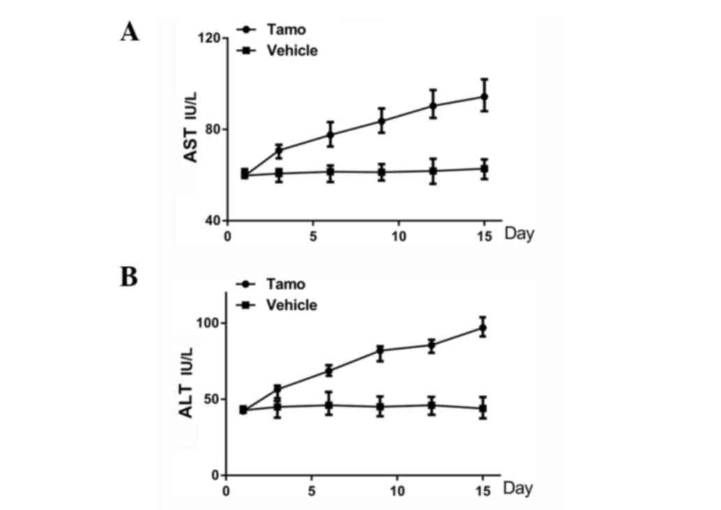
As mice in the experimental group were administrated
with i.p. injection of 6 mg/kg/day TAM, AST and ALT concentrations
in the serum were detected every 3 days, in order to evaluate the
effects of TAM on the metabolic function of the liver. As shown in
Fig. 2, the ALT level began to
increase from the third day compared with the control group and the
results were significant (P<0.05). The AST level began to
increase from day 6 and the results were significant (P<0.01).
At day 15, the total levels of AST and ALT activities were elevated
by 60.8 and 125%, respectively in the TAM-treated group compared to
the saline control group. This occurred mainly due to leakage of
these enzymes from damaged hepatocytes into the bloodstream, which
manifested that a low dose of TAM treatment would lead to the
damage of hepatocytes 2 weeks after the injection.
Morphological changes of liver tissue
under an inverted light microscope
After being treated with 6 mg/kg/day TAM for 2
weeks, small pieces of liver tissue were obtained and processed for
histopathological analysis. Fig. 3A and
B represented the normal structure in the control group.
Fig. 3C and D demonstrated that liver
injury was caused by TAM. The structure of the hepatic lobules
clearly became blurred in certain sections of the regions. Namely,
the folial central vein expanded and engorged. Additionally,
hepatic cells in certain areas swelled into spherical shapes and
fatty metamorphosis was observed fractionally. Nuclei appeared to
be pyknotic and exhibited uneven chromatin distribution.
Furthermore, the cholestasis was also visible.
Liver cell ultrastructural changes
under TEM
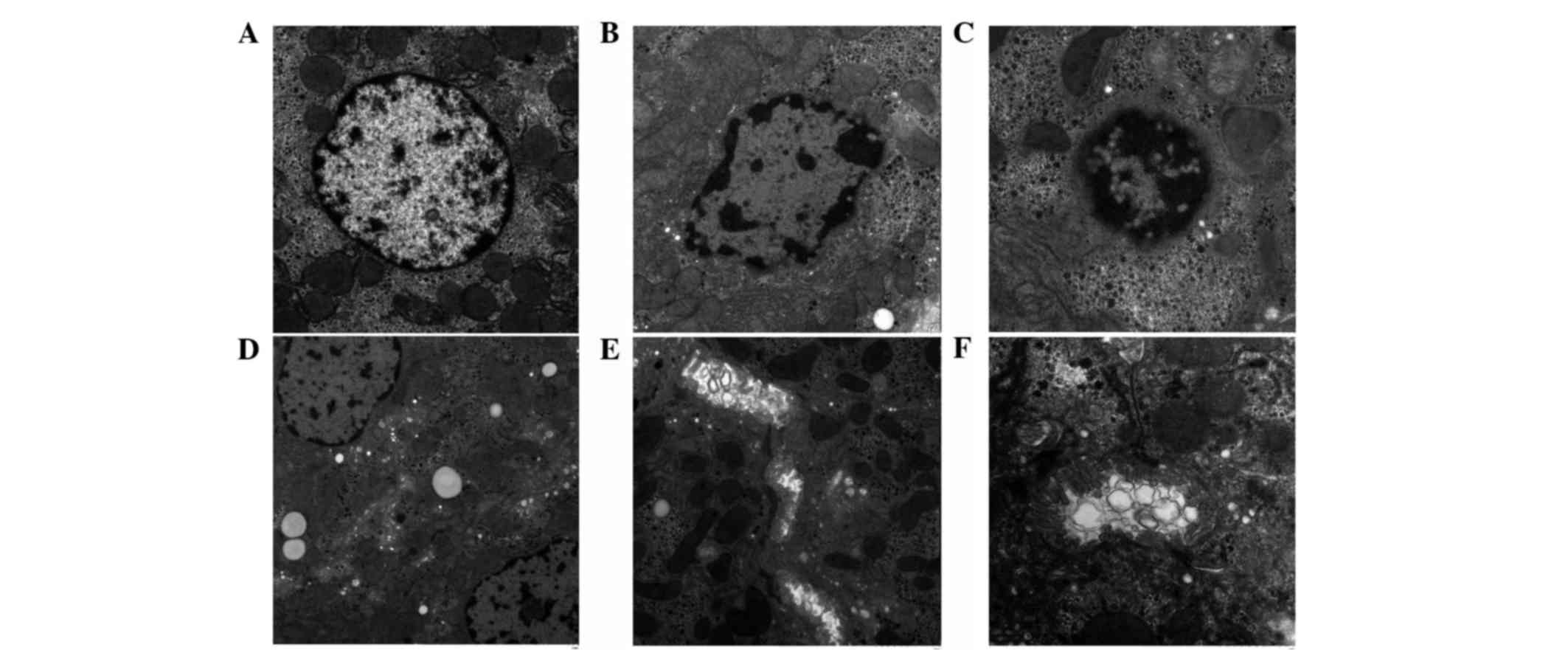
To further explore the ultrastructural changes of
the hepatic cells, TEM was applied to examine its morphological
changes in the TAM-treated group. As shown in Fig. 4, in the control group, normal
hepatocytes appeared to be polygonal in outline, and the central
area had a large nucleus and clear mitochondria (Fig. 4A). However, the nuclei were pyknotic
and unevenly distributed in the TAM-treated group. The majority of
the nuclei exhibited distinct heterochromatin and thick nucleoli.
The chromatins became coarse, granular and agglutinated in the
nuclei (Fig. 4B and C). Hepatocytes
appeared to have undergone vacuolar degeneration with lipid
droplets diffusely distributed in the cytoplasm. In addition,
mitochondrial cristae became vague and disorganized (Fig. 4D). Furthermore, the cholestasis was
also visible under electron microscopy. (Fig. 4E and F).
 | Figure 4.Ultrastructural changes under
transmission electronmicroscopy. (A) The normal liver tissue
structure in the control group (original magnification, ×3,000).
(B) After the administration of tamoxifen for two weeks, the liver
tissue structure in the tamoxifen-treated group (original
magnification, ×4,000). (C-F) The liver tissue structure in the
tamoxifen-treated group [original magnification, (C) ×5,000, (D)
×2,000, (E) ×3,000 and (F) ×7,000]. |
Effects of TAM on caspase-3 expression
level in liver tissues
To further illustrate the specific mechanisms that
were involved in TAM-induced hepatotoxicity, the western blot assay
was used to detect the caspase-3 expression level in the liver
tissues. β-actin served as a loading control. Representative blots
from three independent experiments with similar results are shown
in Fig. 5. The caspase-3 expression
level increased in the TAM-treated group compared with the vehicle
control, and the difference was statistically significant
(P<0.01). As it is well-recognized that caspase-3 is closely
involved in cell apoptosis, these results implicated that one of
the mechanisms involved in TAM-induced hepatotoxicity was
hepatocyte apoptosis.
Discussion
Worldwide, breast cancer is one of the most
prevailing carcinomas in females. In America, breast cancer is the
most common malignancy in females, which accounts for >40,000
fatalities each year (14).
Clinically, based on gene expression profiling [ER, progesterone
receptor (PR) and human epidermal growth factor receptor-2
(HER-2)], breast cancer has been classified into 4 major subtypes:
Luminal A (ER/PR+, HER-2−), luminal B
(ER/PR+, HER-2+), HER-2 overexpressing
(ER−/PR−, HER-2+) and basal-like
(ER−/PR−, HER-2−) (15,16). For the
two-thirds of high-risk breast cancer patients that are positive
for ER and/or PR, endocrine therapy with TAM or aromatase
inhibitors is generally recommended as highly effective (17,18). The
previous studies also provided significant evidence that 5-year
standard TAM therapy would improve their 5-year survival rate,
particularly in postmenopausal women (19). Additionally, the initial results from
the first International Breast Cancer Intervention Study-I revealed
that prophylactic use of TAM reduced the risk of invasive
ER-positive tumors by 31% in women who were at an increased risk
for breast cancer (20).
However, recurrences and side effects have
restricted its long-term use to a large extent. Recent studies have
revealed that long-term used of TAM may cause ranges of side
effects such as hot flashes, night sweats, gynecological symptoms
(vaginal dryness and vaginal discharge), depression, memory loss,
sleep alterations, weight gain and diminished sexual function
(7). Among all these, hepatic injury
or even hepatocarcinoma would be one of the most severe side
effects, which hindered its long-term use (9–11).
Clinically, patients who accept the endocrinotherapy are instructed
to reexamine their liver function every 4 months due to its
hepatotoxicity. Numerous research and clinical studies have
illustrated clearly that TAM causes the inhibition of mitochondrial
β-oxidation and subsequently leads to macrovacuolar steatosis
(21,22). The early symptoms were characterized by
the presence of a single, large lipid vacuole within the cytoplasm
of the hepatocytes (23).
However, Carthew et al (24) reported that treatment of Wistar-AP
female rats with dietary TAM for only 3 months was sufficient to
cause cumulative hepatic DNA damage, which would finally develop
into hepatocarcinoma with or without the promotion of
phenobarbital. In addition, White et al (25) also elucidated that following the
administration of TAM for 7 days (45 mg/kg/day) and extraction of
hepatic DNA, ≤7 radiolabelled adduct spots could be detected, as it
caused a time-dependent increase in the level of adduct detected,
up to a value of ≥1 adduct/106 nucleotides after 7 days
of dosing. These short-term emerged TAM genotoxicity results
instigated the exploration of whether short-term TAM treatment
would cause visible hepatotoxicity at the morphological level or
whether a low dose of TAM would lead to hepatic injury.
Thus, the present study aimed to investigate whether
TAM at a relatively low dose influences liver function at the early
stage of the treatment. At the animal level, 6 mg/kg/day TAM was
selected for 2-week treatment to build a short-term animal model,
while the normal clinical dose for patients is 0.33 mg/kg/day (20
mg/day) and the scientific research dose for mice is 25 mg/kg/day
(26,27). During the process, mice body weights
were detected every day and the AST and ALT levels in serum were
measured every three days. The results revealed that TAM exerted
distinct reducing effects on body weight in the 2-week treatment.
In addition, the levels of ALT and AST in liver tissues were
increased significantly when compared with the control group, and
this is known to occur mainly due to the leakage of these enzymes
from the damaged hepatocytes into the bloodstream. Subsequently,
the morphological changes of hepatocytes were observed in the
TAM-treated group, which changed evidently at the microscopic and
ultrastructural levels. The structure of the hepatic lobules became
blurred in sections of the region. Namely, the folial central vein
expanded and engorged. Additionally, hepatic cells in certain
sections of the regions swelled to spherical shapes and fatty
metamorphosis was observed fractionally. Furthermore, cholestasis
was also visible in certain areas. Under the TEM analysis, it was
observed that the nuclei were pyknotic and unevenly distributed in
TAM-treated group. The majority of the nuclei were endowed with
distinct heterochromatin and thick nucleoli. The chromatins became
coarse, granular and agglutinated in the nuclei. Hepatocytes
appeared to undergo vacuolar degeneration with lipid droplets
diffusely distributed in the cytoplasm. In addition, mitochondrial
cristae became vague and disorganized. Finally, the specific
mechanism that was involved in this hepatotoxicity was explored. A
recent study elucidated that cell apoptosis has an important role
in the process of hepatic cell injury (28). Thus, western blotting was applied to
detect the expression level of caspase-3 in liver tissues, which
had already been proved as one of the core proteins that
participated in cell apoptosis. The results indicated that the
caspase-3 expression level increased significantly in the liver
tissues compared with the control group. This indicated that
apoptosis had a vital role in this TAM-induced liver injury.
In conclusion, the present data showed that a
relatively low concentration of TAM (6 mg/kg/day) for a short time
treatment (2 weeks) would cause hepatotoxicity and change
morphology at the microscopic and ultrastructural levels. Although
the liver function may compensate or reverse the injuries
gradually, the damage that occurred in the short-term TAM therapy
has been shown. Thus, there is a necessity to obtain measures for
monitoring liver function and protection at the early stage of the
TAM endocrinotherapy, prior to apparent and undesirable clinical
symptoms occurring. Furthermore, as DNA damage also occurs at this
early period without clear clinical symptoms, which in the long-run
increases the risk of hepatocarcinoma, exploring alternatives for
TAM in long-term clinical endocrinotherapy is required.
Acknowledgements
The present study was supported by the Fundamental
Research Funds for the Central Universities (grant no.
2014301020203).
References
|
1
|
Weycker D, Edelsberg J, Kartashov A,
Barron R and Lyman G: Risk and healthcare costs of
chemotherapy-induced neutropenic complications in women with
metastatic breast cancer. Chemotherapy. 58:8–18. 2012. View Article : Google Scholar : PubMed/NCBI
|
|
2
|
DeSantis C, Ma J, Bryan L and Jemal A:
Breast cancer statistics, 2013. CA Cancer J Clin. 64:52–62. 2014.
View Article : Google Scholar : PubMed/NCBI
|
|
3
|
Raftopoulou M and Hall A: Cell migration:
Rho GTPases lead the way. Dev Biol. 265:23–32. 2004. View Article : Google Scholar : PubMed/NCBI
|
|
4
|
Hoppe R, Achinger-Kawecka J, Winter S,
Fritz P, Lo WY, Schroth W and Brauch H: Increased expression of
miR-126 and miR-10a predict prolonged relapse-free time of primary
oestrogen receptor-positive breast cancer following tamoxifen
treatment. Eur J Cancer. 49:3598–3608. 2013. View Article : Google Scholar : PubMed/NCBI
|
|
5
|
Jordan VC and O'Malley BW: Selective
estrogen-receptor modulators and antihormonal resistance in breast
cancer. J Clin Oncol. 25:5815–5824. 2007. View Article : Google Scholar : PubMed/NCBI
|
|
6
|
Moerkens M, Zhang Y, Wester L, van de
Water B and Meerman JH: Epidermal growth factor receptor signalling
in human breast cancer cells operates parallel to estrogen receptor
α signalling and results in tamoxifen insensitive proliferation.
BMC Cancer. 14:2832014. View Article : Google Scholar : PubMed/NCBI
|
|
7
|
Lorizio W, Wu AH, Beattie MS, Rugo H, Tchu
S, Kerlikowske K and Ziv E: Clinical and biomarker predictors of
side effects from tamoxifen. Breast Cancer Res Treat.
132:1107–1118. 2012. View Article : Google Scholar : PubMed/NCBI
|
|
8
|
Yeh WL, Lin HY, Wu HM and Chen DR:
Combination treatment of tamoxifen with risperidone in breast
cancer. PLoS One. 9:e988052014. View Article : Google Scholar : PubMed/NCBI
|
|
9
|
Cuzick J, Powles T, Veronesi U, Forbes J,
Edwards R, Ashley S and Boyle P: Overview of the main outcomes in
breast-cancer prevention trials. Lancet. 361:296–300. 2003.
View Article : Google Scholar : PubMed/NCBI
|
|
10
|
Hendrick A and Subramanian VP: Tamoxifen
and thromboembolism. JAMA. 243:514–515. 1980. View Article : Google Scholar : PubMed/NCBI
|
|
11
|
Yang G, Nowsheen S, Aziz K and Georgakilas
AG: Toxicity and adverse effects of Tamoxifen and other
anti-estrogen drugs. Pharmacol Ther. 139:392–404. 2013. View Article : Google Scholar : PubMed/NCBI
|
|
12
|
El-Ashmawy NE and Khalil RM: A review on
the role of L-carnitine in the management of tamoxifen side effects
in treated women with breast cancer. Tumour Biol. 35:2845–2855.
2014. View Article : Google Scholar : PubMed/NCBI
|
|
13
|
Suddek GM: Protective role of thymoquinone
against liver damage induced by tamoxifen in female rats. Can J
Physiol Pharmacol. 92:640–644. 2014. View Article : Google Scholar : PubMed/NCBI
|
|
14
|
Al-Hajj M, Wicha MS, Benito-Hernandez A,
Morrison SJ and Clarke MF: Prospective identification of
tumorigenic breast cancer cells. Proc Natl Acad Sci USA.
100:3983–3988. 2003. View Article : Google Scholar : PubMed/NCBI
|
|
15
|
Perou CM, Sørlie T, Eisen MB, van de Rijn
M, Jeffrey SS, Rees CA, Pollack JR, Ross DT, Johnsen H, Akslen LA,
et al: Molecular portraits of human breast tumours. Nature.
406:747–752. 2000. View
Article : Google Scholar : PubMed/NCBI
|
|
16
|
Goldhirsch A, Wood WC, Coates AS, Gelber
RD, Thürlimann B and Senn HJ: Panel members: Strategies for
subtypes - dealing with the diversity of breast cancer: Highlights
of the St. Gallen International Expert Consensus on the Primary
Therapy of Early Breast Cancer 2011. Ann Oncol. 22:1736–1747. 2011.
View Article : Google Scholar : PubMed/NCBI
|
|
17
|
Howell A, Cuzick J, Baum M, Buzdar A,
Dowsett M, Forbes JF, Hoctin-Boes G, Houghton J, Locker GY and
Tobias JS: ATAC Trialists' Group: Results of the ATAC (Arimidex,
Tamoxifen, Alone or in Combination) trial after completion of 5
years' adjuvant treatment for breast cancer. Lancet. 365:60–62.
2005. View Article : Google Scholar : PubMed/NCBI
|
|
18
|
Mauri D, Pavlidis N, Polyzos NP and
Ioannidis JP: Survival with aromatase inhibitors and inactivators
versus standard hormonal therapy in advanced breast cancer:
Meta-analysis. J Natl Cancer Inst. 98:1285–1291. 2006. View Article : Google Scholar : PubMed/NCBI
|
|
19
|
Tamoxifen for early breast cancer: An
overview of the randomised trials. Early Breast Cancer Trialists'
Collaborative Group. Lancet. 351:1451–1467. 1998. View Article : Google Scholar : PubMed/NCBI
|
|
20
|
Cuzick J, Forbes JF, Sestak I, Cawthorn S,
Hamed H, Holli K and Howell A: International Breast Cancer
Intervention Study I Investigators: Long-term results of tamoxifen
prophylaxis for breast cancer-96-month follow-up of the randomized
IBIS-I trial. J Natl Cancer Inst. 99:272–282. 2007. View Article : Google Scholar : PubMed/NCBI
|
|
21
|
Lee MH, Kim JW, Kim JH, Kang KS, Kong G
and Lee MO: Gene expression profiling of murine hepatic steatosis
induced by tamoxifen. Toxicol Lett. 199:416–424. 2010. View Article : Google Scholar : PubMed/NCBI
|
|
22
|
Murata Y, Ogawa Y, Saibara T, Nishioka A,
Fujiwara Y, Fukumoto M, Inomata T, Enzan H, Onishi S and Yoshida S:
Unrecognized hepatic steatosis and non-alcoholic steatohepatitis in
adjuvant tamoxifen for breast cancer patients. Oncol Rep.
7:1299–1304. 2000.PubMed/NCBI
|
|
23
|
Labbe G, Pessayre D and Fromenty B:
Drug-induced liver injury through mitochondrial dysfunction:
Mechanisms and detection during preclinical safety studies. Fundam
Clin Pharmacol. 22:335–353. 2008. View Article : Google Scholar : PubMed/NCBI
|
|
24
|
Carthew P, Martin EA, White IN, De Matteis
F, Edwards RE, Dorman BM, Heydon RT and Smith LL: Tamoxifen induces
short-term cumulative DNA damage and liver tumors in rats:
Promotion by phenobarbital. Cancer Res. 55:544–547. 1995.PubMed/NCBI
|
|
25
|
White IN, de Matteis F, Davies A, Smith
LL, Crofton-Sleigh C, Venitt S, Hewer A and Phillips DH: Genotoxic
potential of tamoxifen and analogues in female Fischer F344/n rats,
DBA/2 and C57BL/6 mice and in human MCL-5 cells. Carcinogenesis.
13:2197–2203. 1992. View Article : Google Scholar : PubMed/NCBI
|
|
26
|
El-Beshbishy HA: Hepatoprotective effect
of green tea (Camellia sinensis) extract against
tamoxifen-induced liver injury in rats. J Biochem Mol Biol.
38:563–570. 2005. View Article : Google Scholar : PubMed/NCBI
|
|
27
|
El-Beshbishy HA: The effect of dimethyl
dimethoxy biphenyl dicarboxylate (DDB) against tamoxifen-induced
liver injury in rats: DDB use is curative or protective. J Biochem
Mol Biol. 38:300–306. 2005. View Article : Google Scholar : PubMed/NCBI
|
|
28
|
Vince AR, Hayes MA, Jefferson BJ and
Stalker MJ: Hepatic injury correlates with apoptosis, regeneration,
and nitric oxide synthase expression in canine chronic liver
disease. Vet Pathol. 51:932–945. 2014. View Article : Google Scholar : PubMed/NCBI
|


