Pulmonary lymphangioleiomyomatosis in a 46-year-old female: A case report and review of the literature
- Authors:
- Published online on: April 13, 2016 https://doi.org/10.3892/br.2016.652
- Pages: 719-722
Metrics:
Total
Views: 0 (Spandidos Publications: | PMC Statistics:
)
Total PDF Downloads: 0 (Spandidos Publications: | PMC Statistics:
)
Abstract
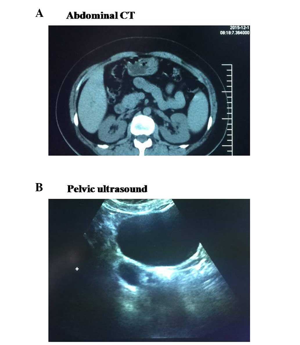
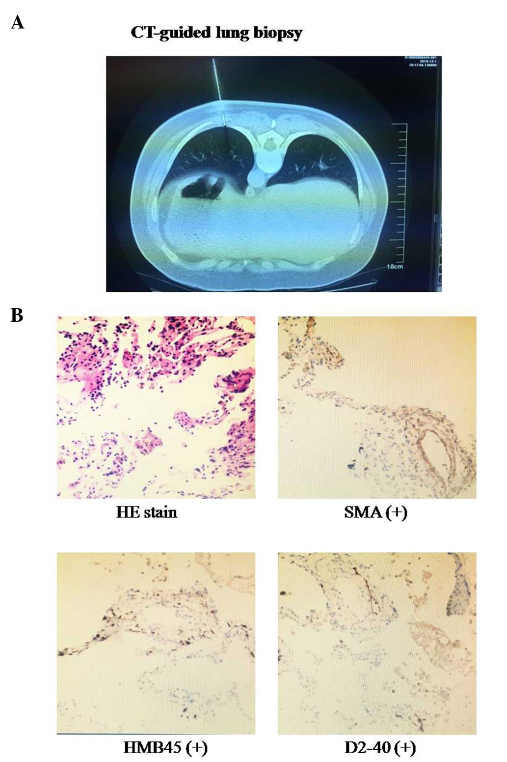
Pulmonary lymphangioleiomyomatosis (PLAM) is a rare disease occurring frequently in women. The present study reported a case of PLAM in a 46-year-old female and reviewed the literature regarding PLAM. The female experienced dyspnea on exertion for 1 month. The chest computed tomography (CT) revealed diffuse thin‑walled cystic lesions, consistent with radiological features of PLAM. CT‑guided lung biopsy showed that characteristic smooth muscle cells were positive for smooth muscle actin, human melanoma black 45 and D2‑40 by histology and immunohistochemistry techniques, resulting in a diagnosis of PLAM. The patient had a hysterectomy for treating uterine myoma 12 years previously. Additionally, lung diffusion function was impaired slightly, abdominal CT showed a right renal cyst and pelvic ultrasound exhibited a right oophoritic cyst. Sirolimus was administered, which improved the dyspnea and pulmonary function, and the patient required long‑term follow‑up. Therefore, this is a rare case of PLAM with renal cyst, oophoritic cyst and uterine myoma in a female. In conclusion, PLAM is a rare proliferative disease in the lung. Chest CT characteristically showed that the cysts were bilateral, diffuse and thin‑walled. Lung biopsy may provide evidence for the diagnosis of PLAM, which was treated with sirolimus.