Introduction
Fungal keratitis is a severe and common ocular
infectious disease that occurs worldwide. This serious condition
may threaten visual acuity and cause blindness (1). The prevalence of fungal keratitis has
been indicated to be as high as 0.015%, which constituted 61.9% of
severe infective keratitis cases in China (1,2).
Clinically, fungal infection slowly and gradually penetrates and
deeply infiltrates the cornea (3).
Typical clinical features may not be observed during the early
stage, which often results in misdiagnosis and a delay in treatment
with antifungal medication (3). The
antifungal agents used for treatment include polyenes, azoles and
fluorinated pyrimidine (4). However,
natamycin is considered to be the only commercially available
broad-spectrum agent (5,6). Due to the high misdiagnosis rate, the
limited number of available antifungal agents and fungal resistance
to treatment, it is usually difficult to control the infection
(1). In ~1/3 of cases, the fungal
infection fails to respond to medical treatment, which may result
in a whole cornea ulcer or corneal perforation (7); in these cases, a therapeutic
penetrating keratoplasty is necessary (8).
Despite receiving an accurate diagnosis and
appropriate treatment, 15–27% of patients with fungal keratitis
require corneal transplantation (9).
Although the complexity and difficulty of keratoplasty are
considerable disadvantages, the cornea donor shortage is an
underlying limitation of this procedure for treating patients with
fungal keratitis (10). There is a
lack of corneal donation despite the high demand in China, with 200
patients registering for corneal donations and only 60 eyes donated
within the past decade, as indicated in a study by Shang and Zhang
in 2010 (11). Therefore,
transitional surgery, such as conjunctival flap covering surgery,
is considered to be another preferable possibility for these
patients (12). The technique of
using a conjunctival flap for the treatment of chronic corneal
ulceration was described by Gundersen (13) in the late 1950s and became a standard
surgical procedure. Historically, this procedure has been used for
bullous keratopathy, bacterial keratitis, viral keratitis and
fungal keratitis as a palliative surgery (14–17). The
advantages of this surgery are as follows: Corneal inflammation and
pain may be well controlled; the flap brings in a blood supply that
promotes healing by increasing corneal access to humoral and
cellular immune mechanisms; and the procedure is usually
cosmetically superior to tarsorrhaphy, and may be performed by the
majority of ophthalmologists without sophisticated instrumentation
or a need for donor tissue (13).
Lingulate, pedicle and bridge flaps have been developed, and the
connected flap has been used for whole corneal ulcers that are not
perforated (18); however,
complications as a result of high tension of the marginal joint,
and difficulty in suturing and fixing the cornea typically lead to
a cracked conjunctival flap and corneal perforation (18).
In the present study, a novel conjunctival flap
covering surgery was developed for treating severe fungal
keratitis. Following surgery, 15/17 patients (88.24%) demonstrated
complete conjunctival re-epithelization and a smooth conjunctival
surface without any complications.
Patients and methods
Patients and clinical
manifestations
A total of 17 patients (1 female and 16 males; age,
50.29±10.92 years) diagnosed with fungal keratitis and a whole
corneal ulcer without perforation were recruited between January
2010 and December 2014 at the Zhongshan Ophthalmic Center, Sun
Yat-Sen University (Guangzhou, China). Medical records were
reviewed, including age, gender, profession, predisposing
associated factors, duration of onset, laboratory tests,
best-corrected visual acuity (BCVA), intraocular pressure (IOP) and
treatment, and all information was used solely for research
purposes. All individuals enrolled in the present study provided
written informed consent prior to their inclusion in the study. The
present study was approved by the Ethics Committee of the Zhongshan
Ophthalmic Center, Sun Yat-Sen University. Furthermore, the study
adhered to the tenets of the Declaration of Helsinki.
Diagnosis
The clinical diagnosis of fungal keratitis was based
on risk factor analysis, corneal features, confocal microscopy
[Heidelberg Retina Tomograph-Rostock Cornea Module (HRT-RCM);
Heidelberg Engineering GmBH, Dossenheim, Germany] and corneal
cultures of fungi. The criterion used for the identification of
fungal filaments on confocal microscopy was the presence of highly
reflective, septate, double-walled filaments varying in size
between 3–8 µm (19). Positivity of
fungal growth from corneal-scraping or the presence of fungal
filaments on confocal microscopy was considered to indicate fungal
keratitis preoperatively. Preoperative B-scan ultrasounds were
performed for all patients to exclude endophthalmitis.
Postoperatively, the scraped necrotic cornea was divided into
several parts for pathological biopsy, hematoxylin and eosin
staining and cultures of bacteria and fungi (20). In H&E staining, Eyes were fixed
in 4% formaldehyde in 0.075 M phosphate buffer for 24 h, dehydrated
in increasing concentrations of ethanol (70–99%) and embedded in
paraffin at 60°C. Sections 5 mm thick were then cut and floated on
deionized water at 45°C, and single sections were mounted on
SuperFrost Plus glass slides (Menzel-Glaser, Braunschweig,
Germany). After rehydrating in alcohol (99–70%) for 3 min in each
concentration, all slides were added to the hematoxylin for 4 min
and eosin for 2 min at 37°C and then subsequently dried at 60°C for
1 h. All sections were visualized with a Zeiss microscope (Zeiss
microscope AXIO Imager A1; Carl Zeiss AG, Oberkochen, Germany) with
a magnification of ×40. In microculture, Sabouraud and potato
dextrose agar were incubated at 25°C to enhance the growth of fungi
and the media containing blood agar, chocolate agar, brain heart
infusion broth and thioglycollate (liquid) for bacteria were
incubated at 37°C (20).
Indication and exclusion criteria for
full-thickness conjunctival flap covering surgery with amniotic
membrane transplantation (FCCS + AMT)
Preoperatively, all patients were prescribed
topically 5% natamycin eye drops (Alcon, Fort Worth, TX, USA) four
times per day, 0.02% fluconazole eye drops (Zhongshan Ophthalmic
Center, Guangzhou, China) every 2 h daily and 0.02% fluconazole
ointment (Zhongshan Ophthalmic Center) once per night for at least
1 month prior to surgery, combined with 300 mg of voriconazole
(Pfizer, New York, NY, USA) taken orally twice daily for at least 2
weeks prior to surgery. Patients who satisfied all of the following
indication criteria underwent FCCS + AMT: i) Poor response to
topical therapy; ii) an increasingly large lesion involving the
entire cornea; iii) gradual thinning of the cornea with no
observable perforation by fluorescein staining; and iv) no
endophthalmitis according to B-scan ultrasounds. Patients meeting
any of the following criteria were excluded from FCCS + AMT: i)
Corneal perforation was present; ii) endophthalmitis was detected
by B-scan ultrasound; iii) hypopyon was present in the eye; and iv)
treatment with antifungal medication was effective.
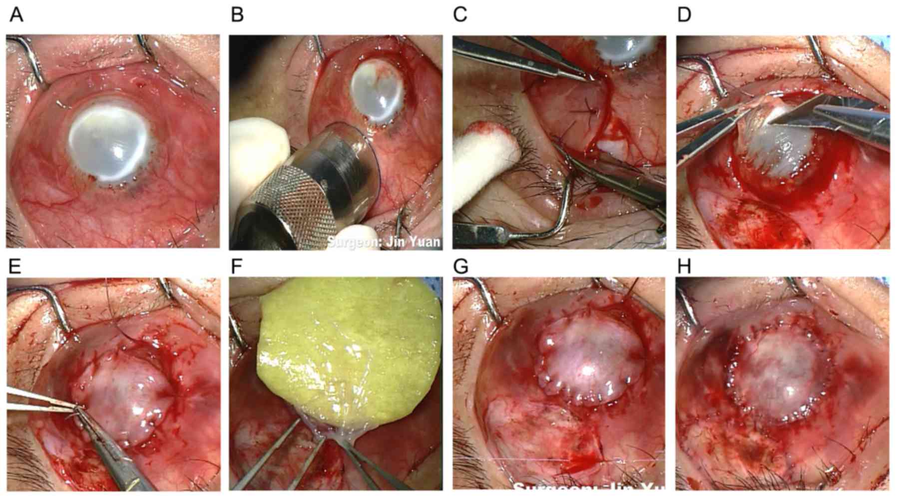
Surgical technique
All surgeries in the present study were performed by
the same surgeon. A trephine with a diameter of 11 mm and a marker
pen were used to mark the margin of the conjunctival flap.
Subsequently, a full-thickness conjunctival flap with Tenon's
capsule was dissected and the suppurative and necrotic cornea was
removed. A regular and smooth surface was cut using a corneal
shaper and cornea fixation forceps to assure the complete
attachment of the conjunctival flap. The conjunctival flap was
secured to the cornea with interrupted 10-0 nylon sutures, and
fixed with the limbal and episcleral tissue of the cornea
peripherally to avoid a conjunctival flap crack or a deficiency of
limbal stem cells postoperatively. The donor bed was transplanted
with the cryopreserved amniotic membrane once thawed, and washed
with phosphate-buffered saline (PBS) in a 20 ml culture dish for
3–5 min with the stromal side facing down to accelerate
conjunctival epithelial migration (Fig.
1). The amniotic membrane was obtained from the Eye Bank of
Guangdong Province (Guangzhou, China), which was acquired from
caesarean deliveries, and was previously confirmed negative of
hepatitis B and C virus or human immunodeficiency virus. The
membrane was prepared and stored sterilely according to previously
reported methods (21). Following
surgery, the removed tissue was cut into 2 µm pieces for
histopathology and fungal cultures (22). Fig. 2
indicated the flow chart displaying the specific treatments
different patients with fungal keratitis received following FCCS +
AMT.
Postoperative treatment and
follow-up
Following FCCS + AMT, all patients were prescribed
the following antifungal agents: Topical 5% natamycin; 0.02%
fluconazole eye drops every 2 h daily; and 0.02% fluconazole
ointment once per night combined with 300 mg of voriconazole for at
least 1 month] and were examined once a month for 3 months. 2
patients developed endophthalmitis with a melted conjunctival flap
and accepted ocular enucleation surgery, whereas 7/15 remaining
patients who satisfied all of the following criteria received
routine sclerokeratoplasty: i) The conjunctival flap did not melt;
ii) severe conjunctival congestion or edema did not occur; and iii)
visual acuity was not less than light perception. The other 8
patients chose the following conservative medication: Topical 5%
natamycin eye drops four times per day, 0.02% fluconazole eye drops
every 2 h daily and 0.02% fluconazole ointment once per night,
combined with 300 mg or 4–6 mg/Kg of voriconazole orally for 3
months after FCCS + AMT immediately due to economic reasons.
Following corneal transplantation, anti-inflammatory
and anti-infectious agents (topically 5% natamycin eye drops four
times per day, 0.2% fluconazole eye drops every 2 h daily, and 0.5%
fluconazole ointment once per night, combined with 300 mg or 4–6
mg/Kg of voriconazole orally for 3 months due to economic reasons)
were prescribed for at least 1 month immediately following
transplantation, combined with 0.05% tacrolimus (FK506) eyedrops
three times a day (Zhongshan Ophthalmic Center) for at least 1 year
to prevent recurrence and graft rejection.
Following sclerokeratoplasty, the following indices
were evaluated at months 1, 6 and 12: BCVA by visual chart; IOP by
the ocular palpation method where index fingers of both hands are
placed on the top of the eyelid, over the cartilage and pressing
the eyeball alternately with one or another finger the doctor may
feel fluctuations (23); and graft
transparency and recurrence rate in which graft failure was defined
as an irreversible loss of graft clarity resulting from any cause,
including persistent epithelial defects, nonendothelial graft
rejection, or interface opacity/vascularization (20). For the ocular palpation method,
Tn=regular intraocular pressure; T+1=slightly increased intraocular
pressure; T+2=rigid eyeball; T+3=hard eyeball, hard as rock;
T-1=slightly softer eyeball; T-2=soft eyeball; T-3=very soft
eyeball (23). Snellen visual acuity
was recorded and approximations for visual acuity worse than 20/400
were determined considering the following: Counting
fingers=20/2,000, hand motions=20/4,000, light perception=20/8,000,
and no light perception=20/16,000. Snellen vision was converted to
logMAR values for statistical analysis (24). IOP and BCVA were also evaluated in
patients that did not receive this surgery at months 1, 6 and 12
following conservative medication (Table II).
 | Table II.BCVA and IOP of patients. |
Table II.
BCVA and IOP of patients.
| Case no. | Preoperative
BCVA | Preoperative
IOP | BCVA following FCCS
+ AMT | IOP following FCCS
+ AMT | BCVA following
sclerokeratoplasty/conservative medication | IOP following
sclerokeratoplasty/conservative medication |
|---|
| P1 | 2.60 | Tn | 2.30 | Tn | 1.00 | Tn |
| P2 | 2.60 | T-1 | 2.30 | Tn | 0.52 | Tn |
| P3 | 2.60 | T+1 | 2.90 | Tn | 0.30 | Tn |
| P4 | 2.30 | Tn | 2.30 | Tn | 2.30 | Tn |
| P5 | 2.60 | Tn | 2.60 | Tn | 2.60 | Tn |
| P6 | 2.30 | T-1 | 2.30 | T-1 | Flap melting and
corneal perforation | Tn |
| P7 | 2.30 | T-1 | 2.00 | T-1 | Flap melting and
corneal perforation | Tn |
| P8 | 2.60 | Tn | 2.60 | Tn | 2.60 | Tn |
| P9 | 2.60 | T-1 | 2.90 | Tn | 2.90 | Tn |
| P10 | 2.60 | T-2 | 2.60 | Tn | 2.60 | Tn |
| P11 | 2.60 | Tn | 2.60 | Tn | 1.00 | Tn |
| P12 | 2.30 | Tn | 2.90 | Tn | 2.90 | Tn |
| P13 | 2.60 | Tn | 2.30 | Tn | 0.30 | Tn |
| P14 | 2.30 | T+2 | 2.90 | Tn | 2.90 | Tn |
| P15 | 2.30 | T+1 | 2.60 | Tn | 1.00 | Tn |
| P16 | 2.30 | T-1 | 2.30 | Tn | 0.70 | Tn |
| P17 | 2.30 | Tn | 2.60 | Tn | 2.60 | Tn |
Statistical analysis
SPSS software version 13.0 (SPSS, Inc., Chicago, IL,
USA) was used for the statistical analysis. Data were presented as
the mean ± standard error of the mean. An unpaired, two-tailed
Student's t-test was used to determine the statistical significance
of BCVA preoperatively and postoperatively. P<0.05 was
considered to indicate a statistically significant difference.
Results
Clinical data and clinical
features
A total of 17 eyes from 17 patients were included in
the present study. From the total 17 patients, 16 patients (94.12%)
were male and 1 patient (5.88%) was female. The mean age was
50.29±10.92 (30–72) years. A total of 11 affected eyes (64.71%)
were right eyes and 6 (35.29%) were left eyes. Of the 17 patients,
the mean disease duration was 2.53±4.66 (0.5–21) months and the
mean diameter of the lesion was 7.71±1.74 (5–10) mm. A
total of 13 (76.47%) patients were farmers; 7 patients experienced
agricultural trauma; 3 patients had a history of a non-agricultural
body entering the eye; 1 patient was hit by an iron wire; and the
other 6 patients had unclear predisposing factors (Table I).
| Table I.Clinical features and clinical
examination. |
Table I.
Clinical features and clinical
examination.
|
|
|
|
| Preoperative |
|
|---|
|
|
|
|
|
|
|
|---|
| Case no. | Risk factor | Duration of
disease, month | Diameter of lesion,
mm | Confocal scan | Corneal-scraping
culture | Postoperative
corneal-scraping culture |
|---|
| P1 | Agricultural
trauma | 2 | 9 | Fungal hyphae | Fusarium
solani | Fusarium
solani |
| P2 | Agricultural
trauma | 1 | 10 | Fungal hyphae | – | – |
| P3 | Agricultural
trauma | 1 | 10 | – | Fusarium
solani | Fusarium
solani |
| P4 | Iron wire
hitting | 1.5 | 7 | Fungal hyphae | – | Fusarium
solani |
| P5 | Unclear | 2 | 10 | Fungal hyphae | – | – |
| P6 | Chalk entering
eye | 21 | 6 | – | Aspergillus
fumigatus | Aspergillus
fumigatus |
| P7 | Agricultural
trauma | 3 | 10 | – | Aspergillus
fumigatus | Aspergillus
fumigatus |
| P8 | Unclear | 1 | 8 | – | Fusarium
solani | Fusarium
solani |
| P9 | Unclear | 2 | 8 | Fungal hyphae | – | Grasses
helminthosporium |
| P10 | Agricultural
trauma | 1 | 9 | Fungal hyphae | – | Aspergillus
flavus |
| P11 | Unclear | 1 | 8 | Fungal hyphae | – | Mucor |
| P12 | Unclear | 1 | 6 | – |
Penicillium |
Penicillium |
| P13 | Agricultural
trauma | 2 | 6 | Fungal hyphae | Fusarium
solani | Fusarium
solani |
| P14 | Pepper sauce
entering eye | 0.5 | 5 | – |
Curvularia |
Curvularia |
| P15 | Insects entering
eye | 1 | 5 | – | Fusarium
solani | Fusarium
solani |
| P16 | Agricultural
trauma | 1 | 6 | Fungal hyphae | – | Aspergillus
flavus |
| P17 | Unclear | 1 | 8 | Fungal hyphae | – |
Cephalosporium |
No cases exhibited perforation preoperatively.
Although these patients exhibited nonspecific clinical signs,
including blurred vision, redness, tearing, photophobia, pain,
foreign body sensation and secretions in the late stage of fungal
keratitis with a whole corneal ulcer, some typical signs, including
a dry and rough corneal surface (10/15 patients), an irregular
feathery-edged infiltrate (8/15 patients), a white ring in the
cornea and a satellite lesion (7/15 patients), were also observed.
Preoperatively, the mean LogMAR BCVA was 2.459±0.037. A total of 3
patients exhibited high IOP (T+1 and T+2), 6 patients had low IOP
(T-1 and T-2) and the remaining 8 patients had normal IOP (Tn)
(Table II).
Confocal microscopy and
microbiology
Prior to FCCS + AMT, fungal hyphae were identified
in 10 cases (58.82%) by confocal microscopy, and corneal scrapings
indicated that 9 cases (52.94%) had positive culture results,
corresponding to 5 cases of Fusarium solani, 2 cases of
Aspergillus fumigatus, 1 case of Curvularia and 1
case of Penicillium (Table
I).
Following FCCS + AMT, positive histopathology and
culture results were revealed in 15 patients (88.24%), including 6
cases of F. solani, 2 cases of A. fumigatus, 2 cases
of A. flavus, 1 case of Grasses helminthosporium, 1
case of Mucor, 1 case of Penicillium, 1 case of
Curvularia and 1 case of Cephalosporium, representing
6 further cases of positive culture results compared with those
observed preoperatively (Table I).
The 2 negative-culture cases had positive confocal scan results
preoperatively.
Prognosis following FCCS + AMT
Following FCCS + AMT, 15 patients (88.24%) achieved
complete conjunctival reepithelization, a vascularized flap with no
inflammation, and a smooth conjunctival surface, whereas 2 patients
(P6 and 7) developed endophthalmitis with a melted conjunctival
flap and corneal perforation at 1 and 2 weeks following FCCS + AMT
surgery. Both of these patients underwent ocular evisceration
surgery. The rate of eyeball preservation was 88.24%. No notable
improvement in BCVA (2.529±0.066) was observed compared with the
preoperative status. IOP was normal in 15 cases (88.24%), as
assessed by the ocular palpation method (23), and the other 2 patients who
experienced a melted flap had low IOP (Table II). No other complications were
encountered in the present study.
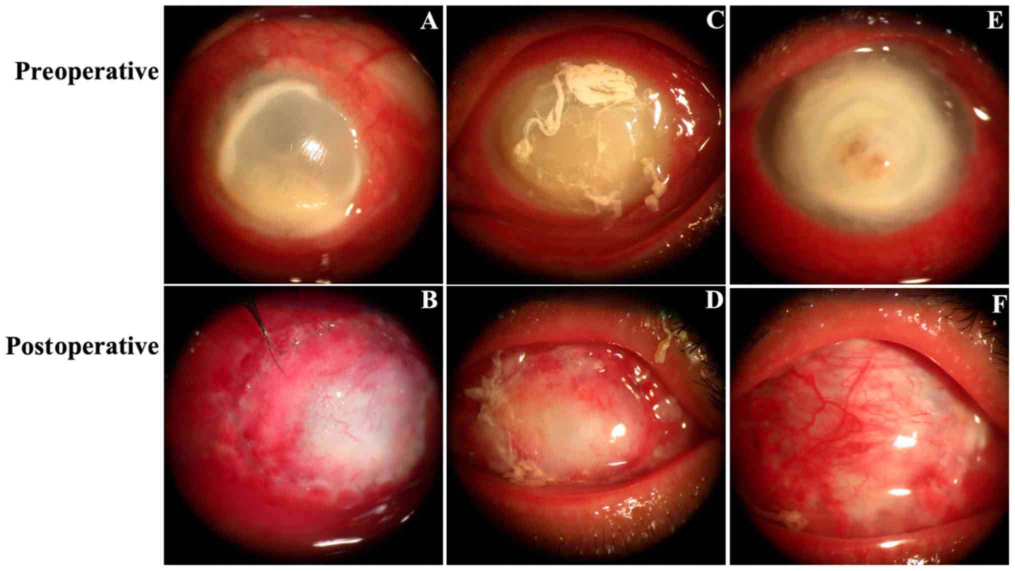
Preoperative and postoperative slit-lamp images were
indicated in Fig. 3. Fig. 3A, C and E demonstrate 3 cases
(patient 1, 2 and 3, respectively) with a conjunctival infection, a
large corneal fungal abscess (lesion diameters were 9, 10 and 10
mm, respectively) without perforation and vision with full light
perception preoperatively. At 1 week following FCCS + AMT (Fig. 3B), the conjunctival flap was fixed
tightly with the corneal limbal tissue, flap edema was observed and
some neovascularization began to appear on the peripheral side. At
2 weeks postoperatively, flap edema was alleviated, the absorbable
suture was absorbed and the peripheral side was smoother when the
flap had begun to connect with the ulcerated cornea (Fig. 3D). At 1 month postoperatively, ocular
surface vascularization and complete conjunctival re-epithelization
was observed, a non-conjunctival inflammatory y reaction was noted
and the conjunctival margins showed stable scarring (Fig. 3F). Fig.
3B, D and F depicted the healing process of the conjunctival
flap from 1 week to 2 weeks and 1 month postoperatively.
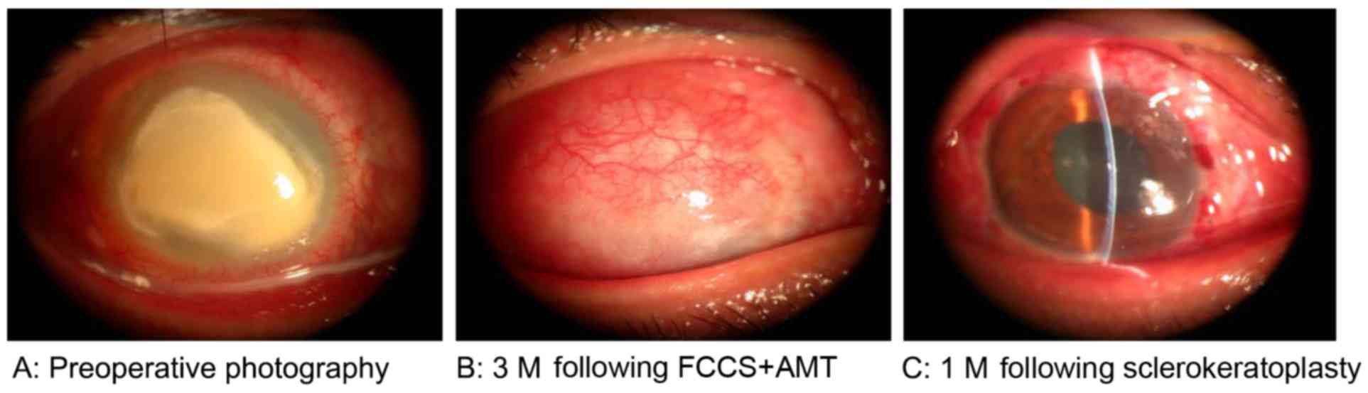
Prognosis following sclerokeratoplasty
surgery
A total of 3 months following FCCS + AMT, 7 patients
satisfied the indication criteria and accepted sclerokeratoplasty
and 8 chose conservative medication due to economic reasons. All
patients were followed up for at least 1 month, and no patients
experienced fungal recurrence or graft rejection. The entire
therapeutic process for 1 case, from preoperative stage to 3 months
following FCCS + AMT and 1 month following sclerokeratoplasty is
demonstrated in Fig. 4. The graft
remained clear through the last follow-up visit. The mean LogMAR
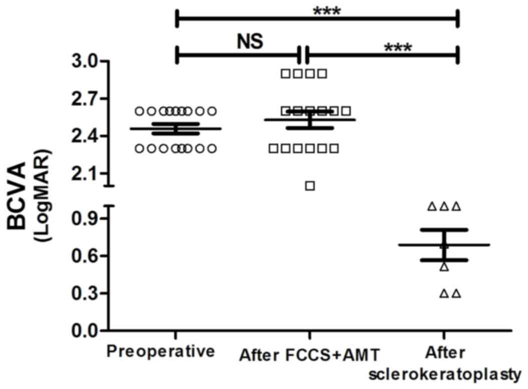
BCVA in the 7 patients at 1 month following sclerokeratoplasty
demonstrated significant improvement (0.689±0.121) compared with
preoperative values (2.459±0.037; P<0.01); similarly, this
improvement was also observed when compared with the BCVA values at
3 months following FCCS + AMT (2.529±0.066; P<0.001; Fig. 5). The mean LogMAR BCVA in the 8
patients who accepted conservative medication was similar to that
at 3 months following FCCS + AMT (Table
II). Furthermore, the IOP for all the patients (via the ocular
palpation method) was normal following sclerokeratoplasty or
conservative medication (Table
II).
Discussion
Fungal keratitis is challenging to overcome as the
incidence has increased, there are a limited number of effective
antifungal agents and there is a shortage of cornea donors for
keratoplasty (4). The perforation
rate is high, particularly when whole corneal ulcers develop during
the late stage (25). Furthermore,
corneal surgery will not repair vision and results in a poor
prognosis in severe cases of fungal keratitis (26). Thus, early diagnosis coupled with
appropriate treatment is critical (27).
Despite ophthalmologists' best efforts, medical
treatment fails in 15–36% of cases (6,28). When
this occurs, therapeutic keratoplasty is necessary (29). However, there is a large discrepancy
between the supply and demand of transplantable organs, despite the
existence of sufficient numbers of potential donors (30). Particularly in developing countries,
the shortage of cornea donors is as high as 52% (10). Therefore, transitional surgeries,
including simple debridement, excisional keratectomy, amniotic
membrane transplantation and conjunctival flap covering surgery,
have an important role in inhibiting disease progression (27).
Surgical debridement may create a smoother surface
for a superficial lesion and promote rapid healing and
re-epithelialization of the cornea; however, it is rarely
successful in severe cases without further surgery and does not
reduce the recurrence of infectious keratitis (31). Amniotic membrane transplantation may
be used as a temporary patch or a permanent graft to promote
healing and decrease inflammation and scarring, but it is not
feasible if the area to be treated is large and the ratio of
dissolution is relatively high in severe fungal keratitis cases
presenting with a whole corneal ulcer (32). Conjunctival flaps have been used to
halt the progression of refractory corneal abscesses, which are
resistant to medical therapy (23).
The flaps provide structural support for necrotic tissue and are
able to control the infection and maintain the integrity of the
globe (33). A study by Abdulhalim
et al (33) reported the
conditions of 20 eyes from 20 patients who underwent bipedicle
conjunctival flap covering surgery as follows: A total of 12 eyes
had fungal keratitis, 7 eyes had bacterial keratitis and 1 eye had
Acanthamoeba keratitis. When considering fungal keratitis with deep
stromal infiltration, a thick flap with Tenon's capsule increased
the resistance to infection and provided stronger mechanical
support for the cornea (27).
Previously, conjunctival flap covering surgery had not been
successful in cases of active fungal corneal ulcers resistant to
topical and systemic antifungal agents (23). The most frequently reported
complications include flap retraction, conjunctival buttonholes and
corneal perforations (34). A study
by Alino et al (34)
indicated that the retraction of a partial flap is likely to be
affected by gravity and blinking action, and the edge of the
partial flap is close to the edge of the defect, which does not
provide enough tension. A study by Sun et al (18) suggested that the major reason for the
failure is incomplete removal of necrotic tissue, which may lead to
a high incidence of corneal perforation. Therefore, in the present
study, the suppurative and necrotic corneas were removed initially,
followed by suturing of a full-thickness conjunctival flap with
Tenon's capsule to the corneal limbal and episcleral tissue
peripherally in a 360-degree manner to increase flap tension.
Furthermore, the donor bed was transplanted with the amniotic
membrane to achieve rapid wound healing.
In the present study, 15/17 patients (88.24%)
demonstrated complete conjunctival re-epithelization and a smooth
conjunctival surface at 3 months postoperatively. Unlike the
studies by Khodadoust and Quinter (35) and Abdulhalim et al (33), no patient experienced conjunctival
retraction or recession of the flap and no other complications
occurred in the present study. We deduced that the advantages of
the technique in the present study were the intact conjunctival
flap, fixed margin and strong mechanical support. However, 2
patients experienced conjunctival flap melting accompanied by
corneal perforation. We suspected that the fungal toxicity of
Aspergillus fumigatus in these 2 patients was high and that
the invasiveness was relatively potent. Additionally, the duration
of disease was 21 and 3 months, respectively, which corresponded to
the late stage of fungal keratitis. The long duration may have
delayed the timely medical and surgical intervention. Thus, prompt
and early diagnosis and surgery is crucial.
The present findings suggested that FCCS + AMT may
be a beneficial choice for promoting wound healing and creating a
greater opportunity for possible corneal transplantation. The
full-thickness conjunctival flap is rich in blood vessels and
lymphatics, which are necessary to transport nutrients to the
corneal surface to help resist infection and provide strong
mechanical support (23).
Additionally, the rich blood supply decreases proinflammatory
mediators and proteases in the microenvironment (23) and the immune modulator wash-out
further decreases the inflammation and aids the healing of
refractory corneal ulcers (36).
Furthermore, the amniotic membrane decreases vascularization and
promotes conjunctival donor healing (32). However, in-depth discussion
concerning FCCS + AMT is required if corneal perforation is
present, particularly when the hole is large. The present results
indicated that 7 patients underwent sclerokeratoplasty surgery and
their visual acuity significantly increased in comparison with the
visual acuity preoperatively; the other 8 patients refused surgery
and chose conservative medication due to economic reasons.
The limitations of the present technique include a
lack of controls and the small sample size. For whole corneal
ulcers in severe fungal keratitis, corneal transplantation is
considered to be an optimal surgery to restore ocular integrity and
FCCS + AMT may be a preferable second choice. In conclusion, FCCS +
AMT may provide a greater opportunity for corneal transplantation,
which may improve the prognosis for patients with fungal
keratitis.
Acknowledgements
The authors would like to thank all the professors
and nurses in the Cornea Department of Zhongshan Ophthalmic Center
(Guangzhou, China). The present study was supported by a grant from
the National Natural Science Foundation of China, received by J.Y.
(grant no. 81270972).
References
|
1
|
Cao J, Yang Y, Yang W, Wu R, Xiao X, Yuan
J, Xing Y and Tan X: Prevalence of infectious keratitis in Central
China. BMC Ophthalmol. 14:432014. View Article : Google Scholar : PubMed/NCBI
|
|
2
|
Xie L, Zhong W, Shi W and Sun S: Spectrum
of fungal keratitis in north China. Ophthalmology. 113:1943–1948.
2006. View Article : Google Scholar : PubMed/NCBI
|
|
3
|
Thomas PA: Fungal infections of the
cornea. Eye (Lond). 17:852–862. 2003. View Article : Google Scholar : PubMed/NCBI
|
|
4
|
FlorCruz NV and Evans JR: Medical
interventions for fungal keratitis. Cochrane Database Syst Rev.
4:CD0042412015.
|
|
5
|
Prajna NV, Krishnan T, Mascarenhas J,
Rajaraman R, Prajna L, Srinivasan M, Raghavan A, Oldenburg CE, Ray
KJ, Zegans ME, et al: The mycotic ulcer treatment trial: a
randomized trial comparing natamycin vs voriconazole. JAMA
Ophthalmol. 131:422–429. 2013. View Article : Google Scholar : PubMed/NCBI
|
|
6
|
Prajna NV, Mascarenhas J, Krishnan T,
Reddy PR, Prajna L, Srinivasan M, Vaitilingam CM, Hong KC, Lee SM,
McLeod SD, et al: Comparison of natamycin and voriconazole for the
treatment of fungal keratitis. Arch Ophthalmol. 128:672–678. 2010.
View Article : Google Scholar : PubMed/NCBI
|
|
7
|
Prajna NV, Krishnan T, Mascarenhas J,
Srinivasan M, Oldenburg CE, Toutain-Kidd CM, Sy A, McLeod SD,
Zegans ME, Acharya NR, et al: Predictors of outcome in fungal
keratitis. Eye (Lond). 26:1226–1231. 2012. View Article : Google Scholar : PubMed/NCBI
|
|
8
|
Li LM, Zhao LQ, Qu LH and Li P: Excimer
laser phototherapeutic keratectomy for the treatment of clinically
presumed fungal keratitis. J Ophthalmol. 2014:9632872014.
View Article : Google Scholar : PubMed/NCBI
|
|
9
|
Henry CR, Flynn HW Jr, Miller D, Schefler
AC, Forster RK and Alfonso EC: Delayed-onset endophthalmitis
associated with corneal suture infections. J Ophthalmic Inflamm
Infect. 3:512013. View Article : Google Scholar : PubMed/NCBI
|
|
10
|
Jiaxu Hong, Weiyun Shi, Zuguo Liu, Roberto
Pineda, Xinhan Cui, Xinghuai Sun and Jianjiang Xu: Limitations of
Keratoplasty in China: A Survey Analysis. PLoS One.
10:e01322682015. View Article : Google Scholar : PubMed/NCBI
|
|
11
|
Shang X and Zhang M: Body and organ
donation in Wuhan, China. Lancet. 376:1033–1034. 2010. View Article : Google Scholar : PubMed/NCBI
|
|
12
|
Sharma A, Mohan K, Sharma R and Nirankari
VS: Repositioning of pedicle conjunctival flap performed for
refractory corneal ulcer. Middle East Afr J Ophthalmol. 21:89–91.
2014. View Article : Google Scholar : PubMed/NCBI
|
|
13
|
Gundersen T: Conjunctival flaps in the
treatment of corneal disease with reference to a new technique of
application. AMA Arch Ophthalmol. 60:880–888. 1958. View Article : Google Scholar : PubMed/NCBI
|
|
14
|
Gao H, Jia Y, Li S, Wang T, Tan Y and Shi
W: Conjunctival flap covering combined with antiviral and steroid
therapy for severe herpes simplex virus necrotizing stromal
keratitis. Scientific World Journal. 2015:5659642015. View Article : Google Scholar : PubMed/NCBI
|
|
15
|
Yang X, Zhou Q and Du S: Conjunctival flap
covering in the treatment of corneal blood staining. Can J
Ophthalmol. 46:442–443. 2011. View Article : Google Scholar : PubMed/NCBI
|
|
16
|
Ding J, Chen T, Hou Z, Qin Y, Hao L and Li
D: Cosmetic shell fitting over a sensitive cornea in mild phthisis
bulbi using total conjunctival flap. Aesthetic Plast Surg.
37:398–401. 2013. View Article : Google Scholar : PubMed/NCBI
|
|
17
|
Yazici B: Use of conjunctiva-Muller muscle
pedicle flap in surgical treatment of necrotizing scleritis.
Ophthal Plast Reconstr Surg. 24:19–23. 2008. View Article : Google Scholar : PubMed/NCBI
|
|
18
|
Sun GH, Li SX, Gao H, Zhang WB, Zhang MA
and Shi WY: Clinical observation of removal of the necrotic corneal
tissue combined with conjunctival flap covering surgery under the
guidance of the AS-OCT in treatment of fungal keratitis. Int J
Ophthalmol. 5:88–91. 2012.PubMed/NCBI
|
|
19
|
Erie JC, McLaren JW and Patel SV: Confocal
microscopy in ophthalmology. Am J Ophthalmol. 148:639–646. 2009.
View Article : Google Scholar : PubMed/NCBI
|
|
20
|
Cruciani F, Cuozzo G, Di Pillo S and
Cavallaro M: Predisposing factors, clinical and microbiological
aspects of bacterial keratitis: A clinical study. Clin Ter.
160:207–210. 2009.PubMed/NCBI
|
|
21
|
Kim JC and Tseng SC: Transplantation of
preserved human amniotic membrane for surface reconstruction in
severely damaged rabbit corneas. Cornea. 14:473–484. 1995.
View Article : Google Scholar : PubMed/NCBI
|
|
22
|
Gopinathan U, Garg P, Fernandes M, Sharma
S, Athmanathan S and Rao GN: The epidemiological features and
laboratory results of fungal keratitis: A 10-year review at a
referral eye care center in South India. Cornea. 21:555–559. 2002.
View Article : Google Scholar : PubMed/NCBI
|
|
23
|
Baum J, Chaturvedi N, Netland PA and
Dreyer EB: Assessment of intraocular pressure by palpation. Am J
Ophthalmol. 119:650–651. 1995. View Article : Google Scholar : PubMed/NCBI
|
|
24
|
The ischemic optic neuropathy
decompression trial (IONDT), . design and methods. Control Clin
Trials. 19:276–296. 1998.PubMed/NCBI
|
|
25
|
Arbelaez JG, Feng MT, Pena TJ, Price MO
and Price FW Jr: A year of cornea in review: 2013. Asia Pac J
Ophthalmol (Phila). 4:40–50. 2015. View Article : Google Scholar : PubMed/NCBI
|
|
26
|
Avunduk AM, Beuerman RW, Varnell ED and
Kaufman HE: Confocal microscopy of Aspergillus fumigatus keratitis.
Br J Ophthalmol. 87:409–410. 2003. View Article : Google Scholar : PubMed/NCBI
|
|
27
|
Eguchi H, Toibana T, Hotta F, Miyamoto T,
Mitamura Y and Yaguchi T: Severe fungal sclerokeratitis caused by
Metarhizium anisopliae: A case report and literature review.
Mycoses. 58:88–92. 2015. View Article : Google Scholar : PubMed/NCBI
|
|
28
|
Anane S, Ben Ayed N, Malek I, Chebbi A,
Lejri S, Bouguila H, Kaouech E, Belhadj S, Kallel K, Ayed S and
Chaker E: Keratomycosis in the area of Tunis: epidemiological data,
diagnostic and therapeutic modalities. Ann Biol Clin (Paris).
68:441–447. 2010.(In French). PubMed/NCBI
|
|
29
|
Yao YF, Zhang YM, Zhou P, Zhang B, Qiu WY
and Tseng SC: Therapeutic penetrating keratoplasty in severe fungal
keratitis using cryopreserved donor corneas. Br J Ophthalmol.
87:543–547. 2003. View Article : Google Scholar : PubMed/NCBI
|
|
30
|
Golchet G, Carr J and Harris MG: Why don't
we have enough cornea donors? A literature review and survey.
Optometry. 71:318–328. 2000.PubMed/NCBI
|
|
31
|
Ozbek Z, Burakgazi AZ and Rapuano CJ:
Rapid healing of vernal shield ulcer after surgical debridement: A
case report. Cornea. 25:472–473. 2006. View Article : Google Scholar : PubMed/NCBI
|
|
32
|
Liu J, Sheha H, Fu Y, Liang L and Tseng
SC: Update on amniotic membrane transplantation. Expert Rev
Ophthalmol. 5:645–661. 2010. View Article : Google Scholar : PubMed/NCBI
|
|
33
|
Abdulhalim BE, Wagih MM, Gad AA, Boghdadi
G and Nagy RR: Amniotic membrane graft to conjunctival flap in
treatment of non-viral resistant infectious keratitis: a randomised
clinical study. Br J Ophthalmol. 99:59–63. 2015. View Article : Google Scholar : PubMed/NCBI
|
|
34
|
Alino AM, Perry HD, Kanellopoulos AJ,
Donnenfeld ED and Rahn EK: Conjunctival flaps. Ophthalmology.
105:1120–1123. 1998. View Article : Google Scholar : PubMed/NCBI
|
|
35
|
Khodadoust A and Quinter AP: Microsurgical
approach to the conjunctival flap. Arch Ophthalmol. 121:1189–1193.
2003. View Article : Google Scholar : PubMed/NCBI
|
|
36
|
Zhou Q, Long X and Zhu X: Improved
conjunctival transplantation for corneal ulcer. Zhong Nan Da Xue
Xue Bao Yi Xue Ban. 35:814–818. 2010.PubMed/NCBI
|