Introduction
Chronic exposure to heavy metals poses a serious
health threat. Among these, mercury, considered by the World Health
Organization as one of the top ten chemicals of major public health
concern (1), has been used for
numerous years for a variety of purposes (2). Exposure mostly occurs through
consumption of fish and fishery products contaminated with organic
mercury, inhalation of mercury vapour from dental amalgams and from
vaccines containing thiomersal (3).
Fish and sea mammals are increasingly becoming a source of mercury
toxicity (4). For a long time,
mercury was principally thought to affect central nervous system,
thus leading to degenerative diseases (5). However, as extensively reviewed by
Fernandes Azevedo et al (6),
mercury also produces profound cardiovascular toxicity. Mercury has
been demonstrated to induce endothelial dysfunction in experimental
models using low doses of mercury (7–11),
attaining the blood mercury concentration just above the safe level
recommended by the Environmental Protection Agency (12). In these studies, reduction of nitric
oxide (NO) bioavailability and increased oxidative stress
consistent with high levels of reactive oxygen species (ROS) were
noted as major causes of endothelial dysfunction observed in
low-dose mercury toxicity. In the light of the above, antioxidants
may have therapeutic potential in the prevention of mercury-induced
endothelial dysfunction. This notion is further supported by a
study by Rizetti et al (9),
which demonstrated that apocynin improves endothelial dysfunction
in aortas of rats exposed to nanomolar concentrations of
mercury.
Ergothioneine (EGT) is an ubiquitous histidine
derivative occurring in higher-order plants and animals (13). In humans, EGT accumulates in cells
and tissues, which are frequently exposed to oxidative stress,
including the liver, bone marrow, lens of the eye, seminal fluid
and blood (14–16). Organic cation transporter, novel,
type 1, encoded by the gene solute carrier family 22, member 4,
mediates the cellular uptake of EGT (17). In contrast to the major tissue
antioxidant glutathione (GSH), EGT is resistant to autoxidation and
does not form disulphides under physiological conditions (18,19).
Several lines of in vitro evidence suggest that EGT is a
potent scavenger of ROS (20–26).
Furthermore, a previous study by our group reported for the first
time that EGT produces relaxation in isolated rat aortas by
inactivating superoxide anions (27). This result and those of further
studies, which indicate a potential role for EGT in the protection
of endothelium (28–30), prompted us to examine its effects on
mercury-induced endothelial dysfunction. The present study was
performed to evaluate the effects of EGT on vascular reactivity in
aortic rings from rats, which were treated with nanomolar
concentrations of mercury chloride.
Materials and methods
Animals
Male Wistar rats (Lemali Ltd., Ankara, Turkey;
weight, 150–175 g; age, 3 months; n=18) were used in the present
study. The protocol for the animal experiment was approved by the
Ethics Committee of Dokuz Eylül University (Izmir, Turkey; approval
no. B.30.2/DEU/0.01/9402). The rats were provided pelleted food and
water ad libitum and were maintained under constant
temperature (22±2°C) and at a relative humidity level of 50% with a
12-h light/dark cycle. Animals were divided into three groups (n=6
in each) and treated for 30 days as follows: i) Control [0.9% NaCl,
0.5 ml administered by intramuscular (IM) injection]; ii) Mercury
chloride (HgCl2) (first dose, 4.6 µg/kg; maintenance
doses, 0.07 µg/kg/day, IM, to make up for daily loss) (10); and iii) HgCl2 + EGT (2
mg/kg, IM).
Reagents
EGT, potassium chloride (KCl), acetylcholine
hydrochloride (ACh), phenylephrine hydrochloride (PE), serotonin
hydrochloride (5-HT) and HgCl2 were obtained from
Sigma-Aldrich (Merck KGaA, Darmstadt, Germany). All drugs were
dissolved in saline (0.9% NaCl).
Preparation of samples
Blood samples were collected by cardiac puncture
under anesthesia with ketamine (100 mg/kg)/xylazine (10 mg/kg)
administered by intraperitoneal injection; the rats were then
sacrificed by decapitation. The thoracic aorta was removed, cleaned
of fat and loose connective tissue, and cut into 2 mm-thick
transverse rings. Aortic rings were suspended between two stainless
hooks in 10-ml organ baths filled with Krebs solution gassed with
95% O2-5% CO2 at 37°C. The composition of
Krebs solution was (in mM): NaCl, 118; KCl, 4.7; CaCl2 ×
2H2O, 2.5; KH2PO4, 1.20;
MgSO4 × 7H2O, 1.17; glucose, 11.1;
NaHCO3, 25. A resting tension of 2 g was applied to the
aortic rings, which were then allowed to equilibrate for 45 min
prior to further experimentation. In this period, tissues were
washed out with Krebs solution every 15 min. Isometrical changes in
tension were processed with MLT0201/RAD force transducers (AD
Instruments, Inc., Colorado Springs, CO, USA) and recorded on
LabChart Pro (version 7.1; AD Instruments, Inc.).
Experimental protocol for vascular
reactivity studies
Concentration-response curves to ACh
(10−9-10−4 M) were recorded in aortic rings
previously contracted with PE (10−6 M). After a washout
period of 45 min, concentration-response curves to PE
(10−9-10−4 M) and to 5-HT
(10−9-10−4.5 M) were recorded,
respectively.
Detection of ROS
Levels of ROS were determined according to the
method described by Wang et al (31), with slight modifications. After a
30-min stabilization period in Krebs solution maintained at 37°C
and gassed with 95% O2 - 5% CO2, aortic rings
were transferred to solid white 96-well plates containing 200 µl
HEPES-buffered Krebs solution (pH 7.4). Following addition of
lucigenin or luminol (final concentration of either, 5 µmol/l), ROS
were quantified using a multi-plate reader (Victor III-1420; Perkin
Elmer, Inc., Waltham, MA, USA). Counts were obtained at 10-second
intervals and corrected for wet tissue weight. Results were
expressed as the area under curve (AUC) for a counting period of 10
min [AUC of relative light units/mg wet tissue].
Tissue homogenization
Aortic tissue was homogenized on ice in PBS (pH 7.4)
using a sonicator (Bandelin Sonopuls, UW 2070; Bandelin, Berlin,
Germany). Homogenate was centrifuged at 10,000 × g for 20 min at
4°C. The supernatant was aliquoted and stored at −80°C for further
evaluation. The protein content was determined by the method of
Lowry et al (32).
Determination of total nitrite
Nitrite levels were determined after conversion of
nitrate to nitrite by nitrate dehydrogenase (33). Aortic supernatant was mixed with an
equal volume of Griess reagent (sulfanilamide 1% w/v;
naphtylethylenediamine dihydrochloride, 0.1% w/v; and
orthophosphoric acid, 25% v/v). Following incubation at 37°C for 10
min, the absorbance was read at 540 nm. Total nitrite levels were
determined from a standard curve with increasing concentrations of
sodium nitrite and normalized to the protein content of the aortic
sample.
Determination of endothelial NO
synthase (eNOS)
Protein levels of eNOS were determined in aortic
supernatants by using an ELISA kit (cat. no. SEA868Ra; Wuhan USCN
Business Co., Ltd., Wuhan, China) according to the manufacturer's
protocols.
Measurement of oxidative stress
markers
The levels of GSH (reduced form) and the ratio of
GSH to oxidized glutathione (GSSG) in blood samples were determined
using the Bioxytech® GSH/GSSG-412 assay (cat. no. 21040;
Oxis International, Inc.; GT Biopharma, Inc., Washington, DC, USA)
according to the manufacturer's protocols. The formation of
malondialdehyde (MDA) and catalase activity in blood samples were
determined using commercially available assay kits
[Bioxytech® MDA-586 (cat. no. 21044) and Catalase-520
assay (cat. no. 21042), respectively; Oxis International, Inc.; GT
Biopharma, Inc.] according to the manufacturer's protocols.
Statistical analysis
All values are expressed as the mean ± standard
error of the mean. Relaxation responses to ACh are expressed as
percentage (%) relaxation of the PE-induced tone. Contractile
responses to PE and 5-HT are expressed as percentage (%) of the
KCl-induced tone. Analysis of variance followed by Tukey's multiple
comparisons test was performed using GraphPad Prism (Version 5.0
for Mac OS X; GraphPad Software, Inc., La Jolla, CA, USA).
P<0.05 was considered to indicate a statistically significant
difference.
Results
Isometric tension recordings
Relaxations
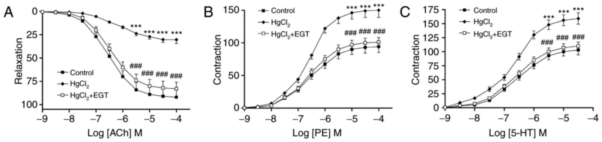
ACh induced concentration-dependent relaxations in
PE-pre-contracted aortic rings from control and treated rats
(Fig. 1A). Treatment with
HgCl2 reduced ACh-induced relaxations by up to 67.1%
compared with the control group (P<0.001) and shifted the
concentration-response curve to the right (Fig. 1A). EGT inhibited the impairment of
ACh-induced relaxation observed in aortic rings from
HgCl2-treated rats (P<0.001; Fig. 1) and significantly increased the
pD2 values (P<0.01; Table
I) compared with HgCl2-treated rats without EGT
treatment.
 | Table I.Effects of EGT on the sensitivity to
ACh, PE and 5-HT in aortas from HgCl2-treated rats. |
Table I.
Effects of EGT on the sensitivity to
ACh, PE and 5-HT in aortas from HgCl2-treated rats.
| Parameter | Control |
HgCl2 |
HgCl2+EGT |
|---|
| Ach | 6.55±0.06 |
6.16±0.07a |
6.46±0.05b |
| PE | 6.54±0.07 | 6.59±0.09 | 6.58±0.05 |
| 5-HT | 6.57±0.08 | 6.64±0.07 | 6.62±0.05 |
Contractions
PE and 5-HT induced concentration-dependent
contractions in aortic rings from control and treated rats
(Fig. 1B and C). Treatment with
HgCl2 significantly increased PE- and 5-HT-induced
contractions compared with the control group (P<0.001; Fig. 1B and C) without affecting the
sensitivity to either agent (Table
I). Co-treatment with EGT prevented the increase in contractile
response to PE and to 5-HT compared with HgCl2-treated
rats without EGT treatment (P<0.001; Fig. 1B and C).
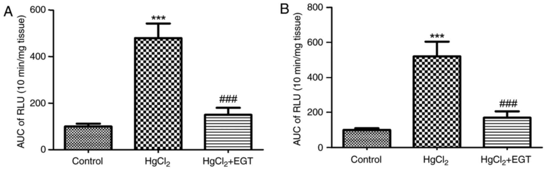
Levels of ROS
Low-dose HgCl2 significantly increased
the levels of ROS in the rat thoracic aortas (Fig. 2A and B). Lucigenin- and
luminol-enhanced chemiluminescence in aortas from
HgCl2-treated rats were ~4.8 and ~5.2 times higher than
in those of control tissues, respectively (for either, P<0.001).
EGT significantly reduced the ROS levels increased by
HgCl2 treatment in HgCl2+EGT-treated rats
compared with HgCl2-treated rats without EGT treatment
(P<0.001; Fig. 2A and B).
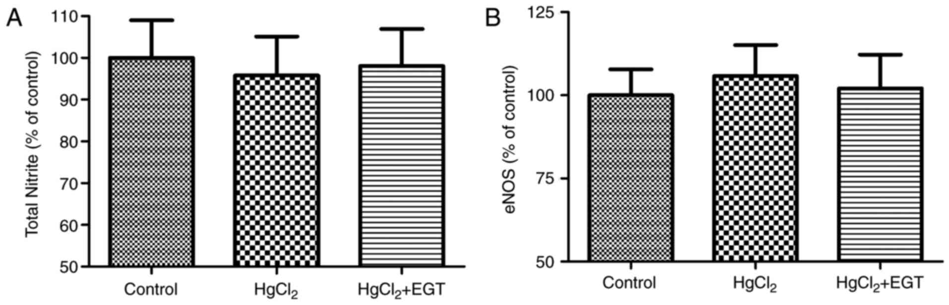
Total nitrite and eNOS levels
Levels of total nitrite and eNOS remained unchanged
among the experimental groups (Fig. 3A
and B).
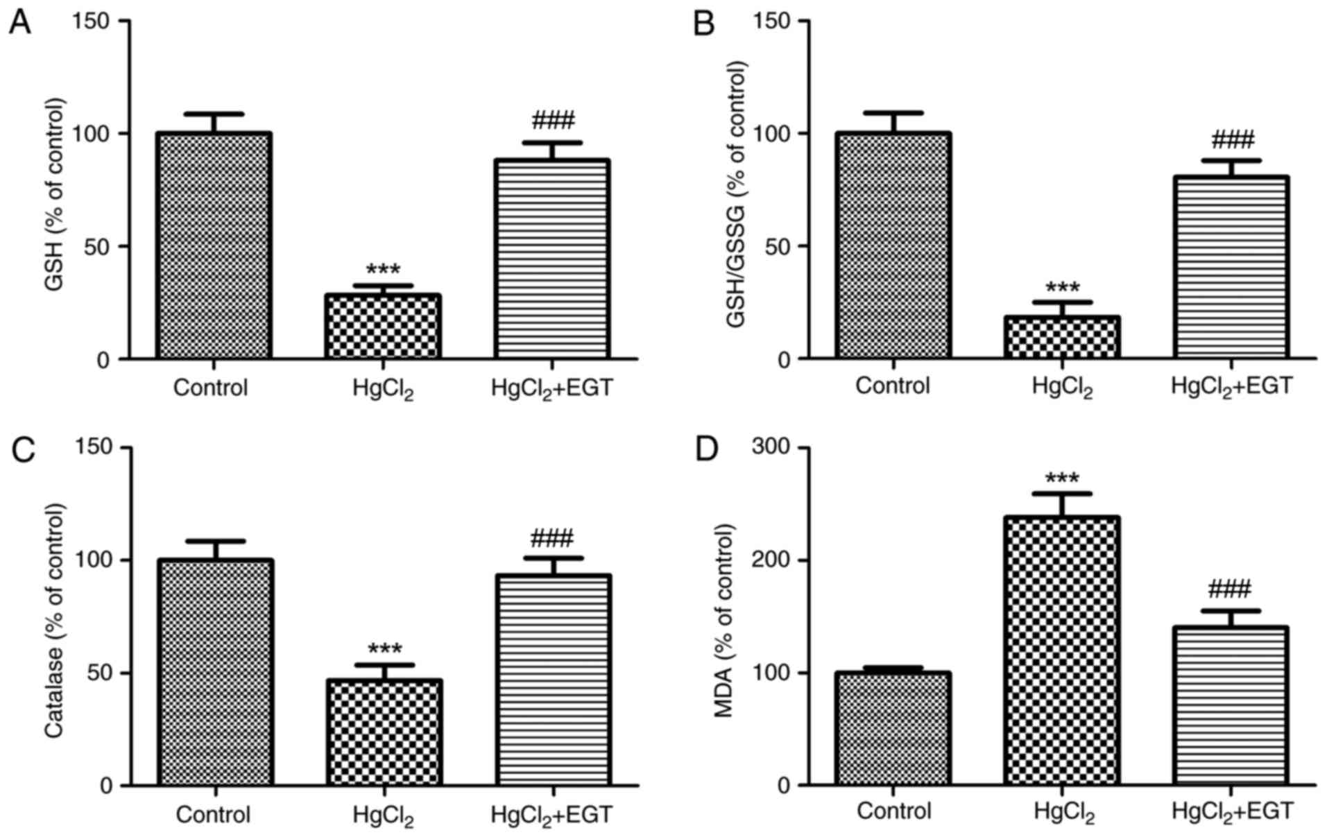
Antioxidant status
Fig. 4 summarizes the
effects of EGT on the antioxidant status in the blood of rats.
Low-dose HgCl2 caused a significant increase in
oxidative stress and lipid peroxidation, and reduced catalase
activity. When compared with the control group, GSH levels and the
GSH/GSSG ratio were significantly lower in HgCl2-treated
rats (P<0.001; Fig. 4A and B).
Similarly, catalase activity decreased by 53.3% in
HgCl2-treated rats compared with the control group
(P<0.001; Fig. 4C). In addition,
lipid peroxidation, as indicated by increased plasma MDA levels,
increased by ~1.3-fold in HgCl2-treated rats compared
with the control group (P<0.001; Fig.
4D). Co-treatment with EGT not only restored the antioxidant
status, but also significantly reduced lipid peroxidation in
HgCl2+EGT-treated rats compared with
HgCl2-treated rats without EGT treatment (P<0.001;
Fig. 4D).
Discussion
Overall health effects of chronic exposure to
mercury are a matter of serious concern and cardiovascular
consequences of mercury toxicity remain an important area of
research. As comprehensively reviewed by Houston (4), exposure to mercury is an underestimated
risk factor for hypertension, coronary heart disease, myocardial
infarction, reduction in heart rate variability, increase in
carotid intima-media thickness and carotid obstruction, generalized
atherosclerosis, renal dysfunction and proteinuria, and an overall
increase in total and cardiovascular mortality. Garcia Gomez et
al (34) reported that the
occurrence of hypertension, stroke and total cardiovascular
mortality in mercury mine workers is increased by 2.78-, 1.17- and
1.51-fold, respectively. It may simply be assumed that these
consequences are most probably due to high levels of occupational
or environmental exposure to mercury. However, evidence from animal
models producing blood mercury levels similar to those of average
human exposure suggest that low-dose mercury promotes endothelial
dysfunction (8–11), a systemic pathological state of the
endothelium, which is widely accepted as an early crucial event in
cardiovascular diseases (35,36). The
present study provides preliminary evidence that EGT, a ubiquitous,
water soluble, sulphur-containing derivative of the amino acid
histidine, prevents low-dose HgCl2-induced endothelial
dysfunction.
The present results may be summarized as follows: i)
Low-dose HgCl2 decreases the maximum value and
sensitivity of the relaxation response to ACh and increases the
maximum value of contractile responses to 5-HT and PE, ii)
HgCl2 increases the levels of ROS in the thoracic aorta,
iii) HgCl2 causes significant reductions in GSH and
catalase levels and decreases the GSH/GSSG ratio, while markedly
increasing MDA formation compared with that in the control group,
and iv) EGT reverses the abovementioned HgCl2-induced
alterations in antioxidant status and vascular reactivity.
In the present study, chronic low-dose
HgCl2 administration to rats caused a marked decline in
the relaxation response to ACh in isolated thoracic aortas by up to
67.1% and significantly decreased the sensitivity, which is
consistent with the results of Wiggers et al (10). The present results indicate that the
HgCl2-associated reduction in relaxant responses and
sensitivity to ACh were almost completely reversed by EGT
treatment. In addition, EGT suppressed the significant increases in
contractile responses to 5-HT and PE in HgCl2-treated
rats. A previous study by our group demonstrated that pre-treatment
with EGT did not affect the ACh-induced relaxation responses in
endothelium-intact rat aortic rings (27). However, in parallel experiments
employing a model of oxidative stress, which is based on inhibition
of endogenous Cu/Zn superoxide dismutase leading to the
accumulation of superoxide anions, EGT recovered the impaired ACh
relaxation (27). In addition, EGT
elicited a concentration-dependent relaxation effect in aortic
rings, which was blunted by endothelial denudation or by inhibition
of NOS (27). Taking the above
results into consideration, the present study first investigated
the possibility that EGT interferes with NO synthesis to increase
Ach-induced relaxation. According to the present results, EGT does
not appear to affect NO synthesis as reflected by similar total
nitrite and eNOS levels among groups. This result gives rise to the
question whether alterations in ROS levels and/or antioxidant
status underlie the ameliorative effects of EGT on NO-dependent
relaxations. Decreased bioavailability of NO due to increased
superoxide anion production by NADPH oxidase is regarded as a
crucial aspect of endothelial dysfunction observed in chronic
exposure to low concentrations of mercury (6,7,37). Superoxide anions not only participate
in endothelial dysfunction, mainly owing to their rapid interaction
with NO, but also produce direct biological effects and serve as a
progenitor for numerous other ROS (38). Several studies revealed that EGT is a
powerful scavenger of ROS and protects cells against a wide range
of stressors (20,26,27).
Indeed, in the present study, high levels of ROS, including
superoxide anions, observed in aortas from HgCl2-treated
rats were significantly diminished by EGT treatment. As is known,
NO rapidly reacts with superoxide anions to form peroxynitrite,
leading to decreased NO bioavailability (39). Furthermore, peroxynitrite, the end
product of this reaction, may also lead to eNOS uncoupling and
cause vasoconstriction (39). Thus,
it is concluded that the superoxide scavenging effects of EGT may
lead to increased NO bioavailability and/or reduced eNOS uncoupling
to restore impaired ACh-induced relaxation without changing eNOS
levels and/or NO production. In addition, EGT improved the
antioxidant status by increasing GSH levels, the GSH/GSSG ratio and
catalase activity, and by reducing lipid peroxidation. Apart from
these results, the protective effects of EGT on vascular reactivity
were also evident in contractile responses. EGT significantly
reduced the increment in PE- and 5-HT-induced contractions. ROS,
particularly superoxide anions, are regarded as endothelium-derived
contracting factors, which have major roles in the regulation of
the arterial tone (40).
Furthermore, superoxide anions and hydrogen peroxide may be
transformed into hydroxyl radicals, which was demonstrated to
increase the synthesis of vasoconstrictor prostanoids (41). In this context, radical scavenging by
and/or the antioxidant activity of EGT may account for the observed
attenuation in contractile responses to PE and 5-HT.
In conclusion, the present study was the first to
report that endothelial dysfunction induced by low doses of
HgCl2 is prevented by EGT. It should be noted that EGT
is acquired by humans through dietary means and accumulates in
cells and tissues that are frequently exposed to oxidative stress
(27). Taking into consideration
that mercury-induced toxicity is often associated with poor
prognosis due to limited treatment options, further studies
evaluating the effects of EGT on the complications of mercury
exposure may provide new insight for therapeutic intervention.
Acknowledgements
Not applicable.
Funding
This study was supported by the Scientific Research
Fund of Ege University, Izmir, Turkey (grant no. 09-ECZ-024).
Availability of data and materials
The analyzed data sets generated during the study
are available from the corresponding author on reasonable
request.
Authors' contributions
GG conceived and designed the experiments. GG and
MZA performed the experiments. GG, MZA and EE analyzed the results
and wrote the paper. All authors have read and approved the final
manuscript.
Ethical approval and consent to
participate
The protocol for the animal experiment was approved
by the Ethics Committee of Dokuz Eylül University (Izmir, Turkey;
approval no. B.30.2/DEU/0.01/9402).
Consent for publication
Not applicable.
Competing interests
The authors declare that they have no competing
interests.
References
|
1
|
World Health Organization, . Mercury and
health. Fact sheet. 2016.http://www.who.int/mediacentre/factsheets/fs361/en/Updated
March 2017.
|
|
2
|
Salonen JT, Seppänen K, Nyyssönen K,
Korpela H, Kauhanen J, Kantola M, Tuomilehto J, Esterbauer H,
Tatzber F and Salonen R: Intake of mercury from fish, lipid
peroxidation, and the risk of myocardial infarction and coronary,
cardiovascular, and any death in eastern Finnish men. Circulation.
91:645–655. 1995. View Article : Google Scholar : PubMed/NCBI
|
|
3
|
Clarkson TW, Magos L and Myers GJ: The
toxicology of mercury-current exposures and clinical
manifestations. N Engl J Med. 349:1731–1737. 2003. View Article : Google Scholar : PubMed/NCBI
|
|
4
|
Houston MC: Role of mercury toxicity in
hypertension, cardiovascular disease, and stroke. J Clin Hypertens
(Greenwich). 13:621–627. 2011. View Article : Google Scholar : PubMed/NCBI
|
|
5
|
Mutter J, Naumann J, Sadaghiani C,
Schneider R and Walach H: Alzheimer disease: Mercury as
pathogenetic factor and apolipoprotein E as a moderator. Neuro
Endocrinol Lett. 25:331–339. 2004.PubMed/NCBI
|
|
6
|
Fernandes Azevedo B, Barros Furieri L,
Peçanha FM, Wiggers GA, Frizera Vassallo P, Ronacher Simões M,
Fiorim J, de Batista Rossi P, Fioresi M, Rossoni L, et al: Toxic
effects of mercury on the cardiovascular and central nervous
systems. J Biomed Biotechnol. 2012:9490482012.PubMed/NCBI
|
|
7
|
Furieri LB, Galán M, Avendaño MS,
García-Redondo AB, Aguado A, Martínez S, Cachofeiro V, Bartolomé
MV, Alonso MJ, Vassallo DV and Salaices M: Endothelial dysfunction
of rat coronary arteries after exposure to low concentrations of
mercury is dependent on reactive oxygen species. Br J Pharmacol.
162:1819–1831. 2011. View Article : Google Scholar : PubMed/NCBI
|
|
8
|
Pecanha FM, Wiggers GA, Briones AM,
Perez-Giron JV, Miguel M, Garcia-Redondo AB, Vassallo DV, Alonso MJ
and Salaices M: The role of cyclooxygenase (COX)-2 derived
prostanoids on vasoconstrictor responses to phenylephrine is
increased by exposure to low mercury concentration. J Physiol
Pharmacol. 61:29–36. 2010.PubMed/NCBI
|
|
9
|
Rizzetti DA, Torres JG, Escobar AG,
Peçanha FM, Santos FW, Puntel RL, Alonso MJ, Briones AM, Salaices
M, Vassallo DV and Wiggers GA: Apocynin prevents vascular effects
caused by chronic exposure to low concentrations of mercury. PLoS
One. 8:e558062013. View Article : Google Scholar : PubMed/NCBI
|
|
10
|
Wiggers GA, Peçanha FM, Briones AM,
Pérez-Girón JV, Miguel M, Vassallo DV, Cachofeiro V, Alonso MJ and
Salaices M: Low mercury concentrations cause oxidative stress and
endothelial dysfunction in conductance and resistance arteries. Am
J Physiol Heart Circ Physiol. 295:H1033–H1043. 2008. View Article : Google Scholar : PubMed/NCBI
|
|
11
|
Wiggers GA, Stefanon I, Padilha AS,
Pecanha FM, Vassallo DV and Oliveira EM: Low nanomolar
concentration of mercury chloride increases vascular reactivity to
phenylephrine and local angiotensin production in rats. Comp
Biochem Physiol C Toxicol Pharmacol. 147:252–260. 2008. View Article : Google Scholar : PubMed/NCBI
|
|
12
|
Rice DC: The US EPA reference dose for
methylmercury: Sources of uncertainty. Environ Res. 95:406–413.
2004. View Article : Google Scholar : PubMed/NCBI
|
|
13
|
Cheah IK and Halliwell B: Ergothioneine;
antioxidant potential, physiological function and role in disease.
Biochim Biophys Acta. 1822:784–793. 2012. View Article : Google Scholar : PubMed/NCBI
|
|
14
|
Melville DB, Horner WH and Lubschez R:
Tissue ergothioneine. J Biol Chem. 206:221–228. 1954.PubMed/NCBI
|
|
15
|
Shires TK, Brummel MC, Pulido JS and
Stegink LD: Ergothioneine distribution in bovine and porcine ocular
tissues. Comp Biochem Physiol C Pharmacol Toxicol Endocrinol.
117:117–120. 1997. View Article : Google Scholar : PubMed/NCBI
|
|
16
|
Shukla Y, Kulshrestha OP and Khuteta KP:
Ergothioneine content in normal and senile human cataractous
lenses. Indian J Med Res. 73:472–473. 1981.PubMed/NCBI
|
|
17
|
Gründemann D, Harlfinger S, Golz S, Geerts
A, Lazar A, Berkels R, Jung N, Rubbert A and Schömig E: Discovery
of the ergothioneine transporter. Proc Natl Acad Sci USA.
102:5256–5261. 2005. View Article : Google Scholar : PubMed/NCBI
|
|
18
|
Melville DB: Ergothioneine. Vitamin Horm.
17:155–204. 1959. View Article : Google Scholar
|
|
19
|
Paul BD and Snyder SH: The unusual amino
acid L-ergothioneine is a physiologic cytoprotectant. Cell Death
Differ. 17:1134–1140. 2010. View Article : Google Scholar : PubMed/NCBI
|
|
20
|
Motohashi N and Mori I: The role of
ergothioneine in the oxidation of reduced nicotinamide adenine
dinucleotide by metmyoglobin or methemoglobin. Chem Pharm Bull
(Tokyo). 31:1702–1707. 1983. View Article : Google Scholar : PubMed/NCBI
|
|
21
|
Reglinski J, Smith WE and Sturrock RD:
Spin-echo 1H NMR detected response of ergothioneine to oxidative
stress in the intact human erythrocyte. Magn Reson Med. 6:217–223.
1988. View Article : Google Scholar : PubMed/NCBI
|
|
22
|
Hartman PE: Ergothioneine as antioxidant.
Methods Enzymol. 186:310–318. 1990. View Article : Google Scholar : PubMed/NCBI
|
|
23
|
Akanmu D, Cecchini R, Aruoma OI and
Halliwell B: The antioxidant action of ergothioneine. Arch Biochem
Biophys. 288:10–16. 1991. View Article : Google Scholar : PubMed/NCBI
|
|
24
|
Aruoma OI, Whiteman M, England TG and
Halliwell B: Antioxidant action of ergothioneine: Assessment of its
ability to scavenge peroxynitrite. Biochem Biophys Res Commun.
231:389–391. 1997. View Article : Google Scholar : PubMed/NCBI
|
|
25
|
Mitsuyama H and May JM: Uptake and
antioxidant effects of ergothioneine in human erythrocytes. Clin
Sci (Lond). 97:407–411. 1999. View Article : Google Scholar : PubMed/NCBI
|
|
26
|
Franzoni F, Colognato R, Galetta F,
Laurenza I, Barsotti M, Di Stefano R, Bocchetti R, Regoli F, Carpi
A, Balbarini A, et al: An in vitro study on the free radical
scavenging capacity of ergothioneine: comparison with reduced
glutathione, uric acid and trolox. Biomed Pharmacother. 60:453–457.
2006. View Article : Google Scholar : PubMed/NCBI
|
|
27
|
Gokce G and Arun MZ: Ergothioneine
produces relaxation in isolated rat aorta by inactivating
superoxide anion. Eur Rev Med Pharmacol Sci. 18:3339–3345.
2014.PubMed/NCBI
|
|
28
|
Sit ASM, Ho EYW, Li RWS, et al:
Ergothioneine shows protective effect on endothelial cells in
oxidative stress. Faseb J. 25:630–633. 2011.
|
|
29
|
Martin KR: The bioactive agent
ergothioneine, a key component of dietary mushrooms, inhibits
monocyte binding to endothelial cells characteristic of early
cardiovascular disease. J Med Food. 13:1340–1346. 2010. View Article : Google Scholar : PubMed/NCBI
|
|
30
|
Li RW, Yang C, Sit AS, Kwan YW, Lee SM,
Hoi MP, Chan SW, Hausman M, Vanhoutte PM and Leung GP: Uptake and
protective effects of ergothioneine in human endothelial cells. J
Pharmacol Exp Ther. 350:691–700. 2014. View Article : Google Scholar : PubMed/NCBI
|
|
31
|
Wang HD, Xu S, Johns DG, Du Y, Quinn MT,
Cayatte AJ and Cohen RA: Role of NADPH oxidase in the vascular
hypertrophic and oxidative stress response to angiotensin II in
mice. Circ Res. 88:947–953. 2001. View Article : Google Scholar : PubMed/NCBI
|
|
32
|
Lowry OH, Rosebrough NJ, Farr Al and
Randall RJ: Protein measurement with the Folin phenol reagent. J
Biol Chem. 193:265–275. 1951.PubMed/NCBI
|
|
33
|
Majithiya JB, Paramar AN and Balaraman R:
Pioglitazone, a PPARgamma agonist, restores endothelial function in
aorta of streptozotocin-induced diabetic rats. Cardiovasc Res.
66:150–161. 2005. View Article : Google Scholar : PubMed/NCBI
|
|
34
|
Gomez Garcia M, Boffetta P, Klink
Caballero JD, Espanol S and Quintana Gomez J: Cardiovascular
mortality in mercury miners. Med Clin (Barc). 128:766–771. 2007.(In
Spanish). View Article : Google Scholar : PubMed/NCBI
|
|
35
|
Gokce N, Keaney JF Jr, Hunter LM, Watkins
MT, Nedeljkovic ZS, Menzoian JO and Vita JA: Predictive value of
noninvasively determined endothelial dysfunction for long-term
cardiovascular events in patients with peripheral vascular disease.
J Am Coll Cardiol. 41:1769–1775. 2003. View Article : Google Scholar : PubMed/NCBI
|
|
36
|
Widlansky ME, Gokce N, Keaney JF Jr and
Vita JA: The clinical implications of endothelial dysfunction. J Am
Coll Cardiol. 42:1149–1160. 2003. View Article : Google Scholar : PubMed/NCBI
|
|
37
|
Massaroni L, Rossoni LV, Amaral SM,
Stefanon I, Oliveira EM and Vassallo DV: Haemodynamic and
electrophysiological acute toxic effects of mercury in
anaesthetized rats and in langendorff perfused rat hearts.
Pharmacol Res. 32:27–36. 1995. View Article : Google Scholar : PubMed/NCBI
|
|
38
|
Guzik TJ and Channon KM: Measurement of
vascular reactive oxygen species production by chemiluminescence.
Methods Mol Med. 108:73–89. 2005.PubMed/NCBI
|
|
39
|
Pacher P, Beckman JS and Liaudet L: Nitric
oxide and peroxynitrite in health and disease. Physiol Rev.
87:315–424. 2007. View Article : Google Scholar : PubMed/NCBI
|
|
40
|
Katusic ZS and Vanhoutte PM: Superoxide
anion is an endothelium-derived contracting factor. Am J Physiol.
257:H33–H37. 1989.PubMed/NCBI
|
|
41
|
Vanhoutte PM: Endothelium-derived free
radicals: For worse and for better. J Clin Invest. 107:23–25. 2001.
View Article : Google Scholar : PubMed/NCBI
|