Introduction
Gastric carcinoma with lymphoid stroma (GCLS) is
rare due to the unique histological features of gastric carcinoma
with a favorable prognosis (1).
GCLS demonstrates a well-defined tumor border and contains several
lymphocytes, plasma cells and lymphoid follicles (1). Lymphoepithelioma-like carcinoma
demonstrates prominent lymphoplasmacytic infiltration and is
commonly associated with Epstein-Barr virus (EBV) infection
(2), which is well-known to be
present in GCLS cancer cells (3–6).
Ezrin is a member of the ezrin/radixin/moesin (ERM)
family, and is characterized by small-sized molecules linking the
plasma membrane and the actin cytoskeleton (7). Ezrin expression and phosphorylation
are also crucial in the regulation of tumor metastasis (8–10).
Ezrin expression is reduced in diffuse-type gastric carcinoma
(11), while ezrin overexpression
is known to be correlated with the progression and poor prognosis
of gastric carcinomas (12,13).
Recently, ezrin phosphorylation was found to be associated with EBV
latent membrane protein 1 (LMP1) and to induce tumor migration in
nasopharyngeal carcinomas (14).
Ezrin expression has been identified in ordinary
gastric carcinomas, whereas ezrin expression in GCLS and its
correlation with EBV infection have not been previously studied.
Therefore, the present study aimed to investigate ezrin expression
and its phosphorylated form in GCLS and non-GCLS.
Materials and methods
Tissue samples
GCLS or without lymphoid stroma (non-GCLS) samples
were surgically resected and diagnosed at the Department of
Anatomic Pathology of the Kyushu University (Fukuoka, Japan)
between 1970 and 2008. The study protocol used was in compliance
with the Ethical Guidelines of the 1975 Declaration of Helsinki.
For strict privacy protection reasons, information identifying the
samples was removed prior to analysis. The GCLS group included 74
males and 26 females with a median age of 62.0 years (range,
37–90), whereas the non-GCLS group included 16 males and 13 females
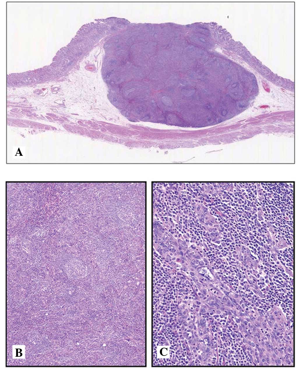
with a median age of 58.4 years (range, 39–82). GCLS was defined as
a well-circumscribed and expansive growth pattern adenocarcinoma
(Fig. 1A) with a dense and diffuse
lymphoid infiltration (Fig. 1B),
and a lace-like or small-nested proliferative pattern of carcinoma
cells with various degrees of cellular pleomorphism (Fig. 1C), according to previously
published studies (1,3,4).
Non-GCLS for the control cases was defined as medullary growth-type
adenocarcinoma lacking frequent lymphoid infiltration or a
glandular component. Subsequently, GCLS (n=104) were collected from
100 patients and non-GCLS (n=29) from 29 patients, based on the
histopathological definition provided above.
In situ hybridization
To test for the presence of EBV, in situ
hybridization (ISH) was performed on paraffin sections with an
EBV-encoded RNA (EBER)-specific peptide nucleic acid (PNA) probe
and a PNA ISH detection kit (DakoCytomation, Carpinteria, CA, USA),
according to the manufacturer’s instructions, as previously
described (15). EBV-positive
nasopharyngeal carcinoma (non-keratinizing undifferentiated
carcinoma) was used as the positive control.
Immunohistochemical staining and
evaluation
Tumor samples were fixed with 10% formaldehyde,
embedded in paraffin, and sectioned into 4-μm slices.
Immunohistochemical staining was performed using the
streptavidin-biotin-peroxidase method (Histofine®
staining kit; Nichirei Co., Tokyo, Japan). The primary antibodies
used in this study were rabbit polyclonal anti-ezrin (dilution,
1:200; Cell Signaling Technology, Inc., Beverly, MA, USA), and
rabbit polyclonal anti-phospho-ezrin (dilution, 1:200; Cell
Signaling Technology, Inc.). The phosphorylated form identified the
endogenous levels of ezrin, radixin and moesin only when
phosphorylated at Thr567 of ezrin, Thr564 of radixin or Thr558 of
moesin, respectively. Following the inhibition of endogenous
peroxidase in a 3% H2O2-methanol solution for
15 min and antigen retrieval microwave irradiation in citrate
buffer (pH 6.0) for the two antibodies, the sections were exposed
to each primary antibody at 37°C overnight. The sections were then
reacted in 3,3′-diaminobenzidine, counterstained with hematoxylin
and mounted. Scoring of the immunohistochemical results was
performed by two pathologists (TT and SA), who were unaware of the
clinical data. Immunohistochemical staining was evaluated in the
carcinoma cell component. The proportion of positive cells was
counted in >1,000 carcinoma cells and was recorded as a
percentage.
Statistical analysis
For the statistical analysis, the expression of
ezrin or p-ezrin in carcinoma cells was identified as a
high-expression group, when the cytoplasm or membrane staining
showed >30% of carcinoma cells. Statistical analysis of group
differences was carried out using the Chi-square, Fisher’s exact
and Student’s-t tests. P<0.05 was considered statistically
significant.
Results
Clinicopathological characteristics of
GCLS and non-GCLS patients
The clinicopathological characteristics of patients
are summarized in Table I. The
GCLS group included 74 males and 26 females with a median age of
62.0 years (range, 37–90), whereas the non-GCLS group included 16
males and 13 females with a median age of 58.4 years (range, 39–82)
(P=0.0381). The tumor size of GCLS was larger compared to that of
non-GCLS (P=0.0084). There were 54, 29 and 21 GCLS cases from the
upper, middle and lower stomach, as well as 7, 15 and 7 non-GCLS
cases from the upper, middle and lower stomach, respectively
(P=0.0194). Lymph node metastasis in GCLS was markedly infrequent
compared to non-GCLS (P=0.0383).
 | Table IClinicopathological characteristics of
patients. |
Table I
Clinicopathological characteristics of
patients.
| Characteristics | GCLS (n=104) | non-GCLS (n=29) | P-value |
|---|
| Age (years) | 62.0 | 58.4 | 0.1605 |
| Gender (M/F) | 78/26 | 16/13 | 0.0381 |
| Tumor size
(cm2) | 4.29 | 5.97 | 0.0084 |
| Location | | | |
|
Upper/middle/low | 54/29/21 | 7/15/7 | 0.0194 |
| Histology W/M/P | 5/25/74 | 0/3/26 | 0.1083 |
| Depth of
invasion | | | |
| M, SM/MP, SS | 48/56 | 18/11 | 0.4306 |
| Lymphatic invasion
+/− | 1/103 | 2/27 | 0.1197 |
| Vessel invasion
+/− | 22/82 | 11/18 | 0.0644 |
| Lymph node +/− | 10/94 | 7/22 | 0.0383 |
| EBER +/− | 78/26 | 0/29 | <0.0001 |
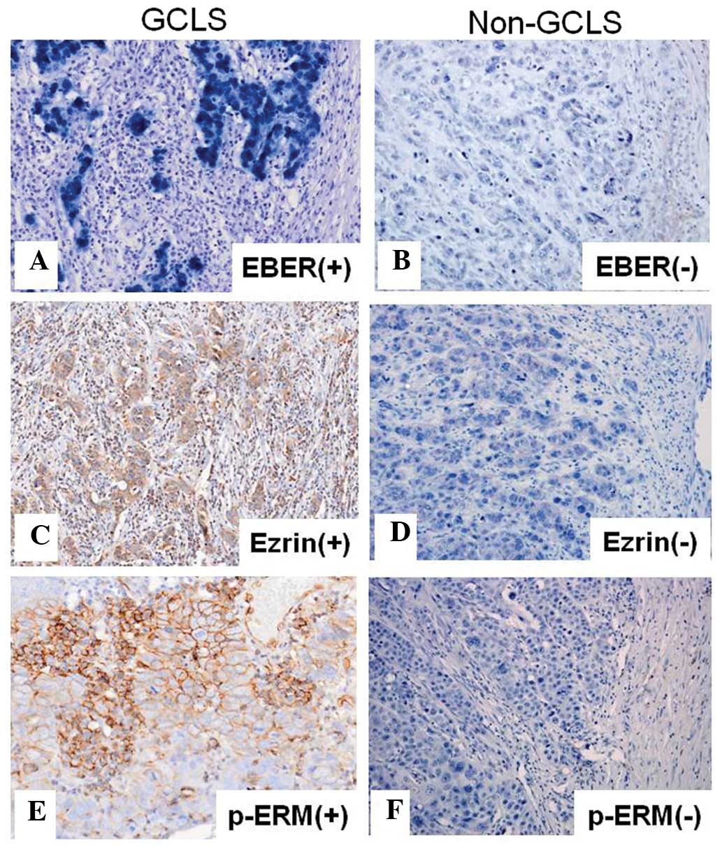
GCLS cases were divided into EBER-positive (n=76)
(Fig. 2A) and -negative cases
(n=28) by in situ hybridization. By contrast, non-GCLS cases
included no EBER-positive cases (Fig.
2B) (P<0.0001).
Ezrin and p-ezrin expression in GCLS and
non-GCLS
Positive ezrin expression was diffusely observed in
the cytoplasm and membrane of carcinoma cells (Fig. 2C), whereas p-ezrin was
predominantly detected in the membrane of carcinoma cells alone
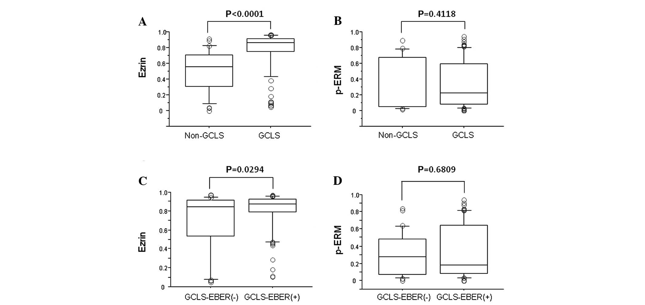
(Fig. 2E). Ezrin expression was
markedly higher in GCLS compared to non-GCLS (P<0.0001)
(Fig. 3A), although there were no
differences in the p-ezrin expression in GCLS and non-GCLS
(Fig. 3B). Ezrin expression was
higher in EBV-positive GCLS compared to -negative GCLS (P=0.0294)
(Fig. 3C), although there was no
difference in p-ezrin expression in EBV-positive and -negative GCLS
(Fig. 3D).
Correlation between ezrin/p-ezrin
expression and clinicopathological characteristics of GCLS
patients
The correlation between the expression of ezrin or
p-ezrin and the clinicopathological patient characteristics is
summarized in Table II. High
expression of ezrin and p-ezrin (>30% of positive carcinoma
cells) of GCLS was observed in 95/104 (91%) and in 45/104 (43%) of
cases, respectively. Forty five of p-ezrin-positive cases included
42 ezrin-positive and 3 -negative cases. No significant differences
were observed between ezrin expression and the clinicopathological
characteristics, however, the high expression of ezrin tended to
correspond to a larger tumor size (P=0.0867). Only p-ezrin in GCLS
was associated with positive lymph node metastasis (P=0.0187).
| Table IICorrelation between ezrin or p-ERM
expression and clinicopathological characteristics. |
Table II
Correlation between ezrin or p-ERM
expression and clinicopathological characteristics.
| Ezrin
| p-ERM
|
|---|
| Characteristics | >30% (n=95) | <30% (n=9) | P-value | >30% (n=45) | <30% (n=59) | P-value |
|---|
| Age (years) | 62.1 | 60.7 | 0.7238 | 60.7 | 62.9 | 0.3407 |
| Gender (M/F) | 70/25 | 8/1 | 0.4444 | 31/14 | 47/12 | 0.2088 |
| Tumor size
(cm2) | 4.446 | 2.656 | 0.0867 | 4.800 | 3.903 | 0.1313 |
| Location | | | | | | |
|
Upper/middle/low | 47/29/19 | 6/0/3 | 0.1412 | 25/15/5 | 28/14/17 | 0.0840 |
| Histology
W/M/P | 4/23/68 | 1/2/6 | 0.6519 | 4/10/31 | 1/15/43 | 0.2329 |
| Depth of
invasion | | | | | | |
| M, SM/MP, SS | 42/53 | 6/3 | 0.2962 | 18/27 | 30/29 | 0.2716 |
| Lymphatic invasion
+/− | 20/75 | 2/7 | 0.9346 | 9/36 | 13/46 | 0.8013 |
| Vessel invasion
+/− | 0/95 | 1/8 | 0.0865 | 1/44 | 0/59 | 0.4327 |
| Lymph node +/− | 10/85 | 0/9 | 0.3059 | 8/37 | 2/57 | 0.0187 |
Discussion
EBV-associated gastric carcinoma accounts for ∼10%
of gastric carcinomas and histologically resembles GCLS (16). Consistent with a previous study,
the GCLS cases of this study demonstrated a male predominance, a
greater tendency towards being located in the fundic or upper
gastric regions and a lower frequency of lymph node metastasis,
compared to the control cases (17). Moreover, 75% of GCLS cases
demonstrated EBER positivity. Taken together, these findings
indicate that the cases examined in this study might be
characterised as typical GCLS cases.
Molecular abnormalities of EBV-associated gastric
carcinomas remain unknown, whereas the CpG island methylation in
the promoter region of PTEN gene has been previously documented
(18). Endo et al (14) have demonstrated EBV LMP1 to be
associated with ezrin phosphorylation in nasopharyngeal carcinoma,
whereas the present is the first study to indicate that ezrin
expression is correlated with GCLS and EBER positivity.
Ezrin is widely expressed in malignant tumors,
including gastric (12,13) and colon cancers (19), hepatocellular carcinoma (20), ovarian (21) as well as breast cancer (22), and is associated with poor
prognosis. Furthermore, ezrin expression is correlated with an
early recurrence of hepatocellular carcinoma (22), invasion of pancreatic
adenocarcinoma (23) and lymph
node metastasis of prostate cancer (24) and nasopharyngeal carcinoma
(25).
According to a comparison between ezrin or p-ezrin
exression and clinicopathological characteristics, only lymph node
metastasis is associated with p-ezrin. In the present study,
anti-ezrin unphosphorylated type and anti-ERM [ezrin
(Thr567)/radixin (Thr564)/moesin (Thr558), Cell Signaling
Technology, Inc.] antibody was used as the anti-ezrin
phosphorylated form at the Thr567 site, also used as the p-ezrin
antibody at Thr567 in previously published studies (26,27).
The phosphorylation of residual Thr567 in ezrin alters the protein
to expose its binding sites (28)
thus resulting in oncogene-induced transformation (29). The phosphorylation at Thr567 may
not be necessary for osteosarcoma metastasis (27). In addition, phosphorylation at
Tyr353, but not at Thr567, is associated with lymph node metastasis
of pancreatic adenocarcinoma (30). However, Tang et al (27) have reported that ezrin
phosphorylation at Thr567 may induce lymph node metastasis of
nasopharyngeal carcinomas. The findings in the present study were
consistent with the hypothesis that p-ezrin at Thr567 is crucial in
lymph node metastasis. Notably, nasopharyngeal carcinoma and GCLS
are commonly associated with EBV infection.
Based on recent studies of targeting for ezrin,
reduction of the ezrin gene by RNA interference (RNAi) may inhibit
the migration and invasion of human gastric cancer cells (31). Berberine also inhibits metastasis
of nasopharyngeal carcinoma cells by targeting Rho kinase-mediated
ezrin phosphorylation at Thr567 (27). These findings suggest that the
molecular target therapy of ezrin and its phosphorylation should be
considered to be EBV-associated GCLS.
In conclusion, ezrin expression is correlated with
GCLS with EBV infection, while the phosphorylation of ezrin is
essential in the lymph node metastasis of GCLS.
References
|
1
|
Watanabe H, Enjoji M and Imai T: Gastric
carcinoma with lymphoid stroma. Its morphologic characteristics and
prognostic correlations. Cancer. 38:232–243. 1976. View Article : Google Scholar : PubMed/NCBI
|
|
2
|
Weiss LM, Movahed LA, Butler AE, et al:
Analysis of lymphoepithelioma and lymphoepithelioma-like carcinomas
for Epstein-Barr viral genomes by in situ hybridization. Am J Surg
Pathol. 13:625–631. 1989. View Article : Google Scholar : PubMed/NCBI
|
|
3
|
Nakamura S, Ueki T, Yao T, et al:
Epstein-Barr virus in gastric carcinoma with lymphoid stroma.
Special reference to its detection by the polymerase chain reaction
and in situ hybridization in 99 tumors, including a morphologic
analysis. Cancer. 73:2239–2249. 1994. View Article : Google Scholar
|
|
4
|
Matsunou H, Konishi F, Hori H, et al:
Characteristics of Epstein-Barr virus-associated gastric carcinoma
with lymphoid stroma in Japan. Cancer. 77:1998–2004. 1996.
View Article : Google Scholar : PubMed/NCBI
|
|
5
|
Oda K, Tamaru J, Takenouchi T, et al:
Association of Epstein-Barr virus with gastric carcinoma with
lymphoid stroma. Am J Pathol. 143:1063–1071. 1993.PubMed/NCBI
|
|
6
|
Burke AP, Yen TS, Shekitka KM, et al:
Lymphoepithelial carcinoma of the stomach with Epstein-Barr virus
demonstrated by polymerase chain reaction. Mod Pathol. 3:377–380.
1990.PubMed/NCBI
|
|
7
|
Vaheri A, Carpén O, Heiska L, et al: The
ezrin protein family: membrane-cytoskeleton interactions and
disease associations. Curr Opin Cell Biol. 9:659–666. 1997.
View Article : Google Scholar : PubMed/NCBI
|
|
8
|
Akisawa N, Nishimori I, Iwamura T, et al:
High levels of ezrin expressed by human pancreatic adenocarcinoma
cell lines with high metastatic potential. Biochem Biophys Res
Commun. 258:395–400. 1999. View Article : Google Scholar : PubMed/NCBI
|
|
9
|
Sarrió D, Rodríguez-Pinilla SM, Dotor A,
et al: Abnormal ezrin localization is associated with
clinicopathological features in invasive breast carcinomas. Breast
Cancer Res Treat. 98:71–79. 2006.PubMed/NCBI
|
|
10
|
Khanna C, Wan X and Bose S: The
membrane-cytoskeleton linker ezrin is necessary for osteosarcoma
metastasis. Nat Med. 10:182–186. 2004. View
Article : Google Scholar : PubMed/NCBI
|
|
11
|
Bal N, Yildirim S, Nursal TZ, et al:
Association of ezrin expression in intestinal and diffuse gastric
carcinoma with clinicopathological parameters and tumor type. World
J Gastroenterol. 13:3726–3729. 2007.PubMed/NCBI
|
|
12
|
Zhao J, Zhang X and Xin Y: Up-regulated
expression of Ezrin and c-Met proteins are related to the
metastasis and prognosis of gastric carcinomas. Histol Histopathol.
26:1111–1120. 2011.PubMed/NCBI
|
|
13
|
Li L, Wang YY, Zhao ZS and Ma J: Ezrin is
associated with gastric cancer progression and prognosis. Pathol
Oncol Res. 17:909–915. 2011. View Article : Google Scholar : PubMed/NCBI
|
|
14
|
Endo K, Kondo S, Shackleford J, et al:
Phosphorylated ezrin is associated with EBV latent membrane protein
1 in nasopharyngeal carcinoma and induces cell migration. Oncogene.
28:1725–1735. 2009. View Article : Google Scholar : PubMed/NCBI
|
|
15
|
Yamamoto H, Kohashi K, Oda Y, et al:
Absence of human herpesvirus-8 and Epstein-Barr virus in
inflammatory myofibroblastic tumor with anaplastic large cell
lymphoma kinase fusion gene. Pathol Int. 56:584–590. 2006.
View Article : Google Scholar : PubMed/NCBI
|
|
16
|
Fukayama M: Epstein-Barr virus and gastric
carcinoma. Pathol Int. 60:337–350. 2010. View Article : Google Scholar : PubMed/NCBI
|
|
17
|
van Beek J, zur Hausen A, Klein Kranenbarg
E, et al: EBV-positive gastric adenocarcinomas: a distinct
clinicopathologic entity with a low frequency of lymph node
involvement. J Clin Oncol. 22:664–670. 2004.PubMed/NCBI
|
|
18
|
Hino R, Uozaki H, Murakami N, et al:
Activation of DNA methyltransferase 1 by EBV latent membrane
protein 2A leads to promoter hypermethylation of PTEN gene in
gastric carcinoma. Cancer Res. 69:2766–2774. 2009. View Article : Google Scholar : PubMed/NCBI
|
|
19
|
Elzagheid A, Korkeila E and Bendardaf R:
Intense cytoplasmic ezrin immunoreactivity predicts poor survival
in colorectal cancer. Hum Pathol. 39:1737–1743. 2008. View Article : Google Scholar : PubMed/NCBI
|
|
20
|
Kang YK, Hong SW, Lee H, et al: Prognostic
implications of ezrin expression in human hepatocellular carcinoma.
Mol Carcinog. 49:798–804. 2010.PubMed/NCBI
|
|
21
|
Moilanen J, Lassus H, Leminen A, et al:
Ezrin immunoreactivity in relation to survival in serous ovarian
carcinoma patients. Gynecol Oncol. 90:273–281. 2003. View Article : Google Scholar : PubMed/NCBI
|
|
22
|
Okamura D, Ohtsuka M, Kimura F, et al:
Ezrin expression is associated with hepatocellular carcinoma
possibly derived from progenitor cells and early recurrence after
surgical resection. Mod Pathol. 21:847–855. 2008. View Article : Google Scholar
|
|
23
|
Kocher HM, Sandle J, Mirza TA, et al:
Ezrin interacts with cortactin to form podosomal rosettes in
pancreatic cancer cells. Gut. 58:271–284. 2009. View Article : Google Scholar : PubMed/NCBI
|
|
24
|
Pang J, Liu WP, Liu XP, et al: Profiling
protein markers associated with lymph node metastasis in prostate
cancer by DIGE-based proteomics analysis. J Proteome Res.
9:216–226. 2010. View Article : Google Scholar : PubMed/NCBI
|
|
25
|
Wang L, Lin GN, Jiang XL, et al:
Expression of ezrin correlates with poor prognosis of
nasopharyngeal carcinoma. Tumour Biol. 32:707–712. 2011. View Article : Google Scholar : PubMed/NCBI
|
|
26
|
Di Cristofano C, Leopizzi M, Miraglia A,
et al: Phosphorylated ezrin is located in the nucleus of the
osteosarcoma cell. Mod Pathol. 23:1012–1020. 2010.PubMed/NCBI
|
|
27
|
Tang F, Wang D, Duan C, et al: Berberine
inhibits metastasis of nasopharyngeal carcinoma 5-8F cells by
targeting Rho kinase-mediated Ezrin phosphorylation at threonine
567. J Biol Chem. 284:27456–27466. 2009. View Article : Google Scholar : PubMed/NCBI
|
|
28
|
Zhu L, Zhou R, Mettler S, et al: High
turnover of ezrin T567 phosphorylation: conformation, activity, and
cellular function. Am J Physiol Cell Physiol. 293:C874–C884. 2007.
View Article : Google Scholar : PubMed/NCBI
|
|
29
|
Tran Quang C, Gautreau A, Arpin M, et al:
Ezrin function is required for ROCK-mediated fibroblast
transformation by the Net and Dbl oncogenes. EMBO J. 19:4565–4576.
2000.PubMed/NCBI
|
|
30
|
Cui Y, Li T, Zhang D and Han J: Expression
of Ezrin and phosphorylated Ezrin (pEzrin) in pancreatic ductal
adenocarcinoma. Cancer Invest. 28:242–247. 2010. View Article : Google Scholar : PubMed/NCBI
|
|
31
|
Wang HJ, Zhu JS, Zhang Q, et al:
RNAi-mediated silencing of ezrin gene reverses malignant behavior
of human gastric cancer cell line SGC-7901. J Dig Dis. 10:258–264.
2009. View Article : Google Scholar : PubMed/NCBI
|