Periosteal chondroma of the distal tibia: Computed tomography and magnetic resonance imaging characteristics and correlation with histological findings
- Authors:
- Published online on: January 22, 2015 https://doi.org/10.3892/mco.2015.492
- Pages: 677-681
Metrics: Total
Views: 0 (Spandidos Publications: | PMC Statistics: )
Total PDF Downloads: 0 (Spandidos Publications: | PMC Statistics: )
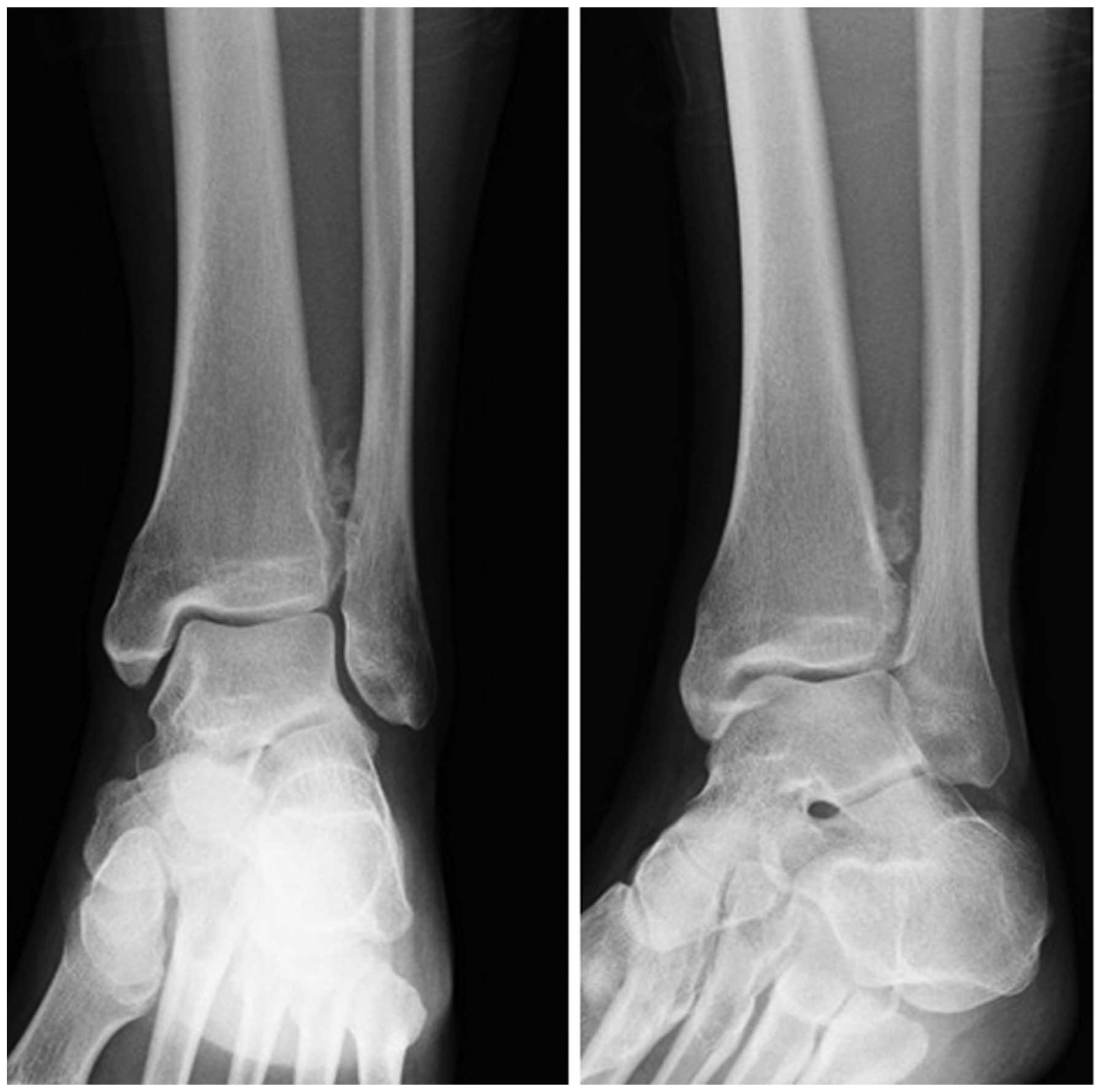
Abstract
Periosteal chondroma is a rare benign hyaline cartilage neoplasm situated on the bone surface. This is the presentation of a unique case of periosteal chondroma arising in the left distal tibial metaphysis of a 25‑year‑old female patient with a history of antecedent trauma. The physical examination revealed swelling and tenderness in the anterolateral aspect of the left distal lower limb. Plain radiographs revealed a discernible soft tissue lesion with peripheral foci of mineralization. Computed tomography scans confirmed the presence of a surface‑based mass with peripheral ossification and a thin rim of calcification. On magnetic resonance imaging, the well‑circumscribed mass exhibited intermediate signal intensity on T1‑weighted sequences and high signal intensity with foci of decreased signal intensity on T2‑weighted sequences. Contrast‑enhanced T1‑weighted sequences revealed predominantly peripheral enhancement without intramedullary involvement. Following an open biopsy, marginal excision with curettage of the underlying bone cortex was performed. Histologically, the tumor consisted of mature hyaline cartilage arranged in distinct lobules. Foci of ossification with mature bone trabeculae forming a thin shell‑like structure were identified in the periphery of the tumor. The mindbomb E3 ubiquitin protein ligase 1 labeling index was <1%. Based on these findings, the tumor was diagnosed as periosteal chondroma. There has been no evidence of local recurrence at 4 months following surgery. Despite its rarity, periosteal chondroma must be considered as a possible diagnosis when confronted with a surface‑based, mineralized lesion in the metaphysis of long bones.