Introduction
Stoma closure is considered a minimally invasive
surgery; however, surgical site infection (SSI) is a frequent
complication. Recently, the number of cases of diverting ileostomy
has increased due to the increase in the number of anal-preserving
surgeries, such as super-low anterior resection (sLAR) and
intersphincteric resection (ISR) for rectal cancer. In this study,
we focused on methods used to decrease the incidence of SSI in
stoma closure.
The incidence of wound infection following stoma
closure ranges between 2 and 41% across different studies (1,2). The most
frequent cause of wound infection is bacterial contamination of the
skin surrounding the ileostomy/colostomy due to prolonged contact
with bowel contents or due to leakage of the ileostomy contents. A
previous study in 1997 introduced a purse-string skin closure
technique (PS) for ileostomy reversal, which reduced the risk of
wound infection and resulted in smaller scars, allowing for better
cosmesis (3).
The number of studies investigating the
effectiveness of PS is currently limited. We previously used a
Penrose drainage tube under the subcutaneous tissue to prevent SSI
and have reported the efficacy of this technique in our hospital
(4).
PS was reported to be better for controlling
superficial incisional SSI (5,6) and we
introduced this technique in June, 2013. In the present study, we
examined surgical techniques by either conventional skin closure
with a drainage tube (CD) or PS and compared the incidence of
superficial incisional SSI between CD and PS in order to assess the
efficacy of the PS in ileostomy and colostomy reversal.
Patients and methods
Patients
We retrospectively reviewed the medical records of
55 consecutive patients who underwent stoma closure. A total of 26
patients who underwent PS between June, 2013 and September, 2014
were compared to 29 patients who underwent CD between October, 2011
and May, 2013 at the Osaka Medical Center for Cancer and
Cardiovascular Diseases, Japan. Medical charts were reviewed for
patient demographics, including age, gender and body mass index;
past medical history, such as the presence of diabetes, chronic
obstructive pulmonary disease, cardiovascular disease and liver
dysfunction; alcohol consumption (regular vs. moderate or
non-drinker), smoking status (smoked within 1 year prior to the
operation), medication records, including perioperative steroid
replacement; American Society of Anesthesiology (ASA) score; and
preoperative serum albumin values for the CD and PS groups.
This study was approved by the Institutional Review
Board of Osaka Medical Center for Cancer and Cardiovascular
Diseases and written informed consent was obtained from all the
patients.
Diagnosis of SSI
Wound infection was defined as the presence of
cellulitis or purulent discharge, with or without positive
bacterial growth, within 30 days after the operation (adopted from
the 1992 Centers for Disease Control and Prevention definition of
SSI) (Table I). The surgical wounds
were routinely observed and evaluated by the surgical team and
surveyed until 30 days after the operation.
 | Table I.Centers for Disease Control and
Prevention guidelines for the diagnosis of superfi cial SSI. |
Table I.
Centers for Disease Control and
Prevention guidelines for the diagnosis of superfi cial SSI.
| Characteristics of
superfcial SSI |
|---|
| 1. | Purulent drainage,
with or without laboratory confrmation, from the superfcial
incision. |
| 2. | Organisms isolated
from an aseptically-obtained culture of fuid or tissue from the
superfcial incision. |
| 3. | At least one of the
following signs or symptoms of infection: Pain or tenderness,
localized swelling, redness or heat and and superfcial incision
deliberately opened by surgeon, unless the incision is
culture-negative. |
| 4. | Diagnosis of
superfcial incisional SSI by the surgeon or attending
physician. |
Surgical technique
All the patients underwent preoperative mechanical
bowel preparation. After the induction of general anesthesia,
prophylactic antibiosis (Cefmetazole, 2 g/day) was administered. An
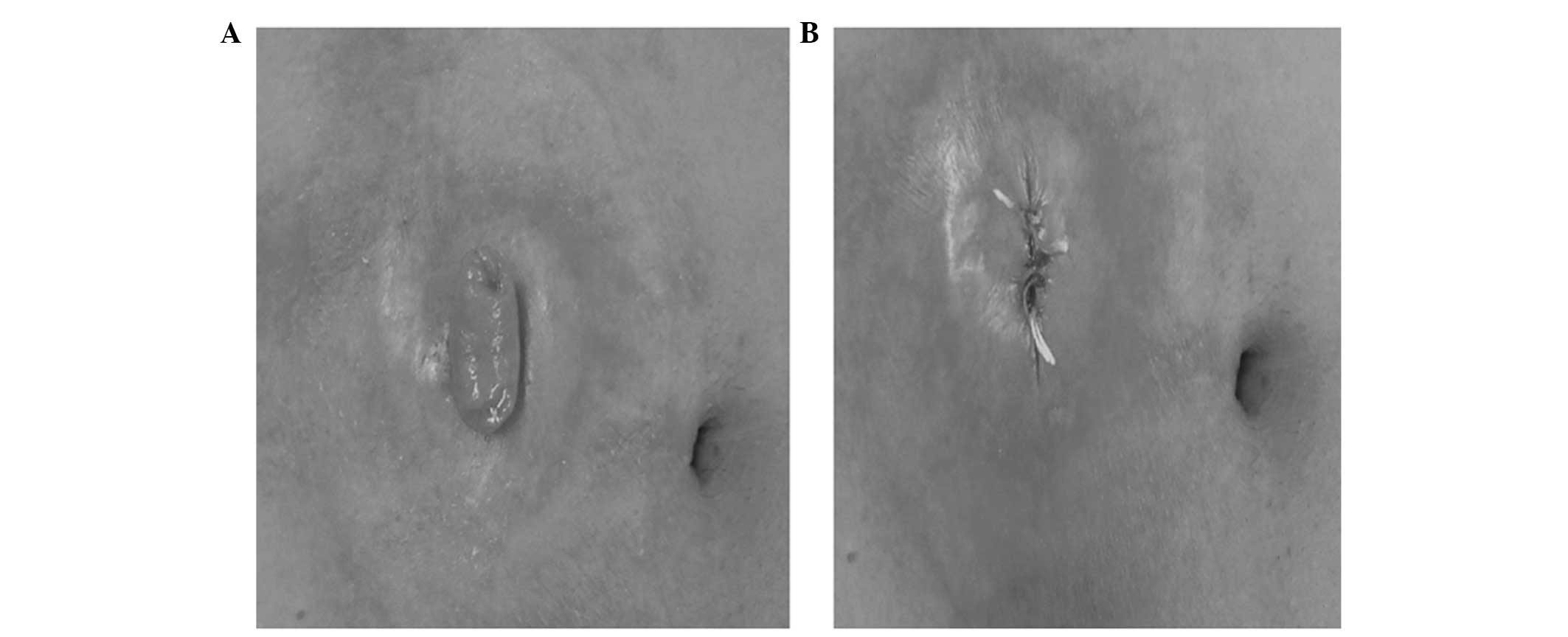
open-ileostomy/colostomy was sutured (Fig. 1A and B) and the wound was irrigated by
povidone iodine and 500 ml of saline. The skin around the sutured
stoma was resected, maintaining a ~3–5 mm margin. The intestinal
tracts leading to the stoma were extracted from the abdominal
cavity and dissection of the sutured stoma was performed, followed
by a functional end-to-end anastomosis or Albert-Lembert
anastomosis. The peritoneum and rectus fascia were closed using
Vicryl 1.0; (Johnson & Johnson Co., New Brunswick, NJ, USA).
Antibiotics were administered for 3 days postoperatively.
In the CD group, the skin site was closed primarily
with a skin stapler. At the stoma site, a Penrose drainage tube was
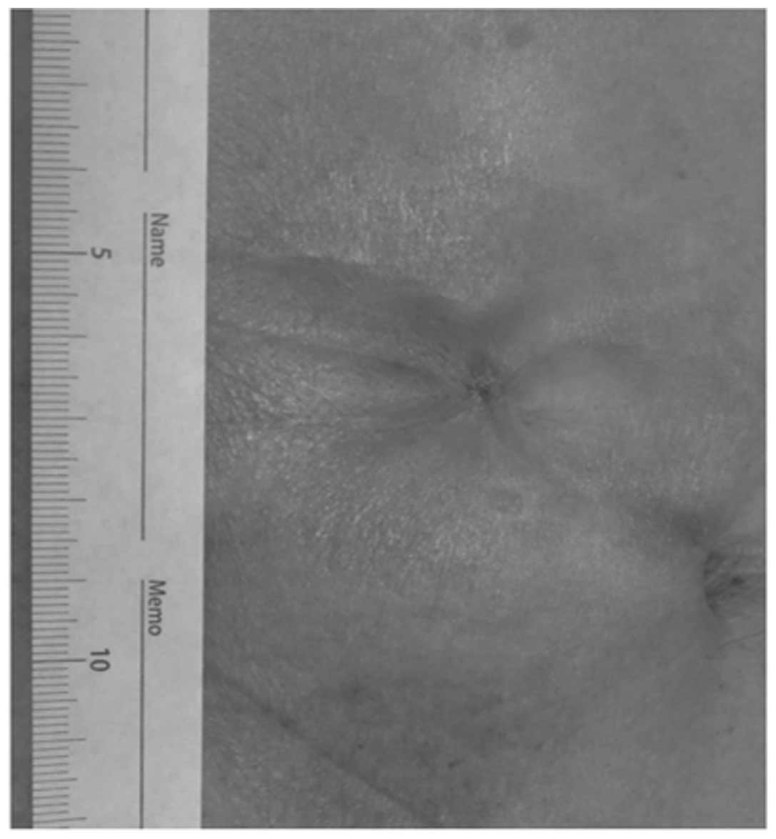
left subcutaneously. In the PS group, the dermis layer was sutured,
drawing a purse-string form using polydioxanone 3.0 suture (Johnson
& Johnson Co.). It is recommended that ~5 mm of the center of
the wound are maintained open to drain any discharge (Fig. 2).
Statistical analysis
The data presented were analyzed using the Pearson's
Chi-square and Fisher's exact tests. For continuous variables, data
were expressed as median (range). The Mann-Whitney U-test was used
for statistical comparison of different groups. P<0.05 was
considered to indicate a statistically significant difference. All
the tests were analyzed using JMP software, version 11.0 (SAS
Institute, Cary, NC, USA).
Results
Patient characteristics
The patient characteristics are summarized in
Table II. There were no significant
differences in patient characteristics, such as age, gender, body
mass index, preoperative comorbidities, ASA scores and preoperative
serum albumin values between the CD and PS groups. The
perioperative factors of the CD and PS groups are summarized in
Table III. The median operative
time of the CD group was 98 min (range, 53–213 min) and that of the
PS group 106 min (range, 60–190 min); this difference was not
statistically significant (P=0.377). In the CD group, the median
blood loss was 15 ml (range, 0–130 ml), whereas in the PS group, it
was 20 ml (range, 0–130 ml) (P=0.203). The median wound length in
the PS group was 0.5 cm, which was significantly shorter compared
to that in the CD group (6 cm) (P<0.001). As regards
postoperative complications, superficial incisional SSI in the CD
group was observed in 4 patients (13.8%); however, in the PS group,
SSI was not observed. Thus, there was a significant difference in
the incidence of superficial incisional SSI between the CD and PS
groups (P=0.049). With regard to other complications, small bowel
obstruction was observed in 2 patients (6.9%) in the CD group and 1
patient (3.8%) in the PS group. In the CD group, postoperative
bleeding was observed in 1 patient (3.8%). Anastomosis leakage was
not observed in any of the two groups. The overall incidence of
complications in the CD and PS groups was not significantly
different (P=0.313). The median postoperative hospital stay was 14
days (range, 9–50 days) in the CD group and 13 days (range, 8–29
days) in the PS group.
| Table II.Patient characteristics (N=55). |
Table II.
Patient characteristics (N=55).
| Characteristics | CD (n=29) | PS (n=26) | P-value |
|---|
| Gender |
|
| 0.738 |
| Male | 20 | 19 |
|
|
Female | 9 | 7 |
|
| Age, years | 58 (34–79) | 65 (27–80) | 0.255 |
| Body mass index | 22 (16–27) | 22 (14–29) | 0.831 |
| ASA score |
|
| NA |
| 1 | 9 | 9 |
|
| 2 | 20 | 14 |
|
| 3 | 0 | 3 |
|
| Preoperative albumin
serum value | 4.1 (3.4–5.7) | 4.1 (3.7–4.9) | 0.799 |
| Preoperative
comorbidities |
|
Diabetes | 1 | 0 | NA |
|
Cardiovascular disease | 2 | 3 | NA |
| COPD | 1 | 0 | NA |
| Steroid
use | 0 | 0 | NA |
| Alcohol
consumption | 7 | 10 | 0.251 |
|
Smoking | 7 | 6 | 0.926 |
| Liver
cirrhosis | 0 | 0 | NA |
|
Ileostomy/colostomy | 26/3 | 22/4 | NA |
| Anastomosis
(FEEA/AL) | 27/2 | 24/2 | NA |
| Table III.Perioperative factors in conventional
skin closure with a drainage tube (CD) and purse-string skin
closure (PS). |
Table III.
Perioperative factors in conventional
skin closure with a drainage tube (CD) and purse-string skin
closure (PS).
| Factors | CD (n=29) | PS (n=26) | P-value |
|---|
| Operative time,
min | 98 (53–213) | 106 (60–190) | 0.377 |
| Blood loss, ml | 15 (0–130) | 20 (0–130) | 0.203 |
| Wound length, cm | 6 (4–8) | 0.5 (0.5–1) | <0.001 |
| Complications | 5 | 2 | 0.313 |
| Wound
infection | 4 | 0 | 0.049 |
| Small
bowel obstructiona | 2 | 1 | NA |
|
Anastomosis
leakagea | 0 | 0 | NA |
|
Postoperative
bleedinga | 0 | 1 | NA |
| Postoperative
hospital stay, days | 14 (9–50) | 13 (8–29) | 0.159 |
Discussion
Wound infection (particularly, superficial
incisional SSI) is the most common complication following stoma
closure, reportedly occurring in one of every three cases (7,8). A
prospective review of the complications of ileostomy closure
reported an SSI rate of 18.3% (9).
The high incidence rate of SSI was considered to be associated with
the primary closure of these wounds. Generally, superficial
incisional SSI is not a fatal complication; however, there is a
risk of severe infection and abdominal wall scar hernia. Therefore,
we reported the utility of the Penrose drain placement under the
subcutaneous tissue for reducing the incidence of SSI (4). However, with this technique, wound
infection may not be sufficiently controlled; thus, we introduced
PS for stoma closure in order to reduce the incidence of SSI.
In the present study, the SSI rate in the CD group
was 8.3%, which is consistent with previous reports (10,11).
However, no patients developed SSI in the PS group. This finding is
in accordance with a previous report (12). PS of stoma reversal is a cosmetically
superior approach (3). A recent study
compared patients treated using PS to those treated using primary
closure (12) and found that no SSI
occurred in the PS group compared to the CD group, in which 40% of
patients developed SSI (P=0.001). These results suggest a lower
risk of SSI when PS is used. To effectively prevent SSI, it is
important to maintain the drainage hole until the discharge from
the wound is reduced (Fig. 2).
Recently, PS has been a focus of investigation with
regard to better cosmetic appearance (Fig. 3) (13,14). In
the present study, the wound length was evaluated in both groups.
PS was performed and ~0.5 cm of the wound was maintained open in
order to drain the discharge. By contrast, the patients in the CD
group required a ~6-cm linear suture. Taken together, the results
suggest that PS allows for better cosmesis in addition to the lower
risk of SSI development.
With the improvement of surgical techniques for anal
preservation by sLAR and ISR in patients with rectal cancer, the
opportunities for primary artificial anus construction have
increased. Stoma closure is associated with a high risk of wound
infection; therefore, it is important to evaluate the usefulness of
PS and open drainage system to prevent severe SSI. We are currently
conducting a randomized trial to confirm our findings and assess
additional end points, including cost-effectiveness and patient
satisfaction.
In conclusion, the present study suggests that PS is
associated with a lower risk of postoperative SSI compared to
primary skin closure, even with the placement of a drainage tube;
it also allows for better cosmesis, including a more favorable
wound length, indicating that PS may be a favorable alternative to
primary skin closure.
References
|
1
|
Hackam DJ and Rotstein OD: Stoma closure
and wound infection: an evaluation of risk factors. Can J Surg.
38:144–148. 1995.PubMed/NCBI
|
|
2
|
Wong KS, Remzi FH, Gorgun E, et al: Loop
ileostomy closure after restorative proctocolectomy: outcome in
1,504 patients. Dis Colon Rectum. 48:243–250. 2005. View Article : Google Scholar : PubMed/NCBI
|
|
3
|
Banerjee A: Pursestring skin closure after
stoma reversal. Dis Colon Rectum. 40:993–994. 1997. View Article : Google Scholar : PubMed/NCBI
|
|
4
|
Imada S, Noura S, Ohue M, et al: Efficacy
of subcutaneous penrose drains for surgical site infections in
colorectal surgery. World J Gastrointest Surg. 5:110–114. 2013.
View Article : Google Scholar : PubMed/NCBI
|
|
5
|
Mirbagheri N, Dark J and Skinner S:
Factors predicting stomal wound closure infection rates. Tech
Coloproctol. 17:215–220. 2013. View Article : Google Scholar : PubMed/NCBI
|
|
6
|
Chow A, Tilney HS, Paraskeva P, Jeyarajah
S, Zacharakis E and Purkayastha S: The morbidity surrounding
reversal of defunctioning ileostomies: a systematic review of 48
studies including 6,107 cases. Int J Colorectal Dis. 24:711–723.
2009. View Article : Google Scholar : PubMed/NCBI
|
|
7
|
Vermulst N, Vermeulen J, Hazebroek EJ,
Coene PP and van der Harst E: Primary closure of the skin after
stoma closure. Management of wound infections is easy without
(long-term) complications. Dig Surg. 23:255–258. 2006. View Article : Google Scholar : PubMed/NCBI
|
|
8
|
Williams LA, Sagar PM, Finan PJ and Burke
D: The outcome of loop ileostomy closure: a prospective study.
Colorectal Dis. 10:460–464. 2008. View Article : Google Scholar : PubMed/NCBI
|
|
9
|
Garcia-Botello SA, Garcia-Armengol J,
Garcia-Granero E, et al: A prospective audit of the complications
of loop ileostomy construction and takedown. Dig Surg. 21:440–446.
2004. View Article : Google Scholar : PubMed/NCBI
|
|
10
|
Parks SE and Hastings PR: Complications of
colostomy closure. Am J Surg. 149:672–675. 1985. View Article : Google Scholar : PubMed/NCBI
|
|
11
|
Pittman DM and Smith LE: Complications of
colostomy closure. Dis Colon Rectum. 28:836–843. 1985. View Article : Google Scholar : PubMed/NCBI
|
|
12
|
Milanchi S, Nasseri Y, Kidner T and
Fleshner P: Wound infection after ileostomy closure can be
eliminated by circumferential subcuticular wound approximation. Dis
Colon Rectum. 52:469–474. 2009. View Article : Google Scholar : PubMed/NCBI
|
|
13
|
Marquez TT, Christoforidis D, Abraham A,
Madoff RD and Rothenberger DA: Wound infection following stoma
takedown: primary skin closure versus subcuticular purse-string
suture. World J Surg. 34:2877–2882. 2010. View Article : Google Scholar : PubMed/NCBI
|
|
14
|
Lee JR, Kim YW, Sung JJ, et al:
Conventional linear versus purse-string skin closure after loop
ileostomy reversal: comparison of wound infection rates and
operative outcomes. J Korean Soc Coloproctol. 27:58–63. 2011.
View Article : Google Scholar : PubMed/NCBI
|