Fibular juxta-articular ganglion: A rare case report and literature review
- Authors:
- Xiaobo Yan
- Zhengliang Zhang
- Nong Lin
- Tao Xie
- Zhaoming Ye
-
View Affiliations
Affiliations: Department of Orthopaedic Oncology, Second Affiliated Hospital, Zhejiang University, Hangzhou, Zhejiang 310058, P.R. China, Department of Orthopaedics, Dongyang People's Hospital, Dongyang, Zhejiang 322100, P.R. China
- Published online on: August 30, 2016 https://doi.org/10.3892/mco.2016.1007
-
Pages:
590-592
Metrics:
Total
Views: 0 (Spandidos Publications: | PMC Statistics:
)
Metrics:
Total PDF Downloads: 0 (Spandidos Publications: | PMC Statistics:
)
This article is mentioned in:
Abstract
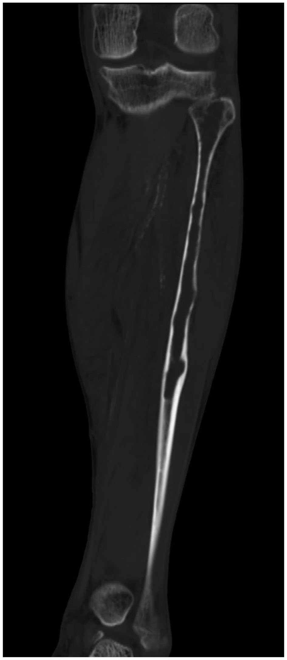
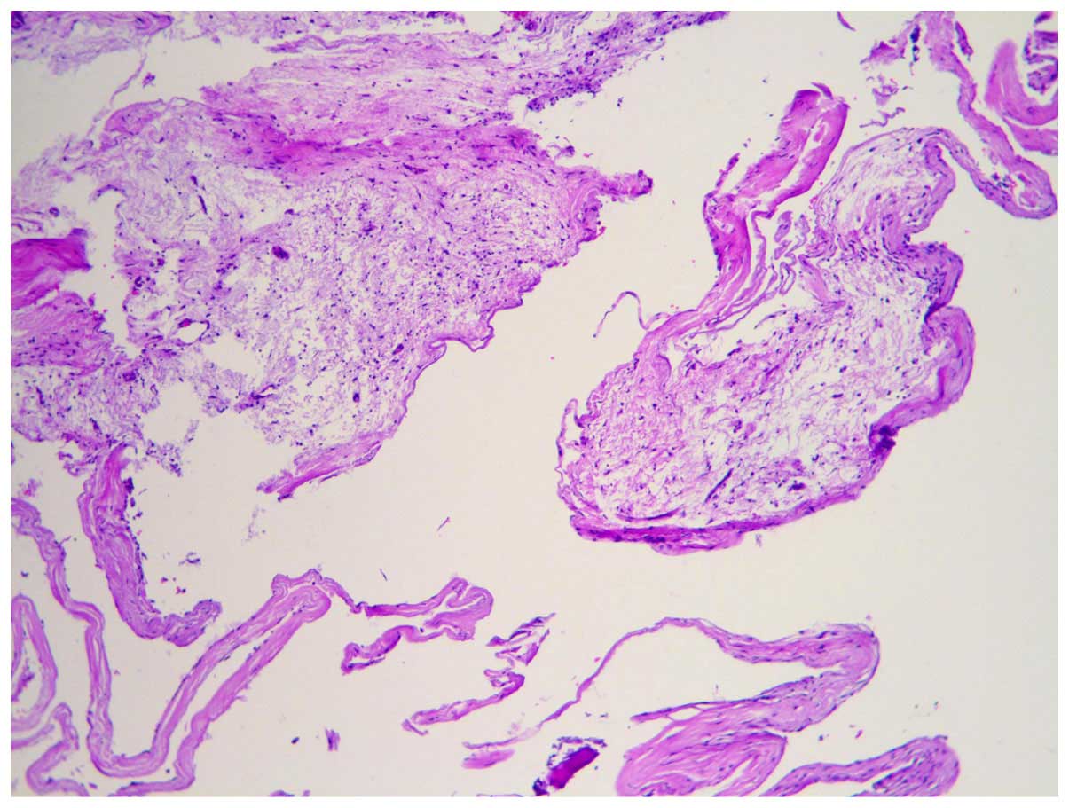
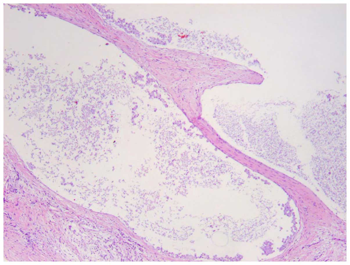
This is the case report of a 65-year-old man who experienced left calf pain after spraining his left ankle. On magnetic resonance imaging (MRI), a well‑defined fluid collection was identified, with low intensity on T1‑weighted images and very high intensity on T2-weighted images. On certain slices, a soft tissue mass in the proximal fibula was identified. Following resection of the fibular lesion, histological examination confirmed a benign tumor with cystic elements, described in the current pathology and radiology literature as juxta‑articular myxoma. This is a rarely encountered but well‑recognised cystic lesion, commonly developing around the knee, that is often misdiagnosed as ganglion cyst, synovial lipoma, lipoma arborescens and pigmented or non‑pigmented villonodular synovitis. Given its more cellular nature and thicker encapsulation, juxta‑articular myxoma may be differentiated from ganglion cyst on MRI with a high index of suspicion, and it is crucial that it is recognized due to its high rate of recurrence. In the present case, the localization of this lesion within the fibula was uncommon, and there was also a soft tissue mass identified outside the bone. Wide resection of the lesion was performed. Recovery was uneventful and the patient remains symptom- and recurrence-free at 8-months follow‑up.
View References
1
|
Aoki Y, Miyamoto K and Harada Y: A case of
ganglion in acetabulum. Kanto Soc Orthop Traumatol. 29:257–261.
1998.
|
2
|
Arao M, Otani T, Funasaki H, Ono N, Katou
T and Serizawa Y: Intraosseous ganglion of the scapula: a case
report. Kanto Soc Orthop Traumatol. 34:239–242. 2003.
|
3
|
Aritomi K, Kusunose K, Kayaoka M, Tomita
Y, Miyazaki H and Hayashi R: Intraosseous ganglion of the scaphoid:
A case report. Orthop Surg. 55:1185–1187. 2004.
|
4
|
Williams HJ, Davies AM, Allen G, Evans N
and Mangham DC: Imaging features of intraosseous ganglia: A report
of 45 cases. Eur Radiol. 14:1761–1769. 2004. View Article : Google Scholar : PubMed/NCBI
|
5
|
Ferkel RD, Field J, Scherer WP, Bernstein
ML and Kasimian D: Intraosseous ganglion cysts of the ankle: A
report of three cases with long-term follow-up. Foot Ankle Int.
20:384–388. 1999. View Article : Google Scholar : PubMed/NCBI
|
6
|
Sedeek SM, Choudry Q and Garg S:
Intraosseous ganglion of the distal tibia: Clinical, radiological,
and operative management. Case Rep Orthop.
2015:7592572015.PubMed/NCBI
|
7
|
Schajowicz F, Sainz M Clavel and Slullitel
JA: Juxta-articular bone cysts (intra-osseous ganglia): a
clinicopathological study of eighty-eight cases. J Bone Joint Surg
Br. 61:107–116. 1979.PubMed/NCBI
|
8
|
Wei XJ, Wang XF, Wang XG, et al:
Juxta-articular bone cyst(intra-osseous ganglion)-report of one
rare multiple case in femur. Orthop J Chin. 10:1048–1050. 2002.
|
9
|
Crane AR and Scarano JJ: Synovial cysts
(ganglia) of bone. Report of two cases. J Bone Joint Surg Am.
49:355–361. 1967.PubMed/NCBI
|